Jan 28, 2022
X-ray Micro Computed Tomography (MicroCT)
- Marta Wawrzyniak1,
- Nathalie Weber1,
- Simon Blanchoud1
- 1University of Fribourg
- Blanchoud lab, UNIFR


Protocol Citation: Marta Wawrzyniak, Nathalie Weber, Simon Blanchoud 2022. X-ray Micro Computed Tomography (MicroCT) . protocols.io https://dx.doi.org/10.17504/protocols.io.b35aqq2e
License: This is an open access protocol distributed under the terms of the Creative Commons Attribution License, which permits unrestricted use, distribution, and reproduction in any medium, provided the original author and source are credited
Protocol status: Working
We use this protocol and it’s working
Created: January 21, 2022
Last Modified: January 28, 2022
Protocol Integer ID: 57218
Keywords: Colonial tunicates, MicroCT, tomography, ascidians, microct for comparative morphology, colonial tunicate, microct, 3d imaging, tissue, simple staining method, micro
Abstract
This protocol has been successfully used with colonial tunicates. It has been adapted to our needs based on the following publication: " MicroCT for comparative morphology: simple staining methods allow high-contrast 3D imaging of diverse non-mineralized animal tissues " by Brian D Metscher.
Materials
4% PFA
PTA
IKI
Ethanol
low melting agarose
fixing paste (e.g. dental wax)
pipette tips 200uL or 1mL
10x PBS, pH 7.2 (store for up to 2 moths at RT)
| A | B | C | |
| Na2HPO4*2H2O | 0.1M | 17.8g | |
| KH2PO4 | 18mM | 2.4g | |
| NaCl | 1.4M | 80g | |
| KCl | 27mM | 2g | |
| water | 1000mL |
10x PBS (amounts calculated for 1L)
Troubleshooting
Fixation
1h 30m
Wash twice in 3.3x PBS for 00:30:00 .
30m
Rinse tree times with 1x PBS and leave in 1x PBS for 01:00:00 .
1h
Staining
4d
Detach the colony with a razor blade and put in a 2mL tube containing 1 mL PTA /0.6 mL IKI/ 0.4 mL Ethanol.
Incubate for 96:00:00 at Room temperature on linear shaker (low speed), protect from light.
4d
Rinse quickly 3-4 times with 1x PBS.
Mounting
Prepare 10 mL of 0.5 % volume low melting agarose.
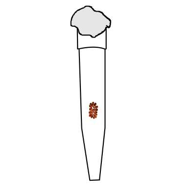
When the agarose has cooled down a bit, pipette 125-150 µL if using a 200ul tip or 750-800 µL if using a 1mL tip (the tip size depends on the animal size).
Carefully place your colony vertically inside the tip (it should be as flat as possible, avoid folds).
Pipette some more agarose in order to cover the animal entirely.
Place the tip into a beaker filled with ice-cubes so that the agarose becomes solid.
Cover the tip with fixing paste.
Store at 4 °C or proceed to the next step.
Mount the tip on the tomograph support using fixing paste in such way that it is perfectly straight.
Place it inside the tomograph.
Follow the tomograph's manual and proceed to scan.
After scanning, the tip with the sample can be stored at 4 °C for several weeks and eventually can be reused.