Protocol Citation: Rajvi Javeri, Naveen Ouellette, Molly Logsdon, Laura Roy, Holly Myers, Judith Baka, Kevin Cao, Andrew Recknagel, Jayaram Chandrashekar 2026. Whole Mouse Brain Delipidation, Immunolabeling, and Expansion Microscopy. protocols.io https://dx.doi.org/10.17504/protocols.io.n92ldpwjxl5b/v2Version created by Avery Weaver
Manuscript citation:
Citation
Glaser A, Chandrashekar J, Vasquez S, Arshadi C, Javeri R, Ouellette N, Jiang X, Baka J, Kovacs G, Woodard M, Seshamani S, Cao K, Clack N, Recknagel A, Grim A, Balaram P, Turschak E, Hooper M, Liddell A, Rohde J, Hellevik A, Takasaki K, Erion Barner L, Logsdon M, Chronopoulos C, de Vries SEJ, Ting JT, Perlmutter S, Kalmbach BE, Dembrow N, Tasic B, Reid RC, Feng D, Svoboda K (2025). Expansion-assisted selective plane illumination microscopy for nanoscale imaging of centimeter-scale tissues. eLife.
License: This is an open access protocol distributed under the terms of the Creative Commons Attribution License, which permits unrestricted use, distribution, and reproduction in any medium, provided the original author and source are credited
Protocol status: Working
We use this protocol and it's working
Created: May 19, 2026
Last Modified: June 08, 2026
Protocol Integer ID: 317481
Keywords: Expansion Microscopy, Whole Brain, Tissue Clearing, Delipidation, Hydrogel, Immunolabeling, Light Sheet, Clearing, Antibody, SPIM, expansion microscopy the mammalian brain, wide single neuron reconstruction, resolution selective plane illumination microscopy, expanded whole mouse brain, selective plane illumination microscopy, contrast imaging of entire brain, isotropic expansion of whole mouse brain, expansion microscopy, whole mouse brain delipidation, whole mouse brain, whole brain data set, mammalian brain, spim microscope without need, complete axonal morphology of individual neuron, spim microscope, neuronal axon, entire brain, description of the brain, individual neuron, imaging, brain area, brain region, wide neural circuit, obtaining brain, tracing complete axonal morphology, neuron, individual axon collateral, contrast imaging, brain, wide neural circuits during normal behavior, distinct neuron type, resolution, spim microscope without the need, complete axonal morphology, mouse
Funders Acknowledgements:
Allen Institute
Abstract
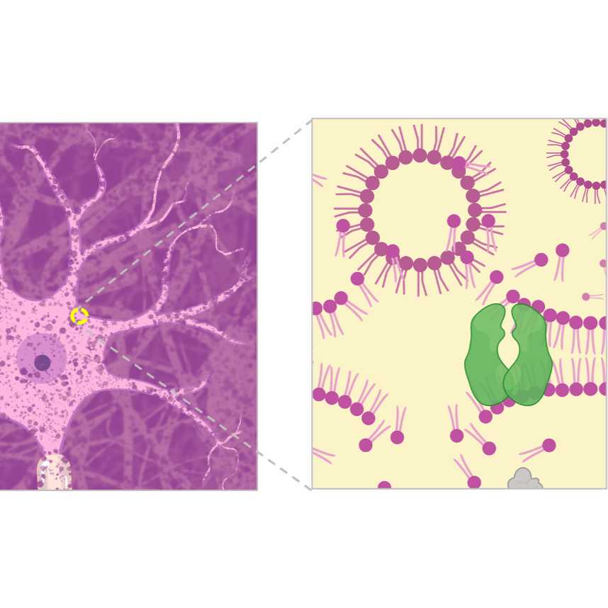
The mammalian brain contains approximately 1,000 brain areas, and each brain area contains multiple (up to 100) cell types. Neurons in one brain region can send projections to dozens of target regions, and distinct neuron types could project to different combinations of target regions. The enumeration and description of the brain’s cell types and their brain-wide connectivity is foundational for understanding how neural activity is routed across brain-wide neural circuits during normal behavior and how these processes are dysregulated in mental disorders.
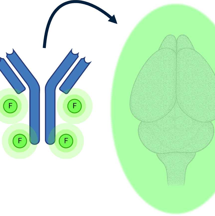
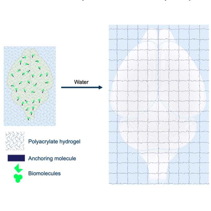
Obtaining brain-wide single neuron reconstructions requires high-resolution, high-contrast imaging of entire brains—neuronal axons travel many centimeters (in the mouse) while individual axon collaterals could be finer than 100nm. Here, we present an integrated protocol for labeling and isotropic expansion of whole mouse brains that results in optically clear specimens ideally suited for high-resolution selective plane illumination microscopy (SPIM) imaging. Pipeline steps are modular, and the protocol is extensible to other large-volume clearing and expansion applications.
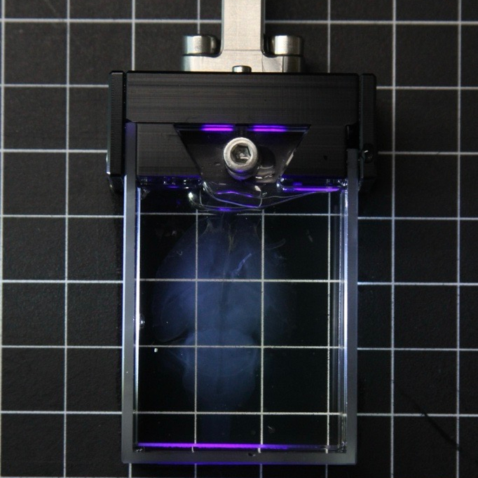
We have imaged expanded whole mouse brains generated using this protocol on our custom-built ExA-SPIM microscope without the need for any tissue slicing. These whole brain data sets are being used for tracing the complete axonal morphology of individual neurons.
DuraDigest enzyme cocktail for brain surface clearing
Combine the following reagents at Room temperature . Prepare a fresh solution before each use.
Reagent
Amount
Final Concentration
Collagenase Type IV
1 mL
0.1 mg/mL
Dispase I
500 uL
0.5 U/mL
10X PBS
850 uL
dd H2O
7.65 mL
Total
10 mL
10 mM Phosphate Buffer pH 8.3
Combine the following reagents, adjust pH to 8.3.
Reagent
Amount
Final Concentration
1M Phosphate Buffer
5 mL
10 mM
Milli-Q water
495 mL
Total
500 mL
SBiP Solution: 0.08% SDS, 16% 2-methyl-2-butanol, 8% 2-propanol, in H2O
Combine the following reagents on ice. Use a fume hood when adding 2-methyl-2-butanol and 2-propanol. Mix On ice until solution is uniform and clear. Store immediately at 4 °C until ready for use. Use each batch within a month for best effect.
Combine the following reagents and stir at Room temperature until fully dissolved, store at4 °C.
Reagent
Amount
Final Concentration
10X PBS
50 mL
1X
Triton X-100
500 uL
0.1%
Tween 20
250 uL
0.05%
5% Sodium azide
4 mL
0.04%
Milli-Q Water
up to 500 mL
Total
500 mL
Post Secondary Staining PTxw: 0.1% Tween 20, 0.1% Triton X-100, 0.04% NaN3, 200mM NaCl in PBS
Combine the following reagents and stir at Room temperature until fully dissolved, store at Room temperature .
Reagent
Amount
Final Concentration
10X PBS
96 mL
1X
5% Sodium Azide
8 mL
0.04%
5M Sodium Chloride Solution
40 mL
200mM
Milli-Q Water
864 mL
Triton X-100
1000 uL
0.1%
Tween 20
1000 uL
0.1%
Total
1000 mL
MBS solution: 100mM MES Buffered Saline, 150mM NaCl, pH 6.0
Combine the following reagents at Room temperature until fully dissolved. Adjust to pH 6. Store for a few months atRoom temperature.
Reagent
Volume
Final Concentration
MES
1 g
100 mM
5M NaCl
1.5 mL
150 mM
10N NaOH
200 uL
Milli-Q Water
48 mL
Total
50 mL
10 mg/mL Acryloyl-X (AcX) in DMSO:
To make 10 mg/mL AcX, add DMSO directly to the bottle of AcX. Vortex to dissolve. Aliquot and store at -20 °C in a desiccated environment. Aliquots should be used within one month.
Reagent
Volume
Final Concentration
Acryloyl X
5 mg
10 mg/mL
Dimethyl Sulfoxide (DMSO)
500 uL
10% (w/v) VA-044 (polymerization initiator):
Combine the following reagents and stir On ice until dissolved. Store at -20 °C up to one month.
Reagents
Volume
Final Concentration
VA-044
1 g
10%
Milli-Q water
10 mL
Stock X (Monomer Solution) Preparation:
To make the Stock X solution, the following stock solutions must be prepared in advance:
50% (w/v) Acrylamide
2% (w/v) N,N Methylene-bis-acrylamide
4.04M Sodium acrylate (2 options: made from acrylic acid or powder form)
50% (w/v) Acrylamide:
Combine the following reagents and stir until dissolved. Store at -20 °C up to one month.
Reagents
Volume
Final Concentration
Acrylamide
5 g
50%
Milli-Q water
10 mL
2% (w/v) N,N Methylene-Bis-Acrylamide
Combine the following reagents and stir until dissolved. Store at -20 °C up to one month.
Reagents
Amount
Final Concentration
N,N Methylene-bis-acrylamide
0.2 g
2%
Milli-Q water
10 mL
4.04M Sodium Acrylate
Add 22.5 mL milli-Q water into a 250 mL glass bottle and cool the solution down to 0 °C on ice. Slowly add in 27.5 mL acrylic acid with stirring until fully mixed. Cover the bottle to the neck with ice and then, with stirring, add 36 mL 10N NaOH over the course of 00:10:00. Make sure to keep the solution at 0 °C.
Insert a pH meter and begin adding 1N NaOH in 1 mL increments until pH 7.6 – 8.0. Keep track of the volume needed to reach this range.
Once desired pH is reached, let the solution warm to Room temperature and check the pH to make sure it is still in correct range. Add water to a final volume of 100mL and store at -20 °C.
Reagents
Amount
14.6M Acrylic acid
27.5 mL
Milli-Q water
22.5 mL + extra to reach 100mL
10N NaOH
36 mL
1N NaOH
~5-10 mL
4.04M Sodium Acrylate (from powder form)
Combine the following reagents and stir until dissolved. Store at -20 °C up to one month.
Reagents
Amount
Final Concentration
Sodium acrylate
18.99 g
4.04 M or 38%
Milli-Q water
50 mL
Note
Powder Sodium Acrylate can be used. However, a yellow solution indicates low purity. If this is observed, discard and use a different batch.
Stock X (Monomer Solution)
Combine the following On ice. Aliquot and store at -20 °C for up to one month.
Combine the following reagents. Aliquot and store at -20 °C for several months.
Reagent
Amount
Final Concentration
1M Tris-HCl pH 8
2 mL
50 mM
10% Triton X-100
2.5 mL
0.5%
5M NaCl
500 uL
50 mM
0.5M EDTA
100 uL
1 mM
10% SDS
1.5 mL
0.3%
Milli-Q Water
42.9 mL
Total
50 mL
0.05X SSC: 20X SSC, Milli-Q Water
Combine the following reagents and stir at Room temperature until fully dissolved, store at Room temperature
A
B
20X SSC
5 mL
Milli-Q Water
1995 mL
Total
2000 mL
10mM Ascorbic Acid, pH 7.0
Combine the following reagents and stir at Room temperature until fully dissolved, adjust pH to 7.0 and then store at Room temperature. Make fresh before each use.
Reagent
Amount
Asorbic Acid
3.52g
Milli-Q Water
2000 mL
Total
2000 mL
Safety warnings
Tetrahydrofuran (THF) and dichloromethane (DCM) are toxic and carcinogenic. THF is flammable. When exposed to air, THF may form explosive peroxides if concentrated by distillation or evaporation. Test for peroxide formation or discard THF after 1 year. Perform the steps that involve these reagents under the fume hood. Dispose of THF and DCM in a hazardous waste stream. Wear lab coat, safety goggles or glasses, and chemical resistant gloves (7.8 MIL). If these solvents contact your gloves, remove immediately and don new gloves.
2-methyl-2-butanol and 2-propanol are corrosive and flammable. Perform the steps that involve these reagents under the fume hood. Dispose of 2-methyl-2-butanol and 2-propanol in a hazardous waste stream. Wear a lab coat, safety goggles or glasses, and gloves.
Sodium azide may be harmful if inhaled. It may cause respiratory tract, skin, and eye irritation and may be fatal if absorbed through skin or swallowed. Sodium azide can react with metal spatulas and metal lab equipment to form shock sensitive salts. Sodium azide reacts with lead, copper, silver, gold and metal halides to form heavy metal azides which are shock sensitive and explosive. Additionally, contact with acids liberates toxic gas. Dispose of sodium azide in a hazardous waste stream. Wear a lab coat, safety goggles or glasses, and gloves.
Acrylamide powders and solutions are toxic if swallowed, inhaled, or absorbed through the skin. It is a mutagen, teratogen and a carcinogen. Dispose of acrylamide and any contaminated consumables in a hazardous waste stream. Wear a lab coat, safety goggles or glasses, and gloves.
PTxw contains harmful chemicals like 5% Sodium Azide. Use appropriate gloves and PPE while handling, avoid contact with skin or clothing, and dispose of it properly in a hazardous waste stream.
Ethics statement
The protocols.io team notes that research involving animals and humans must be conducted according to internationally-accepted standards and should always have prior approval from an Institutional Ethics Committee or Board.
Protocol Overview
10w
This protocol prepares a whole mouse brain for expansion microscopy (ExM). Methods of tissue processing include organic and aqueous delipidation, immunolabeling, ExM (gel embedding and expansion), and mounting the sample in the imaging chamber.
Durotomy and Enzyme Treatment
1h 5m
Durotomy
After perfusion and dissection, brains may contain dura on the cortex and spinal cord. It is essential to remove dura to ensure better antibody penetration into the sample properly and to ensure that no gaps form between the brain surface and the hydrogel during gelation.
Durotomy is performed by teasing apart the dura using fine forceps and angled scissors under a stereoscopic dissection microscope. It is important to keep the brain moist during this procedure. This is done by placing the brain sample in a small petri dish filled with 1x PBS.
30m
Enzyme Treatment
After performing durotomy, brain samples are treated with low concentrations of DuraDigest enzyme cocktail consisting of Collagenase Type IV and Dispase I to clean the brain surface and clear residual debris.
The DuraDigest solution is prepared fresh in 1x PBS before each use.
Transfer the solution to a small petri dish and place an absorbable gelatin sponge in the petri dish for 00:05:00 or until the sponge swells and appears to be saturated.
5m
Place the soaked sponge in another petri dish. Transfer the brain sample onto the sponge and wrap the brain for 00:10:00.
Enzymes are not added directly onto the brain surface to prevent unwanted digestion of the tissue.
10m
Wash the brain thoroughly with 1x PBS 4 times at 00:05:00 intervals, rotating on a nutator at Room temperature for each wash.
20m
The brain sample is now ready for tetrahydrofluran/dichloromethane delipidation.
Tetrahydrofuran / Dichloromethane Delipidation
1w 1d
Reference Tetrahydrofuran and Dichloromethane Delipidation of a Whole Mouse Brain protocol.
After immunolabeling, brains are screened using the MVX10 Stereo Microscope to ensure that the sample is appropriately labelled. Brains are also screened again under the stereoscopic dissection microscope to remove any additional pieces of dura that remained after the durotomy step.
Tissue quality should be assessed thoroughly at this stage, as no changes can be made once the gelation process commences.
Gelation and Digestion
3w
Reference Gelation and Digestion of a Whole Mouse Brain protocol.
Glaser A, Chandrashekar J, Vasquez S, Arshadi C, Javeri R, Ouellette N, Jiang X, Baka J, Kovacs G, Woodard M, Seshamani S, Cao K, Clack N, Recknagel A, Grim A, Balaram P, Turschak E, Hooper M, Liddell A, Rohde J, Hellevik A, Takasaki K, Erion Barner L, Logsdon M, Chronopoulos C, de Vries SEJ, Ting JT, Perlmutter S, Kalmbach BE, Dembrow N, Tasic B, Reid RC, Feng D, Svoboda K. Expansion-assisted selective plane illumination microscopy for nanoscale imaging of centimeter-scale tissues.