Oct 31, 2025
Whole-mount immunofluorescence labelling and optical clearing of spheroids
Peer-reviewed method
- Larisa Tratnjek1,
- Aleksandar Janev1,
- Nataša esnik1,
- Uroš erkvenik1,2,
- Mateja Erdani Kreft1
- 1University of Ljubljana, Faculty of Medicine, Institute of Cell Biology;
- 2University of Ljubljana, Biotechnical Faculty, Department of Biology
- PLOS ONE Lab ProtocolsTech. support email: [email protected]


Protocol Citation: Larisa Tratnjek, Aleksandar Janev, Nataša esnik, Uroš erkvenik, Mateja Erdani Kreft 2025. Whole-mount immunofluorescence labelling and optical clearing of spheroids. protocols.io https://dx.doi.org/10.17504/protocols.io.8epv5kj95v1b/v1
Manuscript citation:
License: This is an open access protocol distributed under the terms of the Creative Commons Attribution License, which permits unrestricted use, distribution, and reproduction in any medium, provided the original author and source are credited
Protocol status: Working
We use this protocol and it's working
Created: August 06, 2025
Last Modified: October 31, 2025
Protocol Integer ID: 224177
Keywords: Spheroids, three-dimensional in vitro model, whole-mount immunofluorescence labelling, optical clearing, confocal microscopy, bladder cancer, protein localization, spatial distribution, mount immunofluorescence labelling of spheroid, spheroids from other cell type, optical clearing of spheroid, small size of spheroid, using spheroid, human urothelial cell line, spatial distribution within intact spheroid, spheroid, intact spheroid, mount immunofluorescence labelling, immunofluorescence labelling, complex tumour microenvironment, tumour microenvironment, confocal microscopy analysis, resolution confocal microscopy analysis, dimensional spheroid model, crucial in cancer research, immunofluorescence, detailed visualization of protein expression pattern, microscopy analysis, cancer research, tissue origin, protein expression pattern
Funders Acknowledgements:
Slovenian Research and Innovation Agency ARIS
Grant ID: P3-0108
Slovenian Research and Innovation Agency ARIS
Grant ID: J7-2594
Slovenian Research and Innovation Agency ARIS
Grant ID: MRIC UL IP-0510
Abstract
Three-dimensional spheroid models are crucial in cancer research as they more accurately recapitulate the complex tumour microenvironment, including oxygen and nutrient gradients and cell–cell interactions, compared to conventional two-dimensional cultures. However, the small size of spheroids poses challenges in handling and processing for microscopic analysis.
This protocol provides a detailed procedure for whole-mount immunofluorescence labelling of spheroids combined with optical clearing of spheroids, enabling high-resolution confocal microscopy analysis. The workflow is demonstrated using spheroids derived from normal (SV-HUC-1) and cancerous (T24) human urothelial cell lines but can be readily adapted to spheroids from other cell types and tissue origins. It allows for detailed visualization of protein expression patterns and their spatial distribution within intact spheroids.
Materials
The following materials and reagents were used:
1 mL and 200 µL micropipette tips – Brand, Germany (Cat. No.: 732012 and 732028)
35 mm glass-bottom dish – MatTek Life Sciences (Cat. No.: P35G-1.5-14-C)
Anti-E-cadherin antibody – BD Biosciences, USA (Cat. No.: 610182)
Anti-Ki-67 antibody – Abcam, United Kingdom (Cat. No.: 16667)
Anti-N-cadherin antibody – Cell Signaling Technology, USA (Cat. No.: 13116)
Benzyl alcohol – Sigma-Aldrich, USA (Cat. No.: 305197)
Benzyl benzoate – Sigma-Aldrich, USA (Cat. No.: B6630)
Bovine Serum Albumin (BSA) – Sigma-Aldrich, USA (Cat. No.: A2153)
Microcentrifuge tubes (0.5 mL and 1.5 mL) – LLG Labware, United Kingdom (Cat. No.: 9409026 and 6.490 852)
Ethanol (100%) – Carlo Erba Reagents, France (Cat. No.: 4146072)
Glutaraldehyde – Serva, Germany (Cat. No.: 23114.01)
Goat anti-mouse Alexa Fluor 488 and 555 – Molecular Probes, Thermo Fisher Scientific, USA (Cat. No.: A11001 and A21422)
Goat anti-rabbit Alexa Fluor 488 and 555 – Molecular Probes, Thermo Fisher Scientific, USA (Cat. No.: A11008 and A21428)
Goat Serum (Normal) – Agilent Technologies, Singapore (Cat. No.: X0907)
Hoechst 33342 – Thermo Fisher Scientific (Cat. No.: 62249)
Micro-slide 8-well glass-bottom slides – Ibidi, Germany (Cat. No.: 80826)
Paraformaldehyde – Sigma-Aldrich, Germany (Cat. No.: 158127)
Pasteur pipette (3 mL) – Brand, Germany (Cat. No.: 747765)
Phosphate-buffered saline (PBS) – prepared from:
Potassium chloride – Merck (Sigma-Aldrich), Germany (Cat. No.: 104936)
Disodium hydrogen phosphate – Merck (Sigma-Aldrich), Germany (Cat. No.: 106580)
Potassium dihydrogen phosphate – Merck (Sigma-Aldrich), Germany (Cat. No.: 104873)
Sodium chloride – Chem-Lab, Belgium (Cat. No.: CL00.1429)
Sterile 96-well ultra-low attachment U-shaped bottom microplates – Corning, USA (Cat. No.: 7007)
Sterile surgical blades (BB510) – B. Braun (Cat. No.: 4512201760)
Triton X-100 – Sigma-Aldrich, USA (Cat. No.: T8787)
Tween-20 – Sigma-Aldrich, USA (Cat. No.: P7949)
The following equipment was used:
Bench-mounted fume cupboard System Delta 30 – Wesemann, Germany
Centrifuge MyFuge Mini – Benchmark Scientific, USA
Confocal microscope LSM900 – Carl Zeiss, Germany
Laminar air-flow cabinet M182 (II) – Iskra Pio, Slovenia
Orbital shaker KS 260 – IKA, Germany
Scale AE 163 – Mettler Toledo, Switzerland
Stereomicroscope SMZ800 – Nikon, Japan
VorTemp 56 Microplate Shaking Incubator – Labnet International, Inc., USA
Protocol for the formation of normal and cancer urothelial spheroids
1w
SV-HUC-1 and T24 spheroids were grown in 96-well ultra-low attachment U-shaped bottom microplates (Corning, New York, NY, USA) and incubated in a humidified incubator at 5% CO2 and 37°C. Seeding densities of 100,000 cells per well (in 200 µL of culture medium) were used to generate spheroids. Spheroids were grown for 7 days prior to their preparation for whole mount immunofluorescence analysis.
Note
During seeding, ensure that pipette tips do not touch the bottom or sides of the wells to avoid damaging the surface coating of the ultra-low attachment U-shaped bottom microplates.
Note
Spheroid loss during washing, solution exchanges, and procedures such as paraffin embedding is expected and correlates with operator experience level and handling technique precision. Always process 20–30% excess spheroids beyond experimental requirements to compensate for potential losses.
1w
Protocol for immunofluorescence labelling and optical clearing for whole-mount spheroid imaging
2d 12h 10m
To optimize fluorescence preservation, we describe a protocol adapted from Smyrek I. and Stelzer EH., 2017, Biomed Opt Express., 2017;8(2):484-499, originally used for light sheet microscopy.
Spheroid transfer and collection
3.1 Use a 1 mL micropipette fitted with tip, or a 3 mL plastic Pasteur pipette to transfer the spheroids from the 96-well low-attachment U-bottom cell culture microplate to a 1.5 mL microcentrifuge tube filled with cold 4% (w/v) formaldehyde in PBS (pH 7.2–7.4).
Note
To prevent spheroids from adhering to the pipette wall and to ensure the maximal transfer, pre-coat the micropipette tips or Pasteur pipette with 1% BSA in PBS by dipping their full length of the tip in 1% BSA in PBS.
Note
Cold fixative (4 °C) was used in the experiments, which improves preservation of phosphorylated proteins and other sensitive antigens in tissue for immunolabelling, compared to warm fixation (protocol adapted from Lerch et al.,2020, Scientific Reports;10(1):2147).
3.2 Quickly aspirate the culture medium and spheroid with the pipette. You should be able to see the spheroid inside the tip. If it is not there, return the medium to the well and aspirate again until you can visually confirm the presence of the spheroid.
3.3 Allow the spheroid to settle at the bottom of the 1 ml tip or Pasteur pipette, then pipette it into the microcentrifuge tube with fixative.
Note
You can transfer several spheroids (3-5) into a single centrifuge tube or work with individual spheroids.
30m
Fixation
4.1 Fix the spheroids with cold 4% (w/v) formaldehyde in PBS (pH 7.2 – 7.4) for 30 minutes at room temperature, ensuring that the spheroids are completely submerged in the fixative.
Note
Test different fixation times (e.g., 15, 30, and 60 minutes) to determine the optimal time for spheroid preservation without over-fixation, which can affect antibody penetration.
4.2 Wash the spheroids with 500 μL of sterile PBS five times for 5 minutes, at room temperature on a shaker set to 800 rpm (1.07 × g, orbital diameter 3 mm).
Note
Ensure that the shaker platform provides uniform motion and a controlled temperature environment. Equivalent orbital shakers from various manufacturers (e.g., VorTemp 56 Microplate Shaking Incubator, Labnet International) can be used, provided the shaking parameters are comparable.
Note
To change the solution in which the spheroid is submerged in a microcentrifuge tube, gently aspirate as much of the solution as possible with a 200 µL/1 mL micropipette fitted with a tip without disturbing the spheroid, and add the appropriate volume of the new solution. However, to prevent accidental spheroid loss, maintain 10-20μL of residual liquid at the container bottom during solution exchanges
Note
When performing solution exchanges, first allow spheroids to settle at container bottom. For accelerated settling: tubes may be gently spun at 2000×g (6000 rpm) for 5 seconds to ensure complete spheroid sedimentation. Visually confirm the presence of spheroids after each step.
1h
Immunofluorescence labelling
The protocol described below includes immunofluorescence labelling of E-cadherin, N-cadherin and Ki-67.
5.1 Block the samples by incubation in 500 μL of blocking buffer (0.1% BSA, 0.2% Triton X-100, 0.05% Tween-20, 10% normal goat serum in PBS) per 1.5 mL microcentrifuge tube for 90 minutes at room temperature on a shaker set to 800 rpm (1.07 × g, orbital diameter 3 mm).
5.2 Remove the blocking buffer and incubate the spheroids with 400 μL of primary antibodies (mouse anti-E-cadherin (1:100), rabbit anti-N-cadherin (1:100) or rabbit anti-Ki-67 (1:100)), diluted in 1% BSA in PBS for 20 hours at 37 °C on a shaker set to 800 rpm (1.07 × g, orbital diameter 3 mm).
Note
Ideally, primary antibodies should be validated in 2D models to ensure compatibility.
Note
For thicker spheroids, consider longer incubation times.
Note
During shaking, ensure the spheroids do not settle at the bottom of the microcentrifuge tube.
5.3 Wash the spheroids with 500 μL of sterile PBS three times for 10 minutes each at room temperature in darkness on a shaker set to 800 rpm (1.07 × g, orbital diameter 3 mm).
Note
Before removing the solution in which spheroids are immersed, spin the microcentrifuge tube with the spheroids briefly for 5 seconds at 2000×g (6000 rpm) to ensure the spheroids settle at the bottom of the tube. Visually confirm the presence of spheroids after each step.
5.4 Incubate the spheroids in 400 μL of secondary antibodies (a mixture of goat anti-rabbit and anti-mouse secondary antibodies (Alexa Fluor 488 and 555; Invitrogen) diluted 1:400 in 1% BSA in PBS for 6 hours at 37 °C in darkness on a shaker set to 800 rpm (1.07 × g, orbital diameter 3 mm).
5.5 Wash the spheroids with 500 μL of sterile PBS three times for 10 minutes each at room temperature in darkness on a shaker set to 800 rpm (1.07 × g, orbital diameter 3 mm).
5.6 For cell nuclei staining, incubate the spheroids in 250 μL of 100 μg/mL Hoechst 33342 solution diluted in 1% BSA in PBS, for 16 hours at 37 °C in darkness on a shaker set to 800 rpm (1.07 × g, orbital diameter 3 mm).
Note
For faster nuclear staining, use a 1× working solution of SYTO™ Deep Red Nucleic Acid Stain (Cat.#S34900, Thermo Fisher Scientific) diluted in 1% BSA in PBS. Incubate spheroids for 2-4 hours at 37 °C in the dark on a shaker set to 800 rpm (1.07 × g, orbital diameter 3 mm).
5.7 Wash the spheroids with 500 μL of sterile PBS three times for 10 minutes each at room temperature in darkness on a shaker set to 800 rpm (1.07 × g, orbital diameter 3 mm).
5.8 Store the spheroids at 4 °C in sterile PBS for up to two months or proceed immediately with the optical clearing process.
Note
Ensure that the storage container is sealed tightly to prevent contamination and dehydration of the spheroids.
1d 22h
Optical clearing
6.1 Prepare increasing concentrations of ethanol in deionized water (30%, 50%, 70%, 90%, 96%, and 100%) and add each solution to separate wells in an 8-well chamber slide (Ibidi μ-slide 8 well).
6.2 Using a 1 mL micropipette fitted with a tip or a 3 mL Pasteur pipette, serially transfer the spheroids into each well of the 8-well chamber slide, allowing them to remain in each solution for a total of 2 minutes per well.
Note
To ensure maximal transfer efficiency, pre-coat micropipette tips or the Pasteur pipette with 1% BSA in PBS.
Note
Dehydration shrinks the spheroids; thus, we recommend using a stereomicroscope to visually confirm their presence in the well/microcentrifuge tube. Add 100 μL of 1:2 solution of BABB to a 35 mm glass-bottom dish (for example MatTek).
6.3 Carefully transfer each spheroid from 100% ethanol into the BABB solution in the dish.
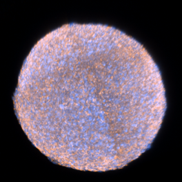
6.4 Incubate the spheroids in the BABB solution until they reach transparency, which may take up to 20 minutes (Fig 1).
Note
Monitor the transparency using a stereomicroscope to ensure optimal clearing.
6.5 Once the spheroids are transparent, proceed with confocal imaging.
Note
Handle the cleared spheroids gently to avoid damage, as they become more fragile after the clearing process.
Fig 1. Optical clearing of spheroids. The spheroids were dehydrated using increasing concentrations of ethanol (EtOH) prior to optical clearing with (1:2, v/v) solution of benzyl alcohol and benzyl benzoate (BABB). It takes up to 20 minutes for the spheroids to become transparent.
40m
Imaging of whole mount spheroids using laser confocal microscopy
7.1 Turn on the confocal microscope system and initialize all required components (lasers, stage, objectives).
7.2 A 20× objective (e.g., Plan-Apochromat 20×/0.8) should be used to provide an optimal balance between resolution and field of view for spheroid imaging. NOTE: Make sure the sample is correctly positioned on the stage.
7.3 Use the live view mode to identify the spheroid's position. Make sure the spheroid is centered in the field of view before proceeding.
7.4 Depending on the spheroid size, use the Tile Region function to select an appropriate number of tiles.
7.5 Typically, a 3×3 tile layout is suitable for imaging spheroids up to 300 μm in diameter.
7.6 Larger spheroids (> 300 μm) may require a 4×3 or larger tiling grid to capture the entire volume.
7.7 Set the Z-Stacks. Adjust the number of Z-stacks to cover the entire spheroid from top to bottom. Set an optimal interval between slices (e.g., ~2-3 μm) to ensure that each slice represents the spheroid structure without missing essential details.
7.8 Adjust the laser power at different depths for optimal image capture. Typically, laser power needs to be:
Lower at the shallowest parts.
Medium power in the middle of the spheroid.
Higher at deeper regions to account for signal attenuation.
Note
Adjust Gain to maintain signal-to-noise ratio.
Note
Keep the pinhole size to approximately 1 AU (adjust based on signal intensity) for optimal resolution and depth penetration.
Note
Verify the imaging setup with live preview and adjust gamma or histogram settings as needed to avoid pixel saturation.
7.9 Acquire Z-Stack Images. Begin the acquisition, ensuring that each slice through the Z-stack is captured with the correct laser intensity and tile setup. The software will automatically generate the 3D image stack.
Note
Acquiring Z-stack images may take several hours.
Note
Ensure the spheroid does not move during imaging (small container, no vibration, constant temperature).
7.10 Once the image acquisition is complete, proceed with necessary image processing steps such as processing and stitching if multiple tiles were used.
12h
Protocol references
Smyrek, I, and E H K Stelzer. “Quantitative three-dimensional evaluation of immunofluorescence staining for large whole mount spheroids with light sheet microscopy.” Biomedical optics express vol. 8,2 484-499. 3 Jan. 2017, doi:10.1364/BOE.8.000484
Lerch, M.L., Kenerson, H.L., Chafin, D. et al. Effect of immediate cold formalin fixation on phosphoprotein IHC tumor biomarker signal in liver tumors using a cold transport device. Sci Rep 10, 2147 (2020). https://doi.org/10.1038/s41598-020-58257-3
Acknowledgements
We thank Sanja Čebraja, Nada Pavlica Dubarič, Marko Radanović and Sabina Železnik for their technical support and expertise in preparing spheroids and samples for electron microscopy. We also thank Urška Dragin Jerman, PhD, for her expertise and assistance with confocal microscopy. The research was done using the Research infrastructure ELIXIR-SI (https://elixir-slovenia.org), funded by the European Regional Development Fund, the Ministry of Science, Education and Sports and by the Slovenian Research and Innovation Agency.