Nov 07, 2025
Whole Brain Embedding for SmartSPIM - EasyIndex with 2% Agarose
- 1Allen Institute for Neural Dynamics;
- 2Boston University
- Allen Institute for Neural Dynamics


Protocol Citation: Holly Myers, Daphne Toglia 2025. Whole Brain Embedding for SmartSPIM - EasyIndex with 2% Agarose. protocols.io https://dx.doi.org/10.17504/protocols.io.3byl4jpn8lo5/v1
License: This is an open access protocol distributed under the terms of the Creative Commons Attribution License, which permits unrestricted use, distribution, and reproduction in any medium, provided the original author and source are credited
Protocol status: Working
We use this protocol and it's working
Created: May 22, 2023
Last Modified: November 16, 2025
Protocol Integer ID: 82289
Keywords: Embedding, Whole brain clearing, Whole brain delipidation, SmartSPIM imaging, Agarose embedding, brain in the agarose solution, agarose block, agarose solution, imperfections in the agarose block, smartspim, imaging, cleared mouse brain, brain specimen, mouse brain, area of the brain, brain, stand during imaging, whole brain
Abstract
This modified LifeCanvas embedding protocol is intended to prepare cleared mouse brains for imaging on the SmartSPIM (see Imaging cleared mouse brains on SmartSPIM). It details mixing and degassing the agarose solution and the technical process of embedding a brain in the agarose solution.
Embedding the brain specimen in a solution of EasyIndex with 2% agarose before SmartSPIM imaging allows the brain to be suspended in the center of a block of index matching solution. The agarose block is glued to the SmartSPIM stand during imaging, sparing any area of the brain from being cut out of imaging. However, any imperfections in the agarose block, like air bubbles, may affect the image.
Materials
Reagents:
EasyIndexLife TechnologiesCatalog #EI-Z1001 (500 mL)
Agarose for ≧1kbp fragment 100gNacalai Tesque Inc.Catalog #01163-76
| Equipment | Product number | |
| Waterbath | Thermofisher Scientific, TSGP2S | |
| StableTemp Vacuum oven | Cole Palmer EW-05053-22 | |
| Plastic embedding mold with mold plunger | Protolabs, customized order (see figure below) | |
| Solid aluminum optical breadboard | ThorLabs, MBH-1224 | |
| Scale | Uline, H-9884 | |
| 20mL glass scintillation vials | Millipore Sigma, Z376817-1PAK | |
| Tape | Uline, S-6748 | |
| Manual single channel pipette P200 | Rainin, 17014390 | |
| Serological pipets | Fisher Scientific, 13-678-11E | |
| Lens paper | Grainger, 799EF3 | |
| Fisherbrand Tissue Path Superfrost Plus Gold Slides | Fisher Scientific, 5158848 | |
| Oven | ThermoScientific, WRB2187511 | |
| Shaker | Benchmark Scientific, WRB3138077 | |
| Phillips head screwdriver | Klein Tools, 9926034 | |
| (1/2") Phillips head screws x4 | Bolt Depot, 6961 | |
| Metal spatula | Cole Parmer, ux-06287-07 | |
| 4c refrigerator | ThermoFisherScientific, 290MC1 | |
| Aluminum foil | Grainger, 4UGE8 | |
| Petri dish | ThermoFisherScientific, 249964 | |
| Benchtop timer | FisherScientific, 06-662-51 |
Note
The plastic embedding mold used in this protocol was custom ordered through Protolabs.
Recipes:
5mL of 2% Agarose + EasyIndex solution:
5mL of 2% agarose solution is sufficient to embed a single mouse brain. The solution must be mixed at least one day prior to embedding in order to hydrate the agarose in EasyIndex.
Measure 0.1g agarose powder on a scale and place in 20mL glass scintillation vial. Using serological pipet, add 5mL EasyIndex. Place on shaker at low speed in 45 °C oven Overnight .
| Reagent | Volume | |
| Agarose powder | 0.1g | |
| EasyIndex | 5mL |
Safety warnings
For proprietary reasons, the reagent contents used in this protocol are not fully disclosed. Perform all steps with gloves on.
Ethics statement
The protocols.io team notes that research involving animals and humans must be conducted according to internationally-accepted standards and should always have prior approval from an Institutional Ethics Committee or Board.
Before start
This protocol is designed for a whole mouse brain that has already been delipidated and index matched using these protocols: Editing Whole Mouse Brain Delipidation - LifeCanvas Active and Refractive Index Matching - EasyIndex.
2% agarose solution should be made at least one day in advance of embedding in order to fully hydrate the agarose powder in EasyIndex.
Prepare Agarose Solution
30m
5mL of 2% agarose solution is required for embedding a single brain specimen. Scale up as needed. Mix the solution in 20 mL glass scintillation vials.
| Reagent | Volume | |
| EasyIndex | 5mL x (number of brains) | |
| Agarose powder | 0.1g x (number of brains) |
Note
Step one is an overnight step and therefore should take place the day before embedding the brain specimens.
Measure 0.1 g x number of brains of agarose powder on scale and place into a glass vial. Each glass vial should contain no more than0.2 g agarose; use multiple vials if needed.
Using serological pipet, transfer (5 mL x number of brains) of EasyIndex into the glass vial(s) containing the agarose power. Each glass vial should contain 5 mL of EasyIndex for every 0.1 g agarose powder.
Gently swirl the glass vial of agarose solution to pre-mix, and place vial on shaker at low speed in a45 °C oven. If this temperature oven is not available, 37 °C oven is sufficient. Leave Overnight .
Setup
1h
Preheat the water bath to 90 °C and preheat the vacuum oven to 80 °C Preheating typically takes 01:00:00 .
1h
Prepare Agarose Solution
30m
Remove the glass container of agarose solution from the oven and aliquot 5 mL of agarose solution into glass vials with a serological pipet. Place glass vials containing agarose solution into preheated 90 °C water bath for 00:30:00 or until agarose solution has thoroughly melted and homogenized. Homogenized agarose solution should be clear with the consistency of water.
30m
Set up Embedding Mold
3h
Assemble an embedding mold secured to a breadboard that will be used to embed delipidated mouse brains in agarose blocks. After assembly, each chamber in the mold can hold a single brain, which can then be filled with molten agarose solution. After the agarose solution hardens, the mold will be disassembled to remove the hardened agarose blocks containing the brains.
Tape two glass slides into the two grooves in the plastic embedding mold.
Note the four holes in the corners of the embedding mold and use these to determine which side of the mold to fasten the slides. On one side of the mold, the holes are surrounded by an indentation to accommodate the head of the screw, and the other side the holes are flush with the mold (see photo examples below). The slides should be taped to the side of the mold where the holes are flush and have no indentation.
Side of embedding mold where the four holes are flush is facing up in this photo, slides are placed in the grooves.
Side of embedding mold where the four holes are surrounded by an indentation is facing up in this photo. Slides should be taped to the opposite side.
Slides have been taped into grooves in embedding mold.
Orient the embedding mold so that the side with the glass slides is flat down against the breadboard, and the side with the indentations around the four holes is facing up. The indentations around the holes will accommodate the heads of the screws.
Embedding mold oriented on breadboard with the holes that have indentations facing up, and slides taped to the reverse side.
Screw four (1/2") Phillips head screws through the four holes in the embedding mold to fasten the mold to the breadboard using a Phillips head screwdriver. This completes the assembly of the embedding mold.
Fully constructed embedding mold; the embedding mold is screwed to the breadboard with four Phillips head screws.
De-gas agarose solution in vacuum oven
Remove glass vial of homogenized agarose solution from water bath, uncap vial, and place in the vacuum oven preheated to 80 °C
Note
If making a larger batch of agarose solution with multiple vials, all vials can be processed together as a batch in step 6.
De-gas the agarose solution using the vacuum oven.
Turn on the "evacuate" setting on the vacuum oven to start the vacuum pump and begin de-gassing the agarose solution.
When the solution starts to bubble, switch the vacuum oven setting to "stop/closed" to shut off the vacuum pump. This will happen once the pressure reaches around0.2 Bar . Allow the agarose solution to bubble for 00:02:00 .
Note
Settings may vary between vacuum ovens. If using a vacuum oven where the unit of measurement is different than Bar, the most important thing is to set the oven to "evacuate" until the agarose starts to bubble, and allowing the solution to bubble for 00:02:00 without bubbling over the top of the vial.
2m
Finally, switch the vacuum oven setting to "vent" to bring the vacuum oven back up to atmospheric pressure.
Degassing is successful if there are no visible bubbles inside the agarose solution. Recap vial and leave vial in the 80 °C vacuum oven with door closed and vacuum off.
If air bubbles are still present inside agarose solution, repeat the degassing process from step 6.
Note
The vacuum setting on the vacuum oven will remain off for the rest of the protocol after the degassing is successfully completed in this step. The vacuum oven will solely be used to keep the agarose solution at an 80c temperature during the rest of the embedding process.
Embedding
6m 50s
This step uses previously cleared adult mouse brain specimens (reference protocols Whole Mouse Brain Delipidation - LifeCanvas Active and Refractive Index Matching - EasyIndex). After following these protocols, the cleared brain specimens will be immersed in 20 mL EasyIndex solution in 50 mL conical tubes.
Remove the whole brain specimen from EasyIndex solution with a metal spatula and place brain onto a piece of lens paper, dorsal side down. Allow the brain to rest for at least 00:00:30 to drain excess EasyIndex.
Retain the conical tube containing EasyIndex solution, storing at Room temperature .
Cleared mouse brain specimen oriented dorsal side down on lens paper.
30s
Use a marker to label one or more chambers in the embedding mold with the letter(s) that corresponds to the sample ID(s) of the brain specimen(s) to be embedded (reference protocol Whole Mouse Brain Delipidation- LifeCanvas step 3.2). This will prevent mix-ups between brain specimens as they are transferred from the conical tube to the embedding mold and back again.
Example: the letter "A" written next to the chamber of the embedding mold (pictured left) corresponds to the "A" written on the lid of the conical tube containing the brain specimen.
Remove vial of agarose from the vacuum oven and let the vial cool at Room temperature until warm but not hot to the touch, about 60 °C .
Note
If processing a larger batch of agarose solution with multiple vials, complete steps 8 - 11 with only one vial at a time, leaving the rest of the vials capped in the vacuum oven.
Uncap the vial and align the mouth of the vial to the corner of one of the eight chambers of the embedding mold while pouring out the agarose solution at an even rate into the chamber. Pour until the embedding chamber is about 1/3 full of agarose solution. Cap vial and place back into the vacuum oven.
Remove any small bubbles that form in the agarose solution in the embedding chamber with a P-200 manual pipette.
After 00:01:20 gently place transfer the brain specimen from the lens paper into the embedding chamber on top of the agarose solution, using either gloved hand or spatula. Keep the brain oriented dorsal side down, so the dorsal surface of the brain is touching the agarose in the chamber. Take care to set the brain gently on top of the agarose solution and not to push it down. The brain should slightly sink into the agarose but ideally should not sink through all of the agarose.
The ventral side of the brain in this photo is facing up. The dorsal side is facing down and touching the metal spatula.
The ventral side of the brain is facing up in this photo. The brain has partially sunk into the agarose solution after being placed into the chamber.
1m 20s
Immediately after placing the brain into the embedding chamber, remove the vial of agarose from the vacuum oven and pour a second layer of agarose solution into the embedding chamber. This layer of agarose should totally cover the ventral surface of the brain.
Stop pouring when the agarose is level with the top edge of the embedding chamber. The surface of the agarose solution should form a flat surface flush with the top surface of the mold. (not convex or concave).
Note
It is important to pour this second layer of agarose immediately after placing the brain into the embedding chamber, so that the first layer of agarose does not cool too much. If the temperature difference between the two layers of agarose is too great during this step, they will consolidate into two distinct layers instead of forming a single structure around the brain.
The ventral surface of the brain is completely submerged in agarose solution during this step. However, the agarose solution is still not flush with the top edge of the embedding chamber.
The top surface of the agarose solution is level with the top of the embedding chamber in this photo, and the brain is centered in the chamber.
5m
Remove any small bubbles that form in the agarose solution in the mold with a P-200 manual pipette. Keep the brain centered in the embedding chamber as the agarose hardens.
Post-embedding
16h
Place embedding block in 4 °C fridge and ensure that block is covered to protect from light. A large plastic petri dish wrapped in aluminum foil may be used as cover for light protection. Leave 16:00:00 .
Use the black mold cutter to remove the agarose block containing the brain specimen.
Unscrew the plastic embedding mold from the breadboard with a Phillips head screwdriver and gently remove the glass slides that are taped to the back of the mold.
Remove the agarose block with the brain embedded inside from the plastic embedding mold using the following directions:
Place the black plastic mold plunger on lab bench and lift the plastic embedding mold so that it is aligned a few inches directly above the black mold plunger. Slowly lower the plastic embedding mold down onto the mold plunger so that the protrusions on the mold plunger fit inside the holes in the embedding mold. Once the embedding mold is completely lowered down onto the mold plunger, the protrusions on the mold plunger will push the agarose block out of the embedding mold.
Embedding mold with agarose blocks held in place with glass slides taped to the back of the mold.
The mold plunger has eight protrusions that fit into corresponding holes in the embedding mold.
The glass slides previously taped to the back of the embedding mold have been removed in this photo, and the embedding mold has been lowered down onto the mold plunger, pushing the agarose blocks out of the mold.
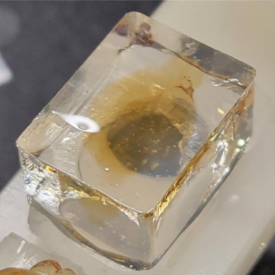
In the finished product, the brains ideally should be centered in the agarose with at least a small margin between the tissue and the edge of the agarose block. The blocks themselves should have smooth, flat surfaces.
Using gloved hand, gently place the agarose block into the conical tube containing 20 mL of EasyIndex solution that was set aside in step 11. Place tube on a shaker at low speed and let sit at least 16:00:00 at Room temperature before it is ready for imaging. Brains may be stored for up to several months at Room temperature in 20 mL of EasyIndex.
16h
Protocol references