Jun 11, 2026
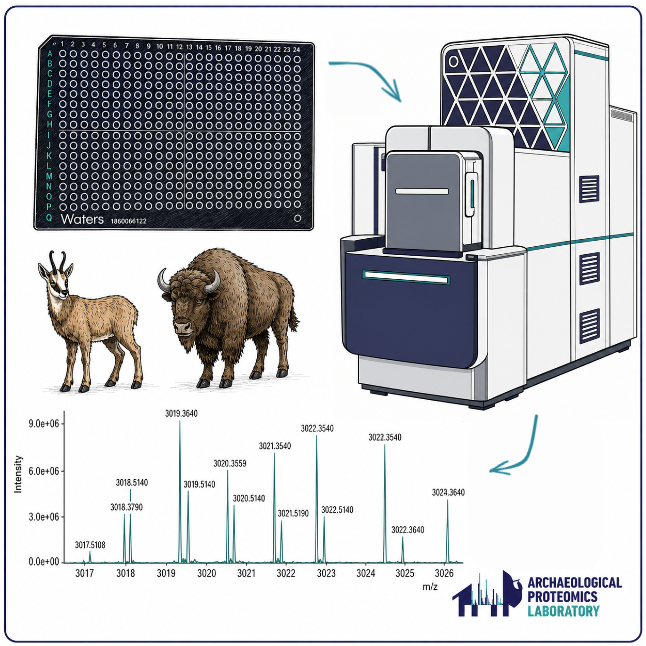
WATERS SELECT SERIES MRT MALDI Spot Analysis for Zooarchaeology by Mass Spectrometry (ZooMS) of collagenous material
- Frankie Tait1,
- Nicholas Michael2,
- Karen Ruebens1
- 1Archaeological Proteomics Laboratory, Department of Archaeology, University of Reading, UK;
- 2Chemical Analysis Facility, University of Reading, UK


Protocol Citation: Frankie Tait, Nicholas Michael, Karen Ruebens 2026. WATERS SELECT SERIES MRT MALDI Spot Analysis for Zooarchaeology by Mass Spectrometry (ZooMS) of collagenous material. protocols.io https://dx.doi.org/10.17504/protocols.io.n2bvjkwbngk5/v1
License: This is an open access protocol distributed under the terms of the Creative Commons Attribution License, which permits unrestricted use, distribution, and reproduction in any medium, provided the original author and source are credited
Protocol status: Working
We use this protocol and it's working
Created: April 21, 2026
Last Modified: June 11, 2026
Protocol Integer ID: 315439
Keywords: ZooMS, Palaeoproteomics, Mass Spectrometry, MALDI-TOF, collagen fingerprinting, Q-ToF, zooarchaeology by mass spectrometry, peptide mass fingerprinting of collagen type, peptide mass fingerprinting, select series mrt maldi spot analysis, peptide peak, mass spectrometry, waters mrt, maldi spot analysis capability, zooarchaeology, taxonomic identification, collagenous material this protocol, mrt, collagen type
Funders Acknowledgements:
UKRI
Grant ID: EP/Y037448/1
Abstract
This protocol gives guidance on how to set up and run a Waters™ Select Series Multi-Reflecting Time-of-Flight (MRT) with a MALDI source attachment for the purposes of Zooarchaeology by Mass Spectrometry (ZooMS) of collagenous material. ZooMS allows for taxonomic identifications to be made based on peptide mass fingerprinting of collagen type 1. The Waters MRT is an ultra-high resolution imaging instrument with MALDI spot analysis capabilities, allowing for high precision ZooMS to be carried out. This instrument has the potential to achieve sub-ppm mass accuracy, therefore, results can give peptide peaks accurate to at least one decimal place allowing for contaminants and non-diagnostic peptide markers to be distinguished.
Equipment
Select Series MRT
NAME
MALDI
TYPE
Waters
BRAND
LINK
355 nm Nd:YAG laser, tightly focused laser beam profile; Quadrupole Multi-Reflecting Time of Flight (Q-ToF)
SPECIFICATIONS
Attachments

_extern.inf
12KB

MALDI_Values.png
259KB

StepWave_Values.png
134KB

MSProfile_Values.png
189KB

IonGuides_Values.png
197KB

Transfer_Values.png
155KB

RF_Values.png
107KB
Sample_A.msd
2.3MB
Sample_B.msd
2.2MB
Guidelines
1. See "Materials" for reagent recipes
2. Mass Spectrometry is sensitive to contaminants eg. detergents, plastics etc.
3. This protocol can be performed in a standard wet chemistry laboratory.
4. Wear relevant PPE as designated by your local laboratory.
5. Follow local lab guidelines regarding sample handling and storage.
6. Follow country and facility specific guidelines regarding the disposal of chemical waste.
7. This protocol is intended to give guidance to mass spectrometry and/or Waters technicians - instrument settings should not be altered without specialist training.
Materials
**Reagents**
0.1% TFA/ 5% Acetonitrile (v/v) in HPLC-MS water: Add 940 µL HPLC-MS water to a 1.5 mL microcentrifuge tube. Add 50 µL ACN. Carefully add 10 µL 10% TFA. Secure cap and vortex briefly.
Conditioning Solution; 0.1% TFA in 50:50 Acetonitrile and HPLC-MS water: for 500ml, add 250 mL ACN to 250 mL LCMS water and add 500 µL 100% TFA.
α-Cyano-4-hydroxycinnamic acid (CHCA) | VWRCatalog #SIALC2020-10G : 10 mg/mL in Conditioning Solution. Ensure powder is fully dissolved either through vortexing or heating (or sonicating if using a glass vessel) - hold up to the light to be sure. Store at Room temperature for no more than 24:00:00 . Store in the dark.
Red Phosphorous (RedP) |Merck MilliporeSigma (Sigma-Aldrich)Catalog #343242-5G 25 mg/mL finely ground suspension in ACN.
Waters™ MassPREP ADH Digestion Standard | WatersCatalog #186002328 Resuspend vial in 1 mL of 0.1% TFA/ 5% ACN (v/v) in LCMS water. This will give a concentration of1 pMol/µL . Aliquots should be stored -20 °C and only thawed immediately before use, do not continuously freeze-thaw.
Methanol (MeOH) |Fisher Scientific UKCatalog #10653963
Water |Fisher Scientific UKCatalog #10777404
Acetonitrile (ACN) |Fisher Scientific UKCatalog #10616653
Equipment
MALDI 480 Target Plate
NAME
Waters
BRAND
405018182
SKU
Polished Stainless Steel
SPECIFICATIONS
Equipment
Select Series MRT
NAME
MALDI
TYPE
Waters
BRAND
LINK
355 nm Nd:YAG laser, tightly focused laser beam profile; Quadrupole Multi-Reflecting Time of Flight (Q-ToF)
SPECIFICATIONS
Protocol materials
Water |Fisher Scientific UKCatalog #10777404
Methanol (MeOH) |Fisher Scientific UKCatalog #10653963
Red Phosphorous (RedP) |Merck MilliporeSigma (Sigma-Aldrich)Catalog #343242-5G
Waters™ MassPREP ADH Digestion Standard | WatersCatalog #186002328
α-Cyano-4-hydroxycinnamic acid (CHCA) | VWRCatalog #SIALC2020-10G
Acetonitrile (ACN) |Fisher Scientific UKCatalog #10616653
Safety warnings
*It is recommended that for MALDI analysis on the MRT, 50 volts is added to the detector voltage that has been obtained via the automatic detector gain setup. This should be done in consultation with the manufacturer.
Before start
1. We recommend using dedicated glassware where possible for preparation and storage of reagents.
2. Peptide extracts for troubleshooting this protocol were produced using an Acid Insoluble ZooMS protocol modified from Buckley et al. 2009 and Welker et al. 2015.
3. We do not recommend spotting the peptide extracts more than 24 hours in advance.
4. Ensure the MALDI laser window, mirror and octopole are clean.
Spotting
Spot Sample 1:1 with α-Cyano-4-hydroxycinnamic acid (CHCA) | VWRCatalog #SIALC2020-10G matrix (see Materials). Spot peptides first, then mix with CHCA by pipetting in a circular motion on the plate1 µL of each
Note
Final volume in each target well should be 2 µL to cover full surface area
Spot the 1 pMol/µL aliquots ofWaters™ MassPREP ADH Digestion Standard | WatersCatalog #186002328 (see Materials) 1:1 with CHCA in the same manner as the peptide extracts. Spot 8 replicates. Each spot has a final concentration of 500 fMol/µL
Allow the plate to dry on a heater block 35-50 °C to encourage crystallisation. Remove as soon as the last spot has dried.
The plate should be kept at room temperature in the dark if not running immediately.
Instrument Calibration
Ensure Red Phosphorous (RedP) |Merck MilliporeSigma (Sigma-Aldrich)Catalog #343242-5G (see Materials) is resuspended before and during this step by shaking gently.
Spot 1 µL of RedP onto a calibrant spot 4-5 times, allowing 00:00:20 in between each dispense for the layer to dry.
Load the plate into the instrument and ensure the vacuum has been established.
Acquire data for the calibration using the following settings
Note
- Positive ; MRT ; Standard ; 1.0s ; 9600
- API Gas flow rate 350 mL/min
- Laser Rate 2500 Hz
- Laser Intensity 300
- Lens Focus 4.00 mm
- Step Size: X 1000 μm and Y 100 μm
- Scan Time 1.0 sec
- Decoding Threshold Automatic
- Function MS
Only accept the calibration if the Mean Prediction Error is ≤ 0.2 ppm.
Instrument Standard and Laser Optimisation
Test three laser intensities: 250, 300 and 350 to confirm optimum settings using the spotted ADH.
Note
The laser intensity needs to be optimised depending on variable environmental factors and laser wear and tear. This will also act as an instrument standard to ensure functionality before and after processing samples.
Acquire data using the following recommended settings
Note
- Positive ; MRT ; Standard ; API Gas ; 1.0s ; 9600
- Laser Rate 2500 Hz
- Lens Focus 4.00 mm
- Well Diameter: 2.15 mm ; Step Size: X 500 μm and Y 150 μm
- Scan Time 1.0 sec
- m/z range 1000 - 3500
- Decoding Threshold Low
- Function MS
Manual Quad Profile:
- Dwell time 20% at 1000 m/z
- Dwell time 40% at 1500 m/z
- Top end mass 3000 m/z
- Ramp time 20%
The RF controls below were applied to allow for higher peptide markers to pass through the quadrupole:
See attachments for full settings and an example run file.
ADH Spectrum
Expected result
Key peptide peaks that should be observed are 2744 m/z and 3369 m/z to ensure higher mass peptides are being appropriately measured.
Sample Data Collection
Use the same settings as for the ADH instrument standard. Set the laser intensity to the optimised setting based on spectral quality of signal-to-noise and TOF MS ES+ counts.
Sample Spectrum
Ensure you set the instrument to acquire all sample data, followed by one ADH spot to confirm consistency throughout the run.
Each spot receives 30 scans which will be summed.
Data processing
The Waters raw data files (.raw) can be converted to mzML format using MSConvert from ProteoWizard (Chambers et al., 2012)
The individual scans can be summed into .txt files using an in-house developed Python script (MZSummer), which we make publicly available here:
MZSummer user interface
See our standard settings in the screenshot above - these settings can be adjusted to suit your needs. (We recommend running this programme overnight as it can take several hours if running a full plate on a laptop)
After summing, any technical replicates can be merged in R using the MALDIquant and MALDIquantForeign packages and published scripts (Gibb and Strimmer, 2012; Mylopotamitaki et al., 2024)
Once you have fully processed the raw data into a workable file, we advise following this separate guide for completing spectral identifications: https://www.protocols.io/view/identifying-zooms-spectra-mammals-using-mmass-kqdg36rppg25/v1. Some modifications may be necessary due to the ultra-high resolution of the spectra (e.g. you may consider unselecting the deisotoping panel for peak picking due to the narrow peaks).
See attachments for example summed and merged MRT spectra: Sample_A and Sample_B
Target Plate Cleaning
Rinse the plate Methanol (MeOH) |Fisher Scientific UKCatalog #10653963 . Visibly check the plate and use a cotton bud, damp with MeOH, to manually wipe any residue that was not easily rinsed away.
Sonicate the plate in 50:50 MeOH and Water |Fisher Scientific UKCatalog #10777404 for 00:15:00
Dry in the fume hood either under regular extraction or with a Nitrogen gas tube.
Protocol references
Buckley, M., Collins, M., Thomas-Oates, J., & Wilson, J. C. (2009). Species identification by analysis of bone collagen using matrix-assisted laser desorption/ionisation time-of-flight mass spectrometry. Rapid Communications in Mass Spectrometry: RCM, 23(23), 3843–3854. https://doi.org/10.1002/rcm.4316
Chambers, M. C., Maclean, B., Burke, R., Amodei, D., Ruderman, D. L., Neumann, S., Gatto, L., Fischer, B., Pratt, B., Egertson, J., Hoff, K., Kessner, D., Tasman, N., Shulman, N., Frewen, B., Baker, T. A., Brusniak, M.-Y., Paulse, C., Creasy, D., … Mallick, P. (2012). A cross-platform toolkit for mass spectrometry and proteomics. Nature Biotechnology, 30(10), 918–920.
Gibb, S. and Strimmer, K., 2012. MALDIquant: a versatile R package for the analysis of mass spectrometry data. Bioinformatics, 28(17), pp.2270-2271.
Mylopotamitaki, D., Fewlass, H., Zavala, E.I., Rougier, H., Sümer, A., Hajdinjak, M., Smith, G.M., Ruebens, K., Sinet-Mathiot, V., Xia, H., Hansen, J., Harking, F., Olsen, J.V., Kirchner, A., Lauer, T., Stahlschmidt, M., Talamo, S., Meller, H., Dietl, H., Orschiedt, J., McPherron, S., Krause, J,, Meyer, M., Welker, F., Schüler, T., Weiss, M., Hublin, J-J. 2024. Homo sapiens reached the higher latitudes of Europe by 45,000 years ago. Nature 626, 341–346.
Samantha Brown 2021. Identifying ZooMS Spectra (mammals) using mMass. protocols.io
https://dx.doi.org/10.17504/protocols.io.bzscp6aw
Welker, F., Soressi, M., Rendu, W., Hublin, J.-J., & Collins, M. (2015). Using ZooMS to identify fragmentary bone from the Late Middle/Early Upper Palaeolithic sequence of Les Cottés, France. Journal of Archaeological Science, 54, 279–286. https://doi.org/10.1016/j.jas.2014.12.010
Acknowledgements
We like to thank Anna Wagner and Geoff M. Smith (University of Reading) in their help testing this protocol on archaeological samples.