Jan 11, 2024
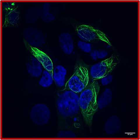
Visualization of LRRK2 filaments in 293T cells
- 1University of California, San Diego


Protocol Citation: Eva Karasmanis, Kathryn Hatch 2024. Visualization of LRRK2 filaments in 293T cells. protocols.io https://dx.doi.org/10.17504/protocols.io.8epv5xpz4g1b/v1
Manuscript citation:
doi: https://doi.org/10.1101/2023.11.14.567123
License: This is an open access protocol distributed under the terms of the Creative Commons Attribution License, which permits unrestricted use, distribution, and reproduction in any medium, provided the original author and source are credited
Protocol status: Working
We use this protocol and it's working
Created: January 11, 2024
Last Modified: May 31, 2024
Protocol Integer ID: 93405
Keywords: ASAPCRN, visualization of lrrk2 filament, lrrk2 filaments in the presence, lrrk2 filament, cell, percentage of cell, lrrk2, visualization
Funders Acknowledgements:
ASAP
Grant ID: ASAP-000519
Abstract
Visualization of LRRK2 filaments in 293T cells
GOAL: Express GFP-LRRK2 with or without DARPin E11 and quantify the percentage of cells with LRRK2 filaments in the presence and absence of MLi-2 in 293T cells.
Image Attribution
Eva Karasmanis
Day 1: fibronectin coating and cell plating
Visualization of LRRK2 filaments in 293T cells
GOAL: Express GFP-LRRK2 with or without DARPin E11 and quantify the percentage of cells with LRRK2 filaments in the presence and absence of MLi-2 in 293T cells.
Constructs needed:
1) CMV-8xHISDaprin C12-FLAG
2) CMV- His-daprin E11-FLAG
3) CMV-GFP-LRRK2
| DMSO | MLi-2 | DMSO | MLi-2 | |
| Replicate 1 | GFP-LRRK2 | GFP-LRRK2 | GFP-LRRK2 DARPin_E11_3xFLAG | GFP-LRRK2 DARPin_E11_3xFLAG |
| Replicate 2 | GFP-LRRK2 | GFP-LRRK2 | GFP-LRRK2 DARPin_E11_3xFLAG | GFP-LRRK2 DARPin_E11_3xFLAG |
| Replicate 3 | GFP-LRRK2 | GFP-LRRK2 | GFP-LRRK2 DARPin_E11_3xFLAG | GFP-LRRK2 DARPin_E11_3xFLAG |
Fibronectin Coating (Sigma cat# F0895, 0.1% solution, 1 mg/ml):
Make 0.01 µg/µL solution of fibronectin, stock @ 1 mg/mL For 6x 35 mm dishes (12 ml fibronectin working stock)- 0.12 mL fibronectin + 11.88 mL 1X PBS
Lay one 22 mm x 22 mm glass coverlsip into each 35 mm dish or 6 well.
Add 2 ml of 10 ug/mL fibronectin per 35 mm dish or 6 well.
Incubate at 37 °C 5% CO2 for 00:45:00
45m
Wash with PBS and let dry for 00:45:00 in the tissue culture hood (no UV)
45m
Plate cells onto Fibronectin coated dishes
Dissociate cells, count and plate 6 well plate with 200K cells /well. For transfection, plate in antibiotic-free media (DMEM+10% FBS)
Day 2 Transfect cells with GFP-LRRK2 and Darpins:
2d 3h 2m
Transfect 800 ng LRRK2 and 400ng Darpin with 5 μL PEI /well.
- cells should be 50-60% confluent
In a sterile tube dilute 800 ng GFP-LRRK2 and 400 ng DARPin E11 plasmid DNA in Optimem (150 µL /well)
Per Well:
a) 150 µL Optimem (prewarmed)+5 µL PEI
b) 150 µL Optimem (prewarmed) +800 ng GFP LRRK2 and 400 ng DARPin E11 DNA.
or
c) 150 µL Optimem (prewarmed) + 800 ng GFP LRRK2
Add PEI to diluted DNA - (Per reaction: 5 µL of 1 µg/µL stock). Mix immediately.
In a sterile tube dilute 800 ng GFP-LRRK2 and 400 ng DARPin E11 plasmid DNA in Optimem (150uL/well)
Per Well:
a) 150 µL Optimem (prewarmed)+ 5 µL PEI
b) 150 µL Optimem (prewarmed) + 800 ng GFP LRRK2 and 400 ng DARPin E11 DNA.
or
c)150 µL Optimem (prewarmed) + 800 ng GFP LRRK2
Add PEI to diluted DNA - (Per reaction: 5 µL of 1 µg/µL stock). Mix immediately.
Incubate at Room temperature for 00:15:00
15m
Add DNA/PEI mixture to cells dropwise.
Swirl the plate to distribute Incubate at 37 °C 5% CO2 for 48:00:00
2d
Day 4 MLi2 or DMSO treatment
2d 3h 2m
Treat Cells with MLi2 or DMSO (5uL per well of a 1 mM stock) for02:00:00 , 37 °C 5% CO2
2h
Fixing and Staining of Cells
2d 3h 2m
Fixing: Prewarm freshly made 3% PFA, 4% sucrose in 1X in PBS 1X. You will need 1 mL per well.·
Aspirate media
Immediately add prewarmed fixation buffer (3% sucrose, 4 % (v/v) PFA in PBS 1X)·
Incubate 00:12:00 Room temperature
12m
2X rinse with PBS 1x
2X Wash with PBS1x for 00:05:00 at Room temperature
5m
2X Wash with PBS 1x for 00:05:00 at Room temperature ·
5m
Quenching: 2X rinse and 2x 00:10:00 of 0.4% NH4Cl 75 millimolar (mM) in PBS 1X
10m
Blocking/Permeabilizing: incubate cells blocking/permeabilizing buffer ( 2% BSA + 0.1 % triton X-100 in PBS 1X) for 00:20:00 Room temperature
During blocking make and spin the antibody (ab) mix. Each coverslip needs ~40-50 uL uL of ab mix. Antibodies are diluted in Blocking buffer ( 2% BSA in PBS 1X ) and spun at 4 °C 00:10:00 m 50000 x g, 4°C to clear aggregates. Remove spun ab and place in new tube. Mix to get even concentration. HERE: we used 1:200 rabbit polyclonal anti-FLAG DYKDDDDK tag – (ptg labs Cat no : 20543-1-AP)
30m
2X Rinse with Wash 2X (00:05:00 , Room temperature ) with blocking buffer
5m
Add primary antibody mix :
Add 40uL on parafilm and flip coverslip on the antibody. Incubate 03:00:00 Room temperature or 4 °C Overnight . If incubating overnight, make a humidity chamber before placing in fridge.
HERE: we used 1:200 rabbit polyclonal anti-FLAG DYKDDDDK tag – (ptg labs Cat no : 20543-1-AP)
3h 5m
Rinse 2X with blocking buffer
Wash 2X 00:05:00 Room temperature with blocking buffer
5m
Add secondary mix in PBS (1:200 Goat a-rabbit Alexa568; SIGMA A-11011 +1:5000 DAPI**) 00:45:00 Room temperature
** DAPI can alternatively be added as a separate step at 1:1000 dilution for 15 min
45m
5X Rinse with PBS 1X
Mount in Fluorsave hard media (Millipore 345789)
Let dry for at least an hour. Store in Fridge 4oC if not imaging immediately. Check coverslip is set with tweezers before imaging.
Imaging and analysis
Blind your mounted slides before imaging to prevent bias during aquisition.
Find areas with transfected cells. Acquire Z stacks by determining top and bottom with a 0.3 um step size. (about 20-25 z stacks) ·
Analysis in Fiji: Go through each image, make max projections, and mark each transfected cells with a ROI (region of interest).
In an excel sheet keep track of each cell you mark and score as 0 if no LRRK2 filaments are present or 1 if some are. ·
Include at least 50 cells/ sample. ·
Calculate % cells with filaments (number of transfected cells with filaments /total number of transfected cells *100) ·
Unblind
Transfer values to prism to generate graph and statistics.