Oct 24, 2020
Visualisation of bacteria around roots
- Andreea S1
- 1University of Groningen
- iGEM Groningen 2020


Protocol Citation: Andreea S 2020. Visualisation of bacteria around roots. protocols.io https://dx.doi.org/10.17504/protocols.io.bjzpkp5n
License: This is an open access protocol distributed under the terms of the Creative Commons Attribution License, which permits unrestricted use, distribution, and reproduction in any medium, provided the original author and source are credited
Protocol status: Other
The protocol was developed based on literature and has not been tested yet.
Created: August 20, 2020
Last Modified: October 24, 2020
Protocol Integer ID: 40719
Keywords: growth of the bacterial biofilm, visualisation of bacteria, bacterial biofilm, roots in the rhizosphere, plant with the bacterial strain, bacteria, bacterial strain, rhizosphere, root, plant
Abstract
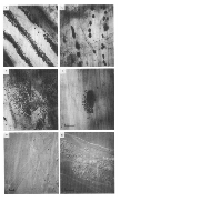
This protocol is carried out to observe the growth of the bacterial biofilm along with the roots in the rhizosphere. In the paper by Bennett, R. and Lynch, J (1981), they inoculated the plant with the bacterial strain and analyzed the growth of the bacterial biofilm around roots.
Citation
LINK
Removal of roots and bacterial counts
Carefully remove the roots from the soil.
Divide the roots into three portions: a) 0 to 2 cm from the tip; b) 2 to 4 cm from the tip; and c) 4 cm from the tip to the seed.
Root samples will have to be examined before and after washing. This is done to determine: a) the pattern and intensity of microbial colonization; b) whether the washing procedure was effective in removing the bacteria from the root surface.
Place each sample in a Erlenmeyer flask containing 10 mL sterile distilled water
Shake for 00:10:00 on a wrist-action flask shaker (speed setting 7)
Prepare serial 10-fold dilutions and subsequently prepare surface colony counts on nutrient agar plates.
Microscopy
There are two ways in which you can observe the roots under the microscope.
Directly mount roots in 0.5 Mass / % volume of aqueous phenol/glacial acetic acid ( 15:4, by volume). Using a
microscope observe the roots.
The other method is where you stain the roots by immersing them for 00:01:00 in 5 % volume aqueous phenol/ 6 Mass / % volume aqueous aniline blue/ glacial acetic acid (15:1:4, by volume). Then carefully rinse and mount in 0.5 Mass / % volume of aqueous phenol/glacial acetic acid ( 15:4, by volume). Observe the samples using a bright field microscope.
Biofilm Formation
Inoculate a plate of Bacillus strain (from -80 °C ) on a BHI agar plate overnight (~18:00:00 ) at 37 °C .
Inoculate5 mL BHI broth from overnight agar plate (one colony) and grow for 04:00:00 with shaking (~200 rpm) at 37 °C .
Inoculate 1:1,000 of broth culture (12 µL ) into 12 mL MSgg media in petri dishes.
Let biofilm form (covered with dish lid without shaking) at 37 °C (usually 3 and 7 days). Ensure that incubator has a water container to ensure the dishes do not dry out.
Citations
R. A. BENNETT AND J. M. LYNCH. Bacterial Growth and Development in the Rhizosphere of Gnotobiotic Cereal Plants
https://pdfs.semanticscholar.org/cf72/4b2b6e05313ad110edc7967590b5fa34ed70.pdf