Aug 27, 2024
Version 2
Viral Enumeration of Water Samples Using Wet Mount Epifluorescence Microscopy V.2
- Madeline Bellanger1,2,
- Pieter Visscher3,
- Richard Allen White III1,2
- 1Department of Bioinformatics and Genomics, North Carolina Research Campus, The University of North Carolina at Charlotte, Kannapolis, North Carolina, USA;
- 2Computational Intelligence to Predict Health and Environmental Risks (CIPHER), The University of North Carolina at Charlotte, Charlotte, North Carolina, USA;
- 3Department of Marine Sciences and Geoscience, University of Connecticut, Storrs, Connecticut, USA


External link: https://journals.asm.org/doi/full/10.1128/aem.01744-23
Protocol Citation: Madeline Bellanger, Pieter Visscher, Richard Allen White III 2024. Viral Enumeration of Water Samples Using Wet Mount Epifluorescence Microscopy. protocols.io https://dx.doi.org/10.17504/protocols.io.q26g7po68gwz/v2
Manuscript citation:
Bellanger M, Visscher P, White RA.2023.Viral enumeration using cost-effective wet-mount epifluorescence microscopy for aquatic ecosystems and modern microbialites. Appl Environ Microbiol89:e01744-23.https://doi.org/10.1128/aem.01744-23
License: This is an open access protocol distributed under the terms of the Creative Commons Attribution License, which permits unrestricted use, distribution, and reproduction in any medium, provided the original author and source are credited
Protocol status: Working
We use this protocol and it's working
Created: February 29, 2024
Last Modified: August 27, 2024
Protocol Integer ID: 95955
Keywords: EFM, epifluorescence microscopy, viral enumeration, microbialite, enumeration, phage, viral enumeration of water sample, gold standard method for environmental viral enumeration, viral enumeration within modern microbialite, effective method for environmental viral enumeration, environmental viral enumeration, aquatic sample, robust method for modern microbialite, water sample, using wet mount epifluorescence microscopy epifluorescence microscopy, wet mount epifluorescence microscopy epifluorescence microscopy, modern microbialite, microbial mat, filtration, aquatic ecosystem, exopolymeric substance, anodisc filter
Funders Acknowledgements:
NASA Astrobiology Institute
Grant ID: NNH22ZDA001N-EXO
Abstract
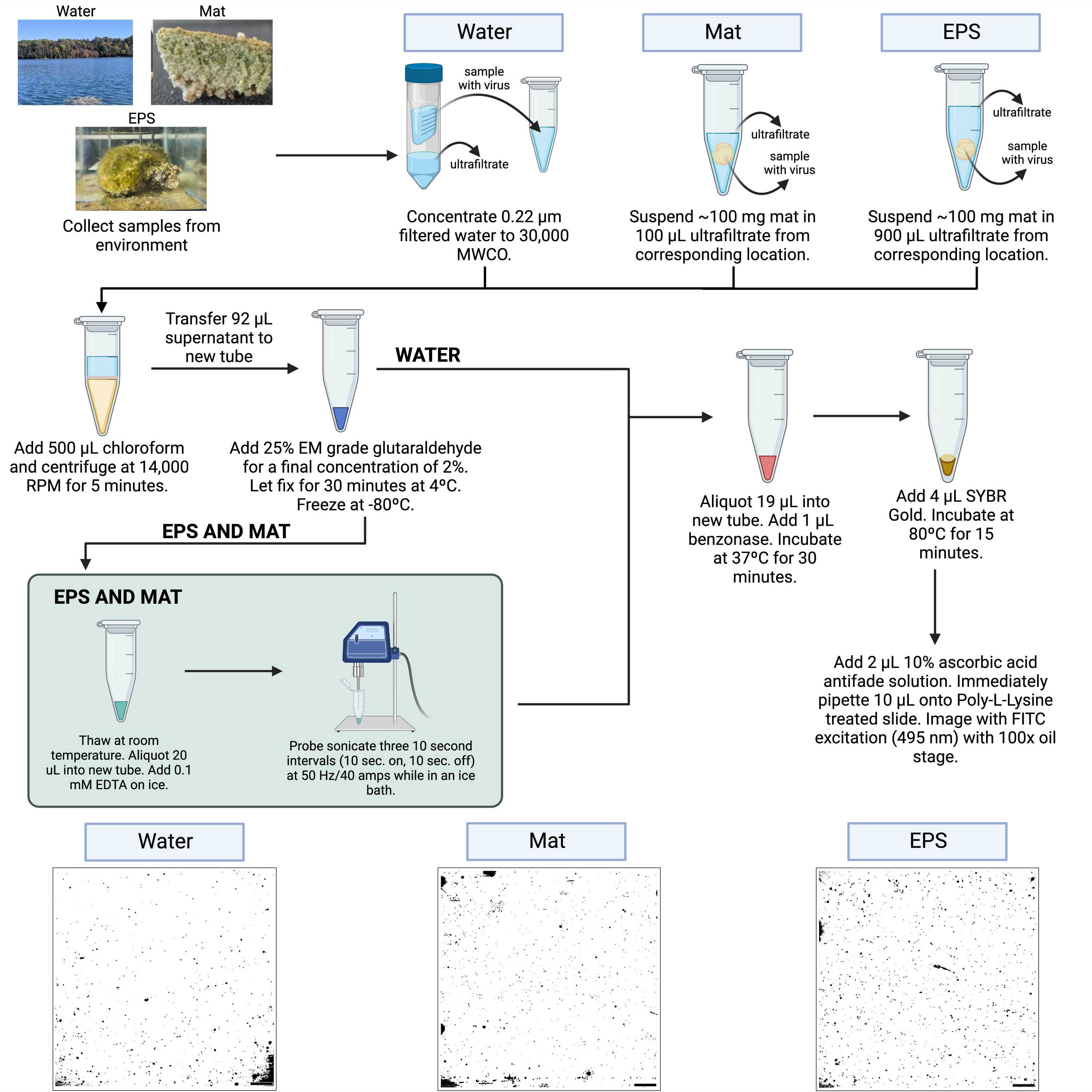
Epifluorescence microscopy (EFM) has been the gold standard method for environmental viral enumeration for over 25 years. Currently, standard EFM methods using the Anodisc filters are no longer cost-effective (>$15 per slide). We present a cost-effective method for environmental viral enumeration from aquatic samples, microbial mats, and exopolymeric substances (EPSs) within modern microbialites using EFM. Our integrated approach, which includes filtration, differential centrifugation, chloroform treatment, glutaraldehyde fixation, benzonase nuclease treatment, probe sonication (EPS and mat only), SYBR Gold staining, wet mounting, and imaging, provides a robust method for modern microbialites and aquatic samples. Our method provides a robust and cost-effective (~$0.75 per sample) viral enumeration within modern microbialites and aquatic ecosystems.
Guidelines
This method is used to enumerate viruses in water samples collected from the environment. See our protocols for microbialite viral enumeration, EPS viral enumeration, and soil viral enumeration!
Materials
Consumables
- Whatman grade 1 qualitative filter paper
- 0.65 µm PVDF Durapore membrane filter (hydrophilic)
- 0.45 µm PVDF Durapore membrane filter (hydrophilic)
- 0.22 µm PVDF Durapore membrane filter (hydrophilic)
- 30 kDa MWCO Millipore centrifuge filters (of desired size)
- 1.5 mL low protein binding, nuclease free microcentrifuge tubes
- P1000 pipette tips
- P200 pipette tips
- P10 pipette tips
- 5 ml Eppendorf tubes
- Microscope slides
- Slide covers
Chemicals
- 70% Ethanol
- 25% EM grade glutaraldehyde
- Chloroform
- EDTA
- Benzonase
- SYBR Gold nucleic acid stain
- Poly-L-Lysine 0.1% w/v
- 1X PBS
- Ascorbic acid
- Microscope immersion oil
Equipment
- P1000 pipette
- P200 pipette
- P10 pipette
- Centrifuge capable of holding 50 ml conical tubes or wide neck bottles (62 mm diameter; 146 mm length, only if using Centricon-70 plus centrifuge filters)
- Microcentrifuge
- Vortexer
- Probe sonicator
- Heat block
- Balance
- Fluorescence microscope equipped with a 100x oil immersion lens and blue excitation light (495 nm)
Protocol materials
Chloroform
Benzonase® NucleaseMerck MilliporeSigma (Sigma-Aldrich)Catalog #E1014 SIGMA
SYBR Gold Nucleic Acid Gel StainCatalog # S-11494
Safety warnings
- Fixation with glutaraldehyde and chloroform treatment needs to be performed in a fume hood with a face shield and proper PPE.
- Needles used to remove glutaraldehyde from serum vials need to be disposed of in a sharps container within the fume hood. Never replace the cap on a needle. Once the needle has been used, immediately deposit it into the sharps container.
Filtration and Cleaning
Record the starting volume of water, this will be used for calculations later.
Filter water through a glass fiber pre filter (Whatman Grade 1 qualitative filter paper).
Collect filtrate and filter through a 0.65 μm PVDF filter (Durapore PVDF membrane filter; hydrophilic).
Collect filtrate again and filter two times through 0.22 μm PVDF filters (Durapore PVDF membrane filter; hydrophilic).
If only filtering samples, . Continue through this section for optional concentration.
Collect filtrate and add to the top portion of 30 kDa MWCO Centricon-70 plus centrifuge filters (Millipore UFC703008).
Note
Centrifuge filters come in a range of volumes (0.5 - 70 mL). These filters can be reused for similar samples. If storing used filters for reuse, after removing concentrate add a small amount of ultrafiltrate or sterile/filtered (0.22 um x2), enough to cover the filter surface, and store at 4ºC.
Centrifuge at 3500 rpm, 00:12:00 (if using smaller volume filters adjust speed and time according to manufacturer instructions).
12m
After centrifugation, collect the ultrafiltrate from the collection cup below the filter and add more 0.22 μm filtered water to the top. Continue to centrifuge as explained in step 7 until all water has passed through the filter.
Note
Centricon-70 plus filters come with retrieval cups, but some smaller sizes do not. Smaller sized filters can be removed and flipped upside down into a 15 or 50 mL conical tube and centrifuged to collect concentrate. Amicon ultra-15 filters do not fit into conical tubes. To collect concentrate use a pipette to remove concentrate from the filter area, careful not to puncture the filter.
After concentrating all the water, attach the retrieval cup to the top of the filter and flip upside down. Centrifuge at 3500 rpm, 00:05:00 .
5m
Collect the concentrate from the retrieval cup and pipette into a 1.5 mL low protein binding nuclease free microcentrifuge tube. Record the volume of concentrate recovered.
Dilute concentrate ~3:10 with ultrafiltrate.
Add 500 µL of Chloroform to the sample.
Safety information
A lab coat, face and eye protection, and double gloves should be worn whenever working with chloroform, in addition to being performed in a fume hood.
Note
A small glass pasteur pipette must be used when working with chloroform. The pasteur pipette’s full volume should be 500 μL.
Centrifuge the samples at 14.000 rpm, 00:05:00 (standard mini-fuge speed).
5m
Use glass pasteur pipette to carefully pipette the sample portion of the supernatant (top part) into a fresh 1.5 mL low protein binding nuclease free microcentrifuge tube, making sure to not get any chloroform.
Aliquot 92 µL of the diluted concentrate into a fresh 1.5 mL low protein binding nuclease free microcentrifuge tube, proceed to the next section: Water Enumeration - Fixation ( ).
Fixation
Add 8 μL of EM grade 25% glutaraldehyde to each of the 92 μL samples (final concentration of 2%). Pipette to mix.
Safety information
Fixation should be done in a fume hood as it requires working with glutaraldehyde Additionally, a lab coat, face and eye protection, and gloves should be worn whenever working with glutaraldehyde.
Allow samples to fix in the dark at 4 °C for 00:30:00 .
30m
OPTIONAL: Flash freeze samples using liquid nitrogen. If liquid nitrogen is not available, putting samples directly in a -80ºC freezer can be done. Samples can be stored in the -80ºC freezer until use.
Note
This is an optional stopping point. Freezing is not necessary and may cause a decrease in viral counts. If you do not have time to proceed to dyeing and imaging, freeze your samples until use.
Preparation for EFM and Imaging
45m
Thaw fixed samples at room temperature (if applicable). During this time, start heating a heat block to 37 °C . Start heating another heat block to 80 °C .
Aliquot 19 µL of sample into a new 1.5 mL low protein binding nuclease free microcentrifuge tube.
Add 1 µL of Benzonase® NucleaseMerck MilliporeSigma (Sigma-Aldrich)Catalog #E1014 SIGMA to each sample. Pipette to mix.
Incubate the samples in a heat block at 37 °C for 00:30:00 .
30m
Add 4 µL SYBR Gold working stock to the sample in the dark and pipette to mix.
Note
The stain is light sensitive so the following steps should be done in the dark. Before preparing working stock, be sure to check if there is already some prepared. Working stocks may be stored in the -20ºC freezer or one working stock at a time may be stored in the 4ºC fridge.
- Thaw commercial stock of SYBR Gold Nucleic Acid Gel StainCatalog # S-11494 at room temperature in the dark.
- Once the commercial stock is thawed, vortex for 10 seconds on medium-high speed, then centrifuge in a microcentrifuge for 5 minutes.
- Dilute the commercial stock 1:10 with autoclaved and filtered (0.22 μm PVDF membrane filters) molecular biology grade water in a fresh 5 mL Eppendorf tube.
- Filter the working stock through a 0.22 μm syringe filter into a fresh 5 mL Eppendorf tube.
- Aliquot 250 μL of the working stock into fresh black or darkened 1.5 mL low protein binding nuclease free microcentrifuge tubes.
- Store the working stocks at -20ºC.
- Working stock that is being used should be stored at 4ºC in the dark. It can work effectively for about a month, but will degrade over time (take note of when working stock is moved to 4ºC).
- Working stock at -20ºC can be stored indefinitely and transferred to 4ºC when ready to use. Avoid freezing and thawing multiple times.
Incubate the sample in a heat block covered in aluminum foil at 80 °C for 00:15:00 .
15m
While the sample is incubating, prepare a 10% ascorbic acid antifade solution.
Note
Ascorbic acid antifade needs to be prepared fresh each time, so only prepare a small amount
as needed.
- Add 1 mL 1X PBS to a fresh 1.5 mL low protein binding nuclease free microcentrifuge tube.
- Add 0.1 g ascorbic acid to the tube.
- Mix thoroughly by vortexing until the ascorbic acid has dissolved completely.
- Filter the mixture twice through 0.22 μm syringe filters.
Remove the sample from the heat block and add 2 µL of ascorbic acid antifade solution. Pipette to mix.
Pipette 5 µL onto a clean, labeled, poly-L-lysine treated slide.
Note
- Thoroughly clean slides with 70% ethanol and allow slides to dry completely.
- Prepare a 10% polylysine solution by diluting Poly-L-Lysine 0.01% w/v 1:10 in autoclaved Nanopure water (18.2 MΩ) (using plastic pipette tips and a plastic container).
- Soak slides in the polylysine solution in a plastic container for 5 minutes (increasing time will not improve performance). Ensure there are no air bubbles on the slides.
- Dry slides in a drying oven at 60ºC for one hour or overnight at room temperature.
- Once dried, slides can be stored in a plastic slide box at room temperature until use.
Gently cover with a cover slide, avoiding creating air bubbles.
Image on a fluorescence microscope under FITC blue excitation light (495 nm) with a 100x oil stage.
Protocol references
Bellanger M, Visscher P, White RA.2023.Viral enumeration using cost-effective wet-mount epifluorescence microscopy for aquatic ecosystems and modern microbialites. Appl Environ Microbiol 89:e01744-23. https://doi.org/10.1128/aem.01744-23