Oct 11, 2021
Vagus Nerve Recordings Using Carbon Fiber Microelectrode Array (CFMA)
- Ahmad Jiman1,
- Elissa Welle1,
- Paras Patel1,
- David Ratze1,
- Elizabeth Bottorff1,
- Julianna Richie1,
- Zhonghua Ouyang1,
- Dongxiao Yan1,
- John Seymour1,
- Cynthia Chestek1,
- Tim Bruns1
- 1University of Michigan
- SPARCTech. support email: [email protected]


Protocol Citation: Ahmad Jiman, Elissa Welle, Paras Patel, David Ratze, Elizabeth Bottorff, Julianna Richie, Zhonghua Ouyang, Dongxiao Yan, John Seymour, Cynthia Chestek, Tim Bruns 2021. Vagus Nerve Recordings Using Carbon Fiber Microelectrode Array (CFMA). protocols.io https://dx.doi.org/10.17504/protocols.io.w37fgrn
License: This is an open access protocol distributed under the terms of the Creative Commons Attribution License, which permits unrestricted use, distribution, and reproduction in any medium, provided the original author and source are credited
Protocol status: Working
We use this protocol and it’s working
Created: January 14, 2019
Last Modified: October 11, 2021
Protocol Integer ID: 19295
Keywords: carbon electrodes, vagus nerve, neural probes, KCl, cfma recordings from the cervical vagus nerve, vagus nerve recording, cervical vagus nerve, carbon fiber microelectrode array, using carbon fiber microelectrode array, obtaining cfma recording, potassium, unti neural activity
Abstract
The carbon fiber microelectrode array (CFMA) has demonstrated promising results in recording single-unti neural activity. This protocol is for obtaining CFMA recordings from the cervical vagus nerve of rats in response to the application of potassium chloride (KCl) on the vagus nerve.
Materials
MATERIALS
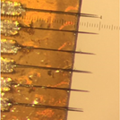
Carbon Fiber Microelectrode Array (CFMA)
Potassium ChlorideCatalog #001772
Carbon Fiber Microelectrode Array (CFMA), NeuroNex MINT
Potassium Chloride, 20 mEq, MWI Animal Health
Potassium Chloride Solution
0.3 mL of potassium chloride (KCl) solution is prepared at a concentration of 2 mEq/mL.
Anesthesia
The animal (Sprague-Dawley female rat) is anesthetised with an intraperitoneal injection of ketamine (90 mg/kg) and xylazine (10 mg/kg). Anesthesia is maintained with a ketamine (30 mg/kg) injection approximately every hour.
Surgical Preparation
A midline cervical incision is made to access the left cervical vagus nerve. Under a dissection microscope, the vagus nerve is isolated (5-7 mm) from the carotid artery and surrounding tissue and placed on a custom 3D-printed nerve holder.
CFMA Insertion
The Carbon Fiber Microelectrode Array (CFMA) is connected to a neural interface processor (Grapevine, Ripple) through a front-end headstage (Nano 2, Ripple). The headstage is controlled by a micromanipulator for accurate insertion of CFMA fibers into the vagus nerve. A small camera (MS100, Teslong) is positioned in the surgical opening to visulaize the alignment and insertion of CFMA fibers into the vagus nerve.
Experiment
Once the CFMA fibers are inserted, a recording trial is intitiated. Forty seconds into the recording trial, 0.3 mL of KCl (2 mEq/mL) is applied on the vagus nerve.
Data Analysis
Recorded signals are sorted for spikes using Plexon Offline Sorter and analyzed using MATLAB to calculates peak-to-peak voltage of spikes, conduction velocity and noise floor.