Feb 12, 2026
Version 2
Usutu virus (Orthoflavivirus usutuense) amplicon sequencing for Illumina (LowCost) V.2
- Gábor Tóth1,
- Marike Petersen1,
- Alexandra Bialonski1,
- Heike Baum1,
- Marcy A. Sikora 1,
- Alex Tomazatos1,
- Balázs Horváth1,
- Jonas Schmidt-Chanasit1,
- Dániel adar1
- 1Bernhard Nocht Institute for Tropical Medicine



Protocol Citation: Gábor Tóth, Marike Petersen, Alexandra Bialonski, Heike Baum, Marcy A. Sikora , Alex Tomazatos, Balázs Horváth, Jonas Schmidt-Chanasit, Dániel adar 2026. Usutu virus (Orthoflavivirus usutuense) amplicon sequencing for Illumina (LowCost). protocols.io https://dx.doi.org/10.17504/protocols.io.eq2lyjormlx9/v2Version created by Gábor óth
License: This is an open access protocol distributed under the terms of the Creative Commons Attribution License, which permits unrestricted use, distribution, and reproduction in any medium, provided the original author and source are credited
Protocol status: Working
We use this protocol and it's working
Created: January 18, 2026
Last Modified: February 12, 2026
Protocol Integer ID: 238852
Keywords: WNV, West Nile virus, Targeted Sequencing, Illumina, iSeq100, Lineage-2, usutu virus, many human pathogenic virus, orthoflavivirus usutuense, ebola virus, widespread arbovirus, methods for usuv, important virus, zika virus, sequencing method, usuv, natural circulation of usuv, illumina amplicon, virus, sequencing, illumina iseq, humans via mosquito bite, iseq100, illumina platform, illumina platform on the market, illumina, mosquito bite, molecular epidemiology, shorter amplicon length
Abstract
Amplicon based targeted sequencing methods proved their importance in case of many human pathogenic viruses (e.g. Zika virus, Ebola virus, SARS-Cov 2). Application and development of this methods for other medically important viruses could be also beneficial as it ensure the possibility of molecular epidemiology.
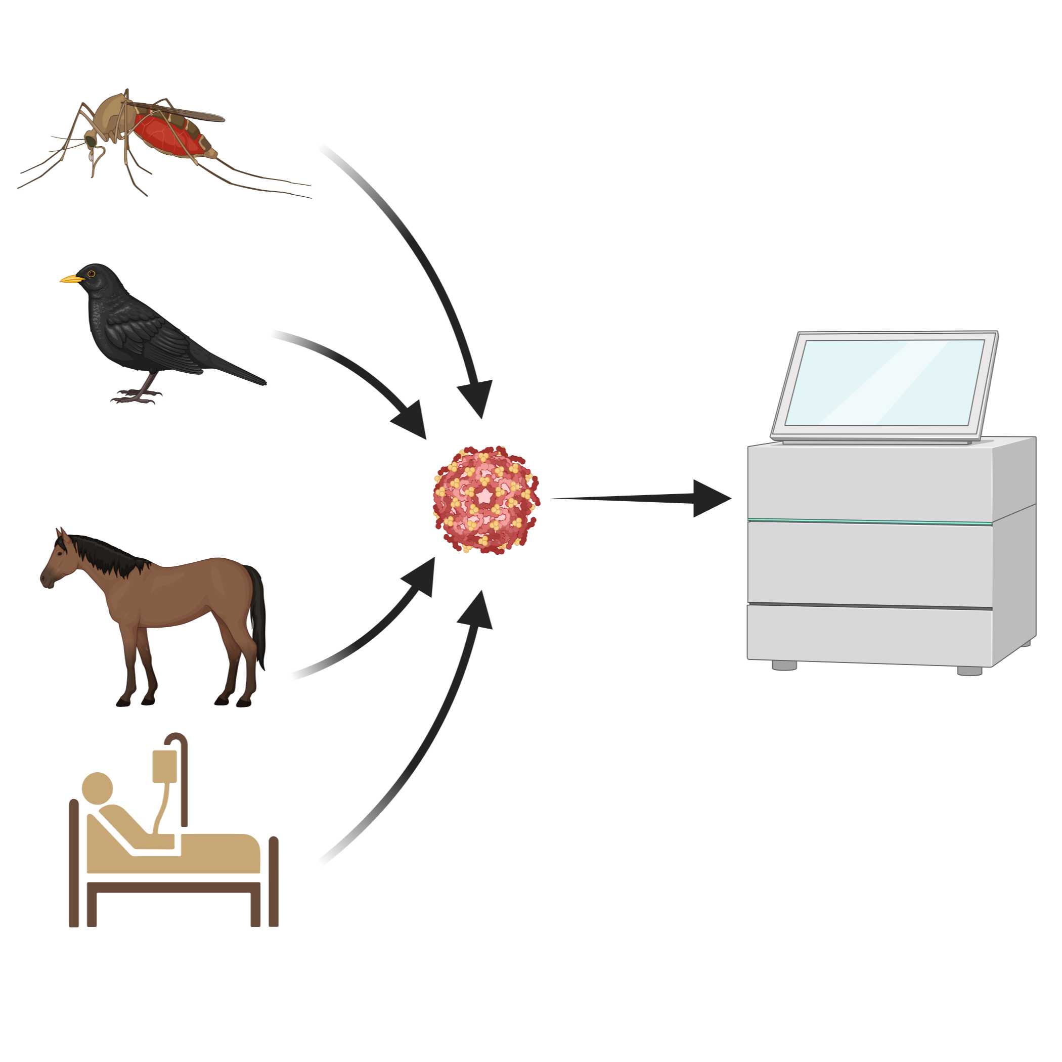
Usutu virus (USUV, Orthoflavivirus usutuense) is widespread arbovirus which is mainly transmitted to humans via mosquito bites. In the natural circulation of USUV multiple hosts are involved which raise the need for sufficient sequencing method.
There are already available, established targeted sequencing methods for USUV, but we wanted to develop an additional one which uses shorter fragment length and optimised for Illumina iSeq 100. Shorter amplicon length works better on low quality samples and iSeq100 is the cheapest (most accessible) Illumina platform on the market.
Materials
| A | B | C | |
| Reagents | |||
| Component | Supplier | Catalog number | |
| QIAamp Viral RNA Mini Kit | Qiagen | 52904 | |
| RealStar® WNV RT-PCR Kit 2.0 | altona Diagnostics GmbH | 322013 | |
| SuperScript IV Reverse Transcriptase | Invitrogen | 18090010 | |
| Random Hexamers (50 μM) | Invitrogen | N8080127 | |
| RNaseOUT™ Recombinant Ribonuclease Inhibitor | Invitrogen | 10777019 | |
| Q5® High-Fidelity DNA Polymerase | New England Biolabs | M0491S | |
| dNTP Mix (10 mM each) | Thermo Scientific™ | R0192 | |
| Individual primers | IDT or Eurofins etc. | ||
| NEBNext® Ultra™ II End Repair/dA-Tailing Module | New England Biolabs | E7546S | |
| QIAseq FX DNA Library UDI Kit (24) | Qiagen | 180477 | |
| Agencourt® AMPure® XP | Beckman Coulter | A63880 | |
| Qubit™ 1X dsDNA High Sensitivity (HS) | Invitrogen | Q33230 | |
| Absolute Ethanol (Molecular Biology Grade) | any | - | |
| Nuclease free water | any | - | |
| SeaKem® LE Agarose | Lonza | 50004 | |
| iSeq 100 i1 Reagent v2 (300-cycle) | Illumina | 20031371 | |
| Consumables | |||
| Qubit™ Assay Tubes | Invitrogen | Q32856 | |
| PCR tubes 0,2 ml | any | - | |
| 1,5 ml tubes | any | - | |
| Pipette tips 0,1-1000 μl | |||
| Equipments | |||
| Magnetic stand or rack | any | - | |
| Pipettes 0,1-1000 μl | any | - | |
| Qubit™ 4 Fluorometer | Invitrogen | Q33238 | |
| End-point PCR machines | any | - | |
| Gel electrophoresis system | any | - | |
| iSeq 100 System | Illumina | ||
Safety warnings
Multiplex PCR based amplification of a targeted pathogen genome could be highly effective, so it could serve as potential contamination source! This and other amplicon based method should be separated from diagnostic units to avoid cross-contamination. The implementation of negative controls are necessary to check the reagents purity.
Nucleic Acid Extraction
RNA extraction with QIAamp Viral RNA kit based on manufacturers recommendation. Other RNA extraction kits can be used as well e.g. Quick-RNA Viral Kit (Zymo Research).
Note
QIAamp‱ Viral RNA Mini Handbook
 HB-0354-008_HB_QA_Viral_RNA_Mini_0623_WW.pdf
 qRT-PCR
In house method, using the primers from Nikolay et. al. and optimised with OneStep RT-PCR Kit (Qiagen, Hilden, Germany).
Component Volume
5x OneStep RT-PCR Buffer 5 µL
OneStep RT-PCR Enzyme Mix 1 µL
dNTP Mix 1 µL
Nuclease Free Water 11.4 µL
UsuFP 0.6 µL
UsuRP 0.6 µL
UsuP 0.4 µL
Sample 5 µL
Final volume 25 µL
Reaction conditions
Step Temperature Time Cycles
Reverse transcription 50 °C 00:30:00 1
Denaturation 95 °C 00:15:00 1
94 °C 00:00:30 45
Amplification 58 °C 00:00:45 45
72 °C 00:01:00 45
Final extension 72 °C 00:10:00 1
Note
The publication for the primers:
Nikolay B, Weidmann M, Dupressoir A, Faye O, Boye CS, Diallo M, et al.
Development of a Usutu virus specific real-time reverse transcription PCR assay based on sequenced strains from Africa and Europe. J Virol Methods. 2014;197.
UsuFP: CAAAGCTGGACAGACATCCCTTAC
UsuRP: CGTAGATGTTTTCAGCCCACGT
UsuP: 6FAM-AAGACATATGGTGTGGAAGCCTGATAGGCA-TMR
OneStep RT-PCR Kit (Qiagen, Hilden, Germany) Handbook:
 EN-QIAGEN-OneStep-RT-PCR-Kit-Handbook.pdf
 Reverse transcription
Reverse transcription is conducted with SuperScript IV Reverse Transcriptase.
Mix the following components in a 0.2 mL tube.
Component Volume
50µM random hexamers 1 µL
10mM dNTPs mix (10mM each) 1 µL
Template RNA 11 µL
Total volume 13 µL
Gently mix by pipetting, and spin down afterwards if needed. Incubate the reaction as follows:
Temperature Time
65 °C 00:05:00
Place on ice for 00:01:00
Note
The master mix should be prepared in a clean cabinet while the sample should be added in another one.
6m
Prepare the following master mix:
Component Volume
SSIV Buffer 4 µL
100mM DTT 1 µL
RNaseOUT RNase Inhibitor 1 µL
SSIV Reverse Transcriptase 1 µL
Total volume 7 µL
Add 7 µL of master mix to the 13 µL annealed template RNA. The final volume is 20 µL . Gently mix by pipetting, and spin down afterwards if needed.
Incubate the reaction as follows:
Temperature Time
25 °C 00:05:00
50 °C 00:50:00
70 °C 00:20:00
Note
The master mix should be prepared in a clean cabinet while the sample should be added in another one.
1h 15m
Primer pool preparation
Primer pool preparation from preordered primers. The primers were designed with the usage of Primalscheme using the sequences from the list below:
 Usuta_primer_set.ods
 The list of designed primers and pool ID (1 or 2) are available in the attached table.
This table contains the necessary amount and ratios of individual primers what should be used for primer pool generation.
Primers should be sorted based on there pool ID and resuspend lyophilised oligos in nuclease free water to 100 µM concentration. We tried to optimise the primer concentrations and the current ratios are written in the table above. Use the mentioned amount (5 µL to 12.5 µL ) from unique primers to generate primer stocks (Pool 1-2).
Dilute the primer stocks (100 µM cc) 1:10 in nuclease free water to get the reaction ready primer solution (10 µM).
Note
Primer pools should be prepared in a clean mastermix cabinet!
For one reaction 2.5 µL reaction ready primer solution (10 µM) is needed. It can be prepared and aliquoted in 100 µL to avoid contamination and degradation.
Multiplex-PCR
Set up the multiplex PCR reaction in 0.2 mL PCR tube.
Reactions with Pool 1 and 2 primers:
Component Pool 1 Pool 2
5X Q5 Reaction Buffer 5 µL 5 µL
10 mM dNTPs 0.5 µL 0.5 µL
Q5 Hot Start DNA Polymerase 0.25 µL 0.25 µL
Primer Pool 1 or 2 (10µM) 2.5 µL 2.5 µL
Nuclease-free water 14.25 µL 14.25 µL
Total volume: 22.5 µL 22.5 µL
Note
The master mix should be prepared in a clean cabinet while the sample should be added in another one.
Add 2.5 µL cDNA directly to each tube and mix well by pipetting. The final volume is 25 µL .
Note
Inside the extraction and sample addition cabinet
Set-up the following program on the thermal cycler:
Step Temperature Time Cycles
Initial Denaturation 98 °C 00:00:30 1
Denaturation 98 °C 00:00:05 20-38
Annealing + Extension 60 °C 00:01:30 20-38
Hold 4 °C Till further processes
Note
The cycle number are determined based on the qPCR result. In our case the Ct number + ~3 cycles formula gave the best result (e.g. The Ct value is 26,54 use 30 cycles). In general, use higher number of cycles than the obtained Ct value.
This is a safe stopping point. The amplicons can be stored at 4°C till further processes.
2m 5s
Amplicon clean-up
Conduct clean-up with Ampure XP Beads.
Sample/Bead ratio = 1:1 (50 µL )
Washing: two times with 200 µL 80% EtOH.
Elution: 28 µL nuclease-free water.
Merge the two pools prior to adding the magnetic beads.
Add 50 µL AMPure XP beads to the sample and mix gently. The sample/magnetic bead ratio is 1:1.
Incubate for 00:05:00 at room temperature.
5m
Place the tube on a magnetic rack for 00:02:00 (until supernatant is completely clear).
2m
Remove supernatant without touching the bead pellet.
Add 200 µL freshly prepared 80% ethanol to the tube. Float the beads through ethanol either by replacing to another magnetic rack or mix via pipetting.
Remove supernatant and discard.
Repeat washing step with 80% ethanol (step 9.6-9.7).
Carefully remove the supernatant and dry the beads for around00:02:00 by leaving the lid open. (Dull pellet indicates dryness)
2m
Remove tube from the magnetic rack and resuspend the pellets in 28 µL nuclease-free water and incubate at room temperature for 00:02:00 .
2m
Place the tube back to a magnetic rack for 00:02:00 until supernatant is completely clear and transfer eluate into clean tube. Make sure that no beads are transferred as well.
2m
Quantification and normalisation
Quantify 1 µL directly from the two different PCR reaction (Pool 1, Pool 2) with the 1x dsDNA HS Assaykit.
Aliquot 199 µL 1x Working solution to Qubit assay tube (equal to the number of your samples). Prepare two extra tubes for the standards with 190 µL 1x Working solution.
Add 1 µL sample for each test tube and 10 µL from the Standards (Standard 1, Standard 2). The final volume is 200 µL .
Mix each sample vigorously by vortexing and pulse centrifuge to collect the liquid.
Incubate at room temperature for 00:02:00 before measuring.
Calibrate Qubit fluorometer with the standards based on manufacturers recommendation.
Read your sample.
Normalization is not required if the concentrations of the two reactions (Pool 1, Pool 2) from the same sample differ by 20 % or less. If the difference exceeds this threshold, use the entire volume of the low concentration pool and add an equivalent concentration from the other pool.
Note
Alternative dsDNA quantification systems can be used as well (e.g.QuantiFluor‱ ONE dsDNA System).
End Repair and dA Tailing
Amplicon end preparation with NEBNext Ultra II End Repair/dA-Tailing Module.
Add the following components to a sterile nuclease-free tube:
Component Volume
NEBNext Ultra II End Prep Reaction Buffer 1.75 µL
NEBNext Ultra II End Prep Enzyme Mix 0.75 µL
50 ng Amplicons x µL x
Nuclease-free water 12.5-x µL
Total volume 15 µL
Note
If the concentration does not reach the 4 ng / µl use the whole amount (25 µL ) from the cleaned up amlicons.
Place sample in a thermal cycler, with the heated lid set to 75 °C , and run the following program:
Temperature Time
20 °C 00:30:00
70 °C 00:15:00
45m
Adapter Ligation
Adapter ligation with QIAseq FX DNA Library Kit.
Component Volume
DNA Ligase Buffer, 5x 10 µL
DNA Ligase 5 µL
UDI Adapter 5 µL
Nuclease-free water 17.5 µL
Previous reaction 15 µL
Final volume 50 µL
Place sample in a thermal cycler without heated lid, set and incubate:
Temperature Time
20 °C 00:15:00
Note
We used QIAseq FX DNA Library Kit as ligation module. Applied concentrations and reaction conditions are optimised for this protocol. Alternative ligase modules could be use after optimisation (e.g. from NEB).
15m
Library clean up
Conduct clean-up with Ampure XP Beads.
Sample/Bead ratio = 1 : 0.6 (30 µL )
Washing: two times with 200 µL 80% EtOH.
Elution: 25 µL nuclease-free water.
Add 30 µL AMPure XP beads to the sample and mix gently. The sample/magnetic bead ratio is 1:0.6.
Incubate for 00:05:00 at room temperature.
Place the tube on a magnetic rack for 00:02:00 (until the supernatant is completely clear).
Remove supernatant without touching the bead pellet.
Add 200 µL freshly prepared 80% ethanol to the tube. Float the beads through ethanol either by replacing to another magnetic rack or mix via pipetting.
Remove supernatant and discard.
Repeat washing step with 80% ethanol (step 12.5-12.6).
Carefully remove the supernatant and dry the beads for around00:02:00 by leaving the lid open. (Dull pellet indicates dryness)
Remove tube from the magnetic rack and resuspend the pellets in 25 µL nuclease-free water and incubate at room temperature for 00:02:00 .
Place the tube back to a magnetic rack for 00:02:00 until the supernatant is completely clear and transfer eluate into clean tube. Make sure that no beads are transferred as well.
Library amplification
Conduct library amplification with the usage of HiFi PCR Master Mix.
Mix the following components in an 0.2 mL tube.
Component Volume
HiFi PCR Master Mix, 2x 12.5 µL
Primer Mix Illumina Library Amp 0.75 µL
Adapter ligated amplicons 11.75 µL
Final volume 25 µL
Step Temperature Time Cycles
Initial Denaturation 98 °C 00:02:00 1
Denaturation 98 °C 00:00:20 4-8
Annealing 60 °C 00:01:30 4-8
Extension 72 °C 00:00:30 4-8
Final extension 72 °C 00:01:00 1
Hold 4 °C Till further processes
Note
Under 0,5 ng the applied cycle number should be 8 while above 4 ng 4 cycles is suitable. Reduced number of PCR cycles can result balanced amplicon distribution. Concentration measurment is recommended at this point.
5m 20s
Final Library clean up
Conduct clean-up with Ampure XP Beads.
Sample/Bead ratio = 1 : 0.7 (17.5 µL )
Washing: two times with 200 µL 80% EtOH.
Elution: 25 µL Elution Buffer.
Add 17.5 µL AMPure XP beads to the sample and mix it gently. The sample/magnetic bead ratio is 1:0.7.
Incubate for 00:05:00 at room temperature.
Place the tube on magnetic rack for 00:02:00 (until the supernatant is completely clear).
Remove supernatant without touching the bead pellet.
Add 200 µL freshly prepared 80% ethanol to the tube. Float the beads through ethanol either by replacing to another magnetic rack or mix via pipetting.
Remove supernatant and discard.
Repeat washing step with 80% ethanol (step 14.5-14.6).
Carefully remove the supernatant and dry the beads for around00:02:00 by leaving the lid open. (Dull pellet indicates dryness)
Remove tube from the magnetic rack and resuspend the pellets in 25 µL elution buffer and incubate at room temperature for 00:02:00 .
Place the tube back to the magnetic rack for 00:02:00 until the supernatant is completely clear and transfer the eluate into clean tube. Make sure that no beads are transferred as well.
Library Size check
Run 5 µL from the cleaned up final library on 1,5% agarose gel. Low concentration of WNV genome in the sample could affect the multiplex PCR effectivity. Missing template genome results high amount of primer dimers. The expected final library size is 400bp to 420 bp. The dimers are appearing around 200 bp.
Note
Examples of clean and primer dimer containing libraries.
Fragment size should be considered in the case of pooling to be sure about the equal sequencing depth of individual samples.Fragment size should be considered in the case of pooling to be sure about the equal sequencing depth of individual samples.Fragment size should be considered in the case of pooling to be sure about the equal sequencing depth of individual samples.Fragment size should be considered in the case of pooling to be sure about the equal sequencing depth of individual samples. Fragment size should be considered in the case of pooling to be sure about the equal sequencing depth of individual samples.Fragment size should be considered in the case of pooling to be sure about the equal sequencing depth of individual samples.
Pooling and loading final libraries
Fragment size should be considered in the case of pooling to be sure about the equal sequencing depth of individual samples.
Pool all samples together in a 1.5 mL Eppendorf Low Binding tube in equimolar concentration.
Dilute the pooled libraries in RSB to 100 pM final concentration. Add PhiX control.
Load 20 µL onto an iSeq 100 2x150 bp flow cell.
Start the sequencing.
Protocol references
Amplicon based targeted sequencing:
Quick, J., Grubaugh, N. D., Pullan, S. T., Claro, I. M., Smith, A. D., Gangavarapu, K., Oliveira, G., Robles-Sikisaka, R., Rogers, T. F., Beutler, N. A., Burton, D. R., Lewis-Ximenez, L. L., de Jesus, J. G., Giovanetti, M., Hill, S. C., Black, A., Bedford, T., Carroll, M. W., Nunes, M., … Loman, N. J. (2017). Multiplex PCR method for MinION and Illumina sequencing of Zika and other virus genomes directly from clinical samples. Nature Protocols, 12(6). https://doi.org/10.1038/nprot.2017.066
ARTIC SARS-CoV-2 sequencing protocol v4 (LSK114) V.4
Koskela von Sydow, A., Lindqvist, C. M., Asghar, N., Johansson, M., Sundqvist, M., Mölling, P., & Stenmark, B. (2023). Comparison of SARS-CoV-2 whole genome sequencing using tiled amplicon enrichment and bait hybridization. Scientific Reports, 13(1). https://doi.org/10.1038/s41598-023-33168-1
Vogels, C. B. F., Hill, V., Breban, M. I., Chaguza, C., Paul, L. M., Sodeinde, A., Taylor-Salmon, E., Ott, I. M., Petrone, M. E., Dijk, D., Jonges, M., Welkers, M. R. A., Locksmith, T., Dong, Y., Tarigopula, N., Tekin, O., Schmedes, S., Bunch, S., Cano, N., … Grubaugh, N. D. (2024). DengueSeq: a pan-serotype whole genome amplicon sequencing protocol for dengue virus. BMC Genomics, 25(1). https://doi.org/10.1186/s12864-024-10350-x
AmpliSeq for Illumina SARS-CoV-2 Research Panel
Usutu virus specific real-time reverse transcription PCR assay
Nikolay B, Weidmann M, Dupressoir A, Faye O, Boye CS, Diallo M, et al. Development of a Usutu virus specific real-time reverse transcription PCR assay based on sequenced strains from Africa and Europe. J Virol Methods. 2014;197.
https://doi.org/10.1016/j.jviromet.2013.08.039
Amplicon based targeted sequencing for USUV:
Oude Munnink BB, Kik M, de Bruijn ND, Kohl R, van der Linden A, Reusken CBEM, et al. Towards high quality real-time whole genome sequencing during outbreaks using Usutu virus as example. Infection, Genetics and Evolution. 2019;73.
Ndione MHD, Diagne MM, Mencattelli G, Diallo A, Ndiaye EH, Di Domenico M, et al. An amplicon-based sequencing approach for Usutu virus characterization. Virol J [Internet]. 2024;21(1):163. Available from: https://doi.org/10.1186/s12985-024-02426-7
Holicki CM, Bergmann F, Stoek F, Schulz A, Groschup MH, Ziegler U, et al. Expedited retrieval of high-quality Usutu virus genomes via Nanopore sequencing with and without target enrichment. Front Microbiol. 2022;13.