Oct 07, 2025
Use of cholera toxin subunit B to label neural projections to major pelvic ganglia
- 1University of Melbourne
- SPARCTech. support email: [email protected]


Protocol Citation: Janet R Keast, Peregrine B Osborne, Nicole Wiedmann, John-Paul Fuller-Jackson 2025. Use of cholera toxin subunit B to label neural projections to major pelvic ganglia. protocols.io https://dx.doi.org/10.17504/protocols.io.36wgq71jovk5/v1
License: This is an open access protocol distributed under the terms of the Creative Commons Attribution License, which permits unrestricted use, distribution, and reproduction in any medium, provided the original author and source are credited
Protocol status: Working
We use this protocol and it's working
Created: June 24, 2022
Last Modified: October 07, 2025
Protocol Integer ID: 65218
Keywords: lower urinary tract, retrograde tracing, neural tracing, major pelvic ganglia, spinal cord, neural projections to major pelvic ganglia, major pelvic ganglia, innervating pelvic visceral organ, preganglionic neurons in the spinal cord, pelvic visceral organ, preganglionic neuron, lower urinary tract, spinal cord, neuron, neural projection, methods of anesthesia, anesthesia
Funders Acknowledgements:
NIH SPARC
Grant ID: OT2OD023872
Abstract
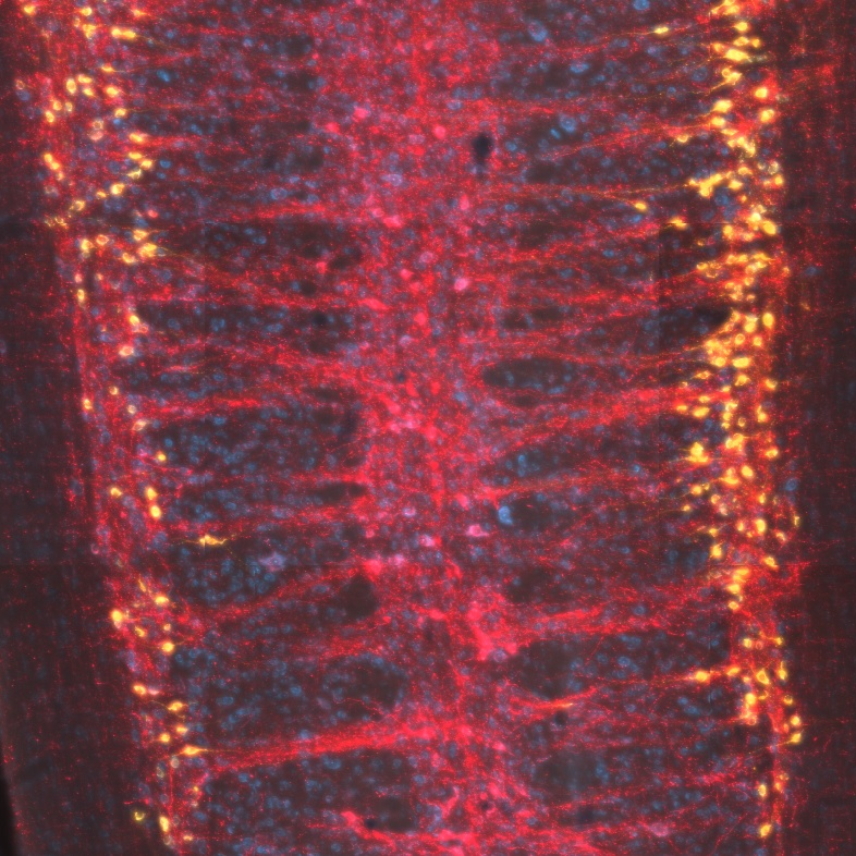
This protocol is used to visualise preganglionic neurons in the spinal cord innervating pelvic visceral organs (e.g., the lower urinary tract) in an experimental adult male or female rat. The protocol is performed under anesthesia and should incorporate all local requirements for standards of animal experimentation, including methods of anesthesia, surgical environment, and post-operative monitoring and care.
Materials
MATERIALS
Parafilm
IsofluraneZoetisCatalog #10015516
LacrilubeEllar Laboratories
Cholera toxin subunit BList LabsCatalog #104
Glass capillaries Warner instrumentsCatalog ##GC100F-15 Sterile Saline (0.9% NaCl) Evans Blue DyeMerck MilliporeSigma (Sigma-Aldrich)Catalog #E2129
Equipment
Picospritzer III Intracellular Microinjection Dispense System
NAME
Injection system
TYPE
Picospritzer
BRAND
052-0500-900
SKU
LINK
100 psi, 2 channel
SPECIFICATIONS
Preparation for surgery
Prepare cholera toxin subunit B solutions: low salt formulation with 0.05% Evans Blue.
Prepare glass pipettes for surgery by prefilling each pulled glass pipette with cholera toxin subunit B
Anesthetise animal (2.5% isoflurane in oxygen, or as required for maintenance)
Apply eye lubricant and place animal on heated pad.
Shave and clean the ventral abdomen.
Surgery
Perform a midline incision in the skin and then the muscle, then gently move organs to visualise the required injection site.
The major pelvic ganglia is located on the dorsal lateral lobe of prostate (male) or cervix (female)
Using fine angled forceps, gently blunt dissect underneath the pelvic ganglia. Once the ganglia is separated from the underlying tissue, slide a sterile 2mm x 2mm piece of parafilm between it and the tissue.
Microinject sterile tracer solution at the selected injection site using a glass pipette attached to a picospritzer. At each injection site, hold the glass pipette in place for ~5 seconds after ejection of the dye, to enable the dye to spread to the underlying tissue. This also minimises leakage. Continue injections until the desired volume is reached.
Wash all injection sites with sterile saline.
For a bilateral injection, perform steps 6-8 on the alternate side.
Close the muscle and skin using approved procedures. Administer analgesics and monitor animal during postoperative period as per local approved procedure.
Tissue harvesting
To analyse tracer distribution in spinal cord or the injection site, 3 days after surgery, anesthetise animals as per local ethical requirements, and perform intra-cardiac perfusion with fixative, then dissect tissues of interest for further study.