Sep 29, 2025
UroMOCA and StimPod Device Implantation
- Dennis Bourbeau1,2,
- Brett Hanzlicek3
- 1MetroHealth Medical Center, Cleveland, OH;
- 2Louis Stokes VA Hospital, Cleveland, OH;
- 3Advanced Platform Technology Center, Louis Stokes VA Hospital, Cleveland, Ohio


Protocol Citation: Dennis Bourbeau, Brett Hanzlicek 2025. UroMOCA and StimPod Device Implantation. protocols.io https://dx.doi.org/10.17504/protocols.io.81wgbxn9olpk/v1
License: This is an open access protocol distributed under the terms of the Creative Commons Attribution License, which permits unrestricted use, distribution, and reproduction in any medium, provided the original author and source are credited
Protocol status: Working
We use this protocol and it's working
Created: September 27, 2023
Last Modified: September 29, 2025
Protocol Integer ID: 88481
Keywords: stimpod device implantation this protocol, implantation procedure of the wireless bladder device, stimpod device implantation, wireless bladder device, wireless bladder, wireless stimulation device, stimpod, uromoca, implantation procedure, implantation, protocol, pig
Abstract
This protocol describes the implantation procedure of the wireless bladder device (UroMOCA) and of the wireless stimulation device (StimPods) into pigs.
Before start
Perform standard surgical prep.
UroMOCA implantation into bladder
Clean the incision site using Betadine solution and isolate the prep area with sterile towels. Sterile instruments will be used.
Place animal in supine position.
Insert single lumen 8 Fr catheter.
Administer analgesic at injection site.
Expose the bladder by making a dorsal midline incision.
Incise bladder dome.
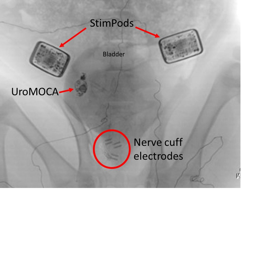
Place UroMOCA device into bladder.
Close bladder incision and evaluate integrity.
Place sterile drape on abdomen for UroMOCA transmission coil.
Verify function of UroMOCA in vivo.
Close abdominal incision.
StimPod Implantation
Place animal in prone position.
Administer analgesic at injection site.
Disinfect and drape lower back.
Make a dorsal midline sacral incision to expose the sacral spine from the second-to-last lumbar dorsal spinal process to the end of the sacrum.
Perform a laminectomy to remove the bone of the spine to expose the sacral roots from S1 to S2.
Implant 4 nerve cuff electrodes, including one each around the sacral root S1 left side, S1 right side, S2 left side and S2 right side.
Place EMG electrodes along the pelvic floor.
All electrodes will be attached to 2 wireless, implantable stimulation devices (StimPod, Micro-Leads, Boston, MA).
Verify function of StimPods in vivo (Power, Data transmission).
Confirm motor response (visual and EMG) to sacral root stimulation on each nerve cuff electrode.
Close back incision.
Monitor animal until recumbent.