Nov 24, 2021
Universal, amplicon-based sequencing method for Canine Distemper Virus (CDV)
Forked from nCoV-2019 sequencing protocol v2 (GunIt)
This protocol is a draft, published without a DOI.
- Gábor Tóth1,
- Zsófia anszki1,
- Gabor Kemenesi1
- 1National Laboratory of Virology (Hungary), University of Pecs
- Szentagothai Research Centre Virology


Protocol Citation: Gábor Tóth, Zsófia anszki, Gabor Kemenesi 2021. Universal, amplicon-based sequencing method for Canine Distemper Virus (CDV). protocols.io https://protocols.io/view/universal-amplicon-based-sequencing-method-for-can-bykwpuxe
License: This is an open access protocol distributed under the terms of the Creative Commons Attribution License, which permits unrestricted use, distribution, and reproduction in any medium, provided the original author and source are credited
Protocol status: In development
We are still developing and optimizing this protocol
Created: September 28, 2021
Last Modified: November 24, 2021
Protocol Integer ID: 53622
Keywords: Canine distemper virus, CDV, NGS, Nanopore, method for canine distemper virus, like lineage of canine distemper virus, canine distemper virus, animal morbillivirus, cdv strain, described cdv strain, distemper virus, portable genome sequencing for ebola surveillance, parvovirus vaccination, vaccines against the virus, new cdv variant, dynamics of cdv epidemiology, cdv epidemiology, veterinary microbiology, canine distemper spillover in domestic dog, veterinary clinics of north america, literature of cdv, vaccine, sequencing method, canine distemper spillover, multihost pathogen, other carnivorous families like mustilidae, cdv, hyena, genome sequencing, aetiology of canine, veterinary clinic, other carnivorous family, portable genome sequencing, canine, novel strain, pathogen, hyanidae, comparative immunology, efficacy of the vaccine, ebola surveillance, pathogens in europe, infectious disease, domestic dog, other virus, virus, universal amplicon, infectious respiratory disease complex, immunisation failure, di
Abstract
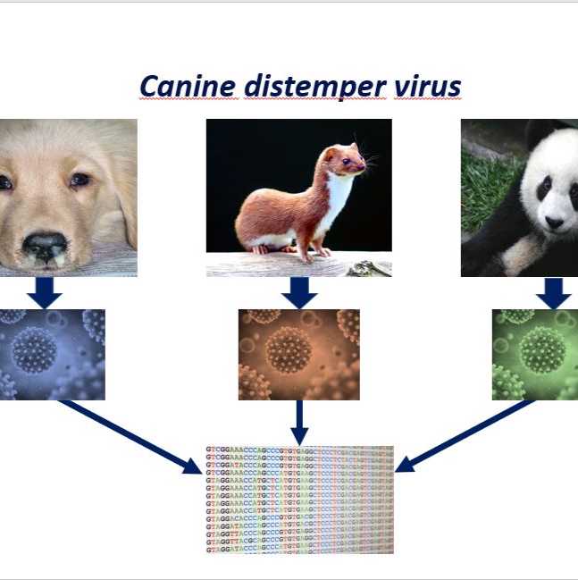
Canine distemper virus is a multihost pathogen wich mostly affects family Caniade (dog, fox, coyote, wolf) but it is also occur in other carnivorous families like Mustilidae (ferret, skunk, badger, mink, weasel, otter), Procyonidae (racoon, lesser panda, kinkajou), Hyanidae (hyenas) or Ursidae (bear).
Increasing surveillance needed to identify new CDV variants and to understand the dynamics of CDV epidemiology . There are available vaccines against the virus, but it is important to asses the efficacy of the vaccine against these novel strains. To maintain these follow up efforts we developed an universal amplicon based sequencing method which is capable to generate data from the previously described CDV strains.
The protocol was forked and designed by the original work of Josh Quick (https://www.protocols.io/view/ncov-2019-sequencing-protocol-v2-bdp7i5rn?version_warning=no).
Now this method is in experimental phase therefore it should be widely tested to evaluate it's efficiency.
Some literature of CDV:
-Martella, V., Elia, G. and Buonavoglia, C. (2008) ‘Canine Distemper Virus’, Veterinary Clinics of North America: Small Animal Practice, 38(4), pp. 787–797. doi: 10.1016/J.CVSM.2008.02.007.
-Kapil, S. and Yeary, T. J. (2011) ‘Canine Distemper Spillover in Domestic Dogs from Urban Wildlife’, Veterinary Clinics of North America: Small Animal Practice, 41(6), pp. 1069–1086. doi: 10.1016/J.CVSM.2011.08.005.
-Day, M. J. et al. (2020) ‘Aetiology of Canine Infectious Respiratory Disease Complex and Prevalence of its Pathogens in Europe’, Journal of Comparative Pathology, 176, pp. 86–108. doi: 10.1016/J.JCPA.2020.02.005.
-Takeda, M. et al. (2020) ‘Animal morbilliviruses and their cross-species transmission potential’, Current Opinion in Virology, 41, pp. 38–45. doi: 10.1016/J.COVIRO.2020.03.005.
-Decaro, N., Buonavoglia, C. and Barrs, V. R. (2020) ‘Canine parvovirus vaccination and immunisation failures: Are we far from disease eradication?’, Veterinary Microbiology, 247, p. 108760. doi: 10.1016/J.VETMIC.2020.108760.
-Koç, B. T., Akkutay-Yoldar, Z. and Oğuzoğlu, T. Ç. (2021) ‘New members to Arctic-like lineage of canine distemper virus from Turkey’, Comparative Immunology, Microbiology and Infectious Diseases, 78, p. 101678. doi: 10.1016/J.CIMID.2021.101678.
-Chang, Z. et al. (2021) ‘Spatiotemporal dynamics for an impulsive eco-epidemiological system driven by canine distemper virus’, Applied Mathematics and Computation, 402, p. 126135. doi: 10.1016/J.AMC.2021.126135.
Literature of the amplicon-based sequencing:
-Quick, Joshua et al. “Real-time, portable genome sequencing for Ebola surveillance.”Naturevol. 530,7589 (2016): 228-232. doi:10.1038/nature16996
-Quick, Joshua et al. “Multiplex PCR method for MinION and Illumina sequencing of Zika and other virus genomes directly from clinical samples.”Nature protocolsvol. 12,6 (2017): 1261-1276. doi:10.1038/nprot.2017.066
Materials
Primers IDT
Extraction kits; Zymo Quick-RNA Viral Kit Zymo R1034 or
QIAamp Viral RNA Mini Qiagen 52904
SuperScript IV (50 rxn) Thermo 18090050
dNTP mix (10 mM each) Thermo R0192
Random Hexamers (50 µM) Thermo N8080127
RNase OUT (125 rxn) Thermo 10777019
Q5 Hot Start HF Polymerase NEB M0493S
NEBNext Ultra II End-prep NEB E7546S
NEBNext Quick Ligation Module NEB E6056S
Native Barcoding Expansion Kit 1-12 Nanopore EXP-NBD104
Native Barcoding Expansion Kit 13-24 Nanopore EXP-NBD114
Sequencing Auxiliary Vials Nanopore EXP-AUX001
Short Fragment Buffer Expansion kit Nanopore EXP-SFB001
Flow Cell Priming Kit Nanopore EXP-FLP002
R9.4.1 flow cells Nanopore FLO-MIN106
cDNA preparation
Mix the following components in an 0.2mL 8-strip tube;
Component Volume
50µM random hexamers 1 µL
10mM dNTPs mix (10mM each) 1 µL
Template RNA 11 µL
Total 13 µL
Note
Viral RNA input from a sample should be between Ct 18-35. If Ct is between 12-15, then dilute the sample 100-fold in water, if between 15-18 then dilute 10-fold in water. This will reduce the likelihood of PCR-inhibition.
Note
A mastermix should be made up in the mastermix cabinet and aliquoted into PCR strip tubes. Tubes should be wiped down when entering and leaving the mastermix cabinet.
Gently mix by pipetting and pulse spin the tube to collect liquid at the bottom of the tube.
Incubate the reaction as follows:
65 °C for 00:05:00
Place on ice for 00:01:00
Add the following to the annealed template RNA:
Component Volume
SSIV Buffer 4 µL
100mM DTT 1 µL
RNaseOUT RNase Inhibitor 1 µL
SSIV Reverse Transcriptase 1 µL
Total 20 µL
Note
A mastermix should be made up in the mastermix cabinet and added to the denatured RNA in the extraction and sample addition cabinet. Tubes should be wiped down when entering and leaving the mastermix cabinet.
Gently mix by pipetting and pulse spin the tube to collect liquid at the bottom of the tube.
Incubate the reaction as follows:
50 °C 00:50:00
70 °C 00:10:00
Hold at 5 °C
Primer pool preparation
If required resuspend lyophilised primers at a concentration of 100µM each
Note
Universal CDV primers for this protocol were designed using Primal Scheme and generate overlapping 1000 and 2000 nucleotide amplicons. Primer names and dilutions are listed in the table below.
The list of high quality genomes which were used to design the primers:
Universal CDV primers for 1000 basepair long amplicon set:
Universal CDV primers for 2000 basepair long amplicon set:
Generate primer pool stocks by adding 5 µL of each odd region primer to a 1.5 mL Eppendorf labelled “Pool 1 (100µM)” and each even region primer to a 1.5 mL Eppendorf labelled “Pool 2 (100µM)”. These are your 100µM stocks of each primer pool.
Number of primers: Final volume:
CDV_1000_pool_1: 63 315 µL
CDV_1000_pool_2: 50 250 µL
CDV_2000_pool_1: 32 160 µL
CDV_2000_pool_2: 27 135 µL
Note
Primers should be diluted and pooled in the mastermix cabinet which should be cleaned with decontamination wipes and UV sterilised before and after use.
Dilute this primer pool 1:10 in molecular grade water, to generate 10µM primer stocks. It is recommend that multiple aliquots of each primer pool are made to in case of degradation or contamination. Our recommendation is that yous should use 50 µL from the stock solution and add 450 µL nuclease-free water to generate the working solution of the primers.
Note
Primers need to be used at a final concentration of 0.015µM per primer.
Multiplex PCR
In the mastermix hood set up the multiplex PCR reactions as follows in 0.2mL 8-strip PCR tubes:
Reactions with the 1000 bp amplicon set:
Component Pool 1 Pool 2
5X Q5 Reaction Buffer 5 µL 5 µL
10 mM dNTPs 0.5 µL 0.5 µL
Q5 Hot Start DNA Polymerase 0.25 µL 0.25 µL
V3 Primer Pool 1 or 2 (10µM) 2.5 µL 2.0 µL
Nuclease-free water 14.25 µL 14.75 µL
Total 22.5 µL 22.5 µL
Reactions with the 2000 bp amplicon set:
Component Pool 1 Pool 2
5X Q5 Reaction Buffer 5 µL 5 µL
10 mM dNTPs 0.5 µL 0.5 µL
Q5 Hot Start DNA Polymerase 0.25 µL 0.25 µL
V3 Primer Pool 1 or 2 (10µM) 2.0 µL 2.0 µL
Nuclease-free water 14.75 µL 14.75 µL
Total 22.5 µL 22.5 µL
Note
A PCR mastermix for each pool should be made up in the mastermix cabinet and aliquoted into PCR strip tubes. Tubes should be wiped down when entering and leaving the mastermix cabinet.
In the extraction and sample addition cabinet add 2.5 µL cDNA to each tube and mix well by pipetting.
Note
The extraction and sample addition cabinet should should be cleaned with decontamination wipes and UV sterilised before and after use.
Pulse centrifuge the tubes to collect the contents at the bottom of the tube.
Set-up the following program on the thermal cycler:
Step Temperature Time Cycles
Heat Activation 98 °C 00:00:30 1
Denaturation 98 °C 00:00:15 25-35
Annealing 64 °C 00:05:00 25-35
Hold 4 °C Indefinite 1
Note
Cycle number should be 25 for Ct 18-21 up to a maximum of 35 cycles for Ct 35
Clean up
Label a 1.5 mL Eppendorf tube for each sample and assemble the following PCR dilution for each sample:
Component Volume
Pool 1 PCR reaction 25 µL
Pool 2 PCR reaction 25 µL
SPRI Beads 25 µL
70% EtOH 2X100 µL
Nuclease-free water 25 µL
In a new 1.5 ml Eppendorf tube pool all 25 µl PCR product from the same primer set (1000 pool 1-2 or 2000 pool 1-2)reactions together.
Add 0.5x volume of SPRI beads to the sample tube and mix gently by either flicking or pipetting. If you were performed 25 µL reactions it should be also 25 µL .
Pulse centrifuge to collect all liquid at the bottom of the tube.
Incubate for 00:05:00 at room temperature.
5m
Carefully remove and discard the supernatant, being careful not to touch the bead pellet.
Add 100 µL 70% EtOH and resuspend beads completely by pipette mixing.
Pulse centrifuge to collect all liquid at the bottom of the tube.
Remove supernatant and discard.
Repeat steps 14.6-14.9 to perform a second EtOH wash.
Pulse centrifuge to collect all liquid at the bottom of the tube and carefully remove as much residual ethanol as possible using a P10 pipette.
With the tube lid open incubate for 00:02:00 or until the pellet loses it's shine (if the pellet dries completely it will crack and become difficult to resuspend).
2m
Resuspend pellet in 25 µL nuclease-free water, mix gently by either flicking or pipetting and incubate for 00:02:00 .
2m
Place on magnet and transfer sample to a clean 1.5mL Eppendorf tube ensuring no beads are transferred into this tube.
Quantification and normalisation
Label another 1.5 mL Eppendorf tube for each sample.
Note
Input to the one-pot native barcoding reaction is 125 ng per sample in the case of 1000bp set and 250 ng with the 2000bp set . Process at least 6 samples plus one negative control per library in order to have sufficient material to load on the sequencer at the end.
You should measure the concentration and dilute the sample to appropriate concentration for the end-prep and barcoding reactions.
1000bp_set: 25 ng / 1 µL
2000bp_set: 50 ng / 1 µL
Note
If the concentration is under the targeted one, you can increase the input volume pcr product in the End-prep reaction if you replacing the water.
PCR pruduct: 5 µL + Nuclease free water: 7.5 µL = 12.5 µL
Native barocoding
Barcode the amplicon pools using the one-pot native barcoding approach.
Set up the following reaction for each sample:
Component Volume
PCR dilution from previous step 5 µL
Nuclease-free water 7.5 µL
Ultra II End Prep Reaction Buffer 1.75 µL
Ultra II End Prep Enzyme Mix 0.75 µL
Total 15 µL
Incubate at room temperature for 00:10:00
Incubate at 65 °C f for 00:10:00
Incubate on ice for 00:01:00
In a new 1.5mL Eppendorf tube set up the following reaction:
Component Volume
Previous reaction mixture 1.5 µL
Nuclease-free water 5.7 µL
NBXX barcode 2.5 µL
Ultra II Ligation Master Mix 10 µL
Ligation Enhancer 0.3 µL
Total 20 µL
Note
Use one native barcode from the EXP-NBD104 (1-12) or EXP-NBD114 (13-24) per sample. Use from 6 to 24 barcodes in a library, any fewer and there will be insufficient total material to achieve good yields.
Incubate at room temperature for 00:20:00
Incubate at 65 °C for 00:10:00
Incubate on ice for 00:01:00
Note
The 65°C incubation is to inactivate the DNA ligase to prevent barcode cross-ligation when reactions are pooled in the next step.
In a new 1.5 ml Eppendorf tube pool all 20 µL one-pot barcoding reactions together.
Add 0.4x volume of SPRI beads to the sample tube and mix gently by either flicking or pipetting. For example add 96 µL SPRI beads to 240 µL 12-plex pooled one-pot native barcoding reactions.
Note
0.4x volume of SPRI will only bind 400 bp amplicons in the presence of ligation buffer as in a one-pot reaction, do not use 1x as this will result in excessive native barcode carryover.
Pulse centrifuge to collect all liquid at the bottom of the tube.
Incubate for 00:05:00 at room temperature.
Place on magnetic rack and incubate for 00:02:00 or until the beads have pelleted and the supernatant is completely clear.
Carefully remove and discard the supernatant, being careful not to touch the bead pellet.
Add 700 µl SFB and resuspend beads completely by pipette mixing.
Note
SFB will remove excess adapter without damaging the adapter-protein complexes. Do not use 70% ethanol as in early clean-ups.
Pulse centrifuge to collect all liquid at the bottom of the tube.
Remove supernatant and discard.
Repeat steps 11-13 to perform a second SFB wash.
Pulse centrifuge and remove any residual SFB.
Note
You do not need to allow to air dry with SFB washes.
Add 200 µl of room-temperature 70 % volume ethanol to bathe the pellet.
Carefully remove and discard ethanol, being careful not to touch the bead pellet.
Note
Only perform 1x 70% ethanol wash
Pulse centrifuge to collect all liquid at the bottom of the tube and carefully remove as much residual ethanol as possible using a P10 pipette.
With the tube lid open incubate for 00:01:00 or until the pellet loses it's shine (if the pellet dries completely it will crack and become difficult to resuspend).
Resuspend pellet in 30 µL Elution Buffer (EB), mix gently by either flicking or pipetting and incubate for 00:02:00 .
Place on magnet and transfer sample to a clean 1.5mL Eppendorf tube ensuring no beads are transferred into this tube.
Quantify 1 µL of the barcoded amplicon pool using the Quantus Fluorometer using the ONE dsDNA assay.
Remove Lambda DNA 400 ng/µL standard from the freezer and leave on ice to thaw. Remove ONE dsDNA dye solution from the fridge and allow to come to room temperature.
QuantiFluor(R) ONE dsDNA System, 500rxnPromegaCatalog #E4870
Set up two 0.5 mL tubes for the calibration and label them 'Blank' and 'Standard'
Add 200 µL ONE dsDNA Dye solution to each tube.
Mix the Lambda DNA standard 400 ng/µL standard by pipetting then add 1 µL to one of the standard tube.
Mix each sample vigorously by vortexing for 00:00:05 and pulse centrifuge to collect the liquid.
Allow both tubes to incubate at room temperature for 00:02:00 before proceeding.
Selection 'Calibrate' then 'ONE DNA' then place the blank sample in the reader then select 'Read Blank'. Now place the standard in the reader and select 'Read Std'.
Set up the required number of 0.5 mL tubes for the number of DNA samples to be quantified.
Note
Use only thin-wall, clear, 0.5mL PCR tubes such as Axygen #PCR-05-C
Label the tubes on the lids, avoid marking the sides of the tube as this could interfere with the sample reading.
Add 199 µL ONE dsDNA dye solution to each tube.
Add 1 µL of each user sample to the appropriate tube.
Note
Use a P2 pipette for highest accuracy.
Mix each sample vigorously by vortexing for 00:00:05 and pulse centrifuge to collect the liquid.
Allow all tubes to incubate at room temperature for00:02:00 before proceeding.
On the Home screen of the Quantus Fluorometer, select `Protocol`, then select `ONE DNA` as the assay type.
Note
If you have already performed a calibration for the selected assay you can continue, there is no need to perform repeat calibrations when using ONE DNA pre diluted dye solution. If you want to use the previous calibration, skip to step 11. Otherwise, continue with step 9.
On the home screen navigate to 'Sample Volume' and set it to 1 µL then 'Units' and set it to ng/µL.
Load the first sample into the reader and close the lid. The sample concentration is automatically read when you close the lid.
Repeat step 16 until all samples have been read.
The value displayed on the screen is the dsDNA concentration in ng/µL, carefully record all results in a spreadsheet or laboratory notebook.
Set up the following AMII adapter ligation and clean-up with SFB.
Set up the following AMII adapter ligation reaction:
Component Volume
End-repaired amplicon pools 30 µL
NEBNext Quick Ligation Reaction Buffer (5X) 10 µL
Adapter Mix (AMII) 5 µL
Quick T4 DNA Ligase 5 µL
Total 50 µL
Note
There will be some variation in clean-up efficiencies but expect to carry around 80% through a clean-up.
Incubate at room temperature for 00:20:00
Add 50 µL (1:1) of SPRI beads to the sample tube and mix gently by either flicking or pipetting.
Note
Vortex SPRI beads thoroughly before use to ensure they are well resuspended, the solution should be a homogenous brown colour.
Pulse centrifuge to collect all liquid at the bottom of the tube.
Incubate for 00:05:00 at room temperature.
Place on magnetic rack and incubate for 00:02:00 or until the beads have pelleted and the supernatant is completely clear.
Carefully remove and discard the supernatant, being careful not to touch the bead pellet.
Add 250 µL SFB and resuspend beads completely by pipette mixing.
Note
SFB will remove excess adapter without damaging the adapter-protein complexes. Do not use 70% ethanol as in early clean-ups.
Pulse centrifuge to collect all liquid at the bottom of the tube.
Remove supernatant and discard.
Repeat steps 14-16 to perform a second SFB wash.
Pulse centrifuge and remove any residual SFB.
Note
You do not need to allow to air dry with SFB washes.
Add 15 µL EB and resuspend beads by pipette mixing.
Incubate at room temperature for 00:02:00 .
Place on magnetic rack.
Transfer final library to a new 1.5mL Eppendorf tube.
Quantify 1 µL of the final library using the Quantus Fluorometer using the ONE dsDNA assay.
Note
Final library can be now be stored in 10 mM Tris pH 8 at 4°C for up to a week if needed otherwise proceed directly to MinION sequencing.
Remove Lambda DNA 400 ng/µL standard from the freezer and leave on ice to thaw. Remove ONE dsDNA dye solution from the fridge and allow to come to room temperature.
QuantiFluor(R) ONE dsDNA System, 500rxnPromegaCatalog #E4870
Set up two 0.5 mL tubes for the calibration and label them 'Blank' and 'Standard'
Add 200 µL ONE dsDNA Dye solution to each tube.
Mix the Lambda DNA standard 400 ng/µL standard by pipetting then add 1 µL to one of the standard tube.
Mix each sample vigorously by vortexing for 00:00:05 and pulse centrifuge to collect the liquid.
Allow both tubes to incubate at room temperature for 00:02:00 before proceeding.
Selection 'Calibrate' then 'ONE DNA' then place the blank sample in the reader then select 'Read Blank'. Now place the standard in the reader and select 'Read Std'.
Set up the required number of 0.5 mL tubes for the number of DNA samples to be quantified.
Note
Use only thin-wall, clear, 0.5mL PCR tubes such as Axygen #PCR-05-C
Label the tubes on the lids, avoid marking the sides of the tube as this could interfere with the sample reading.
Add 199 µL ONE dsDNA dye solution to each tube.
Add 1 µL of each user sample to the appropriate tube.
Note
Use a P2 pipette for highest accuracy.
Mix each sample vigorously by vortexing for 00:00:05 and pulse centrifuge to collect the liquid.
Allow all tubes to incubate at room temperature for00:02:00 before proceeding.
On the Home screen of the Quantus Fluorometer, select `Protocol`, then select `ONE DNA` as the assay type.
Note
If you have already performed a calibration for the selected assay you can continue, there is no need to perform repeat calibrations when using ONE DNA pre diluted dye solution. If you want to use the previous calibration, skip to step 11. Otherwise, continue with step 9.
On the home screen navigate to 'Sample Volume' and set it to 1 µL then 'Units' and set it to ng/µL.
Load the first sample into the reader and close the lid. The sample concentration is automatically read when you close the lid.
Repeat step 16 until all samples have been read.
The value displayed on the screen is the dsDNA concentration in ng/µL, carefully record all results in a spreadsheet or laboratory notebook.