Oct 19, 2023
Transfection by electroporation of GFP-LRRK2 and Immunofluorescent imaging of MEFs VPS35 (D620N) mutants stably expressing LysoTag
- Rotimi Y. Fasimoye1,
- Dario R. Alessi1
- 1Medical Research Council Protein Phosphorylation and Ubiquitylation Unit, School of Life Sciences, University of Dundee, Dow Street, Dundee DD1 5EH, UK


Protocol Citation: Rotimi Y. Fasimoye, Dario R. Alessi 2023. Transfection by electroporation of GFP-LRRK2 and Immunofluorescent imaging of MEFs VPS35 (D620N) mutants stably expressing LysoTag. protocols.io https://dx.doi.org/10.17504/protocols.io.n2bvj3dwxlk5/v1
License: This is an open access protocol distributed under the terms of the Creative Commons Attribution License, which permits unrestricted use, distribution, and reproduction in any medium, provided the original author and source are credited
Protocol status: Working
We use this protocol and it's working
Created: September 25, 2023
Last Modified: May 31, 2024
Protocol Integer ID: 88587
Keywords: ASAPCRN, mouse embryonic fibroblast, lysotag transfection of foreign dna, expressing lysotag transfection, subcellular localisation of protein, embryonic fibroblast, immunofluorescent imaging of mefs vps35, lrrk2 into mef, colocalization of gfp, transfection by electroporation, subcellular localisation, immunofluorescent imaging, protein, microscopy, immunofluorescent, molecular biology, tagged lrrk2, expressed gfp, such as lrrk2, transfection, cell, foreign dna, primary cell, large sized protein, lysosomal, 250kd protein, low transfection efficiency, electroporation
Funders Acknowledgements:
Aligning Science Across Parkinson's
Grant ID: ASAP-000463
Disclaimer
DISCLAIMER – FOR INFORMATIONAL PURPOSES ONLY; USE AT YOUR OWN RISK
The protocol content here is for informational purposes only and does not constitute legal, medical, clinical, or safety advice, or otherwise; content added to protocols.io is not peer reviewed and may not have undergone a formal approval of any kind. Information presented in this protocol should not substitute for independent professional judgment, advice, diagnosis, or treatment. Any action you take or refrain from taking using or relying upon the information presented here is strictly at your own risk. You agree that neither the Company nor any of the authors, contributors, administrators, or anyone else associated with protocols.io, can be held responsible for your use of the information contained in or linked to this protocol or any of our Sites/Apps and Services.
Abstract
Transfection of foreign DNA and Immunofluorescent (IF) microscopy are powerful tools used in cellular and molecular biology to monitor subcellular localisation of proteins. Mouse embryonic fibroblasts (MEFs) are primary cells notorious for their low transfection efficiency by mostly available chemical methods. This efficiency become even lower if the aim is to express a large sized protein, such as LRRK2, a 250kD protein. Here, we described a method where we used electroporation to transiently transfect GFP-tagged LRRK2 into MEFs. We also used IF microscopy to visualise the subcellular localisation of the transiently expressed GFP-LRRK2. Furthermore, we investigated the colocalization of GFP-LRRK2 with a lysosomal localised TMEM192-3xHA.
Attachments

855-2211.docx
422KB
Materials
Materials
Cell lines
- Mouse Embryonic Fibroblast VPS35 WT (stably expressing TMEM192-3xHA)
- Mouse Embryonic Fibroblast VPS35 D620N (stably expressing TMEM192-3xHA)
Plasmids
- GFP-LRRK2 (DU13363). Plasmid available at MRCPPU depository at [email protected]
Antibodies
Table 1: List of primary antibodies
| Antibody | Company | Cat. number | Host species | |
| GFP | Abcam | AB13970 | Chicken | |
| HA | Roche | 47877600 | Rat |
Table 2: List of fluorophore-conjugated secondary antibodies
| A | B | C | D | E | |
| Antibody | Conjugated Fluorophore | Company | Cat. number | Host Species | |
| anti-Chicken | Alexa 488 | Invitrogen | A11039 | Goat | |
| anti-Rat | Alexa 594 | Invitrogen | 21209 | Donkey |
Media and Reagents
- Growth Media:
| A | B | |
| Dulbecco’s Modified Eagle’s Medium (DMEM) (GIBCO 11960-085) | ||
| Foetal Bovine Serum (FBS) (Sigma F7524 Batch# BCBW6817) | 10% | |
| L-Glutamine (GIBCO 25030024) | 1% | |
| Penicillin-Streptomycin (GIBCO 15140122) | 1% | |
| NEAA (GIBCO 11140-035) | 1X | |
| Sodium Pyruvate (GIBCO 11360-039) | 1mM |
- Transfection media: Growth media without Penicillin-Streptomycin (Pen-Step)
- Dulbecco's phosphate-buffered saline (PBS) (GIBCO 14190169)
- Bovine Serum Albumin, BSA (Roche, 10735094001)
- Sodium Azide (Sigma, S2002).
- Hoechst 33342 solution (Thermo, 62249)
- VECTASHIELD antifading Mounting media (|Vector Laboratories, H1000)
Equipment
- NEPA21 Super Electroporator with electroporator chamber (SONIDEL)
- 2mm gap Cuvette with individual pipette (SONIDEL, EC-002S)
- Incubator with FPI-sensor system and display controller MB1 (BINDER GmbH. Model: CB150. Power Output: 1.40kW, 230V, 6.1 Amp). This incubator has CO2 and O2 control.
- Leica TCS SP8 MP Multiphoton Microscope.
- Super Premium microscope slides (Frosted on one side) (VWR, 631-0114)
- Borosilicate Glass square coverslips (VWR, 631-0125)
- DeNovix‱ CellDrop Brightfield cell counter
Consumables
- 6-well tissue culture Petri Dishes (ThermoFisher. Catalog# 140675).
- Standard 1ml and 200 µl Pipette tips (Greiner bio-one. Catalog# 686271 and 685261 respectively).
DMEM, high glucose, no glutamineThermo FisherCatalog #11960085
L-Glutamine (200mM)Thermo Fisher ScientificCatalog #25030024
Penicillin-Streptomycin (10,000 U/mL)Thermo Fisher ScientificCatalog #15140122
MEM Non-Essential Amino Acids Solution (100X)Thermo FisherCatalog #11140035
DPBS no calcium no magnesiumGibco - Thermo Fisher ScientificCatalog #14190169
Bovine Serum Albumin Fraction VMerck MilliporeSigma (Sigma-Aldrich)Catalog #10735094001
Sodium azideMerck MilliporeSigma (Sigma-Aldrich)Catalog #S2002
Hoechst 33342 Solution (20 mM)Thermo FisherCatalog #62249
VECTASHIELD® Antifade Mounting Medium Vector LaboratoriesCatalog #H-1000
Nunc™ Cell-Culture Treated Multidishes, 6 wellThermo FisherCatalog #140675
PIPETTE TIPS 100- 1000 µL BLUE SUITABLE FOR EPPENDORF STERILE 60 PIECES PER RACKgreiner bio-oneCatalog #686271
PIPETTE TIP 10 - 100 µL SUITABLE FOR EPPENDORF 96 PIECES / ST RACKgreiner bio-oneCatalog #685261
Anti-GFP antibody (ab13970)AbcamCatalog #ab13970
Goat anti-Chicken IgY (H L) Secondary Antibody, Alexa Fluor 488 Thermo Fisher ScientificCatalog #A11039
Transfection of cells with GFP-LRRK2 plasmid by electroporation
18h
Place coverslips in 6 well plate (one coverslip per well) and add 2 mL of media. Place the plate in an incubator.
Pellet cells from 100% confluent 10cm plate and resuspend in 1 mL media.
Count cells and resuspend in media in a way that there are 30000-40000 cells per 10 µL .
Power on Electroporator and plug in the electroporator chamber to the output socket.
Set the following Poring Pulse parameters:
- Voltage: 200V
- Length: 5ms
- Interval: 50ms
- Number of cycles: 2
- Decay rate: 10%
- Polarity: +
Set Transfer Pulse parameters:
- Voltage: 20V
- Length: 50ms
- Interval: 50ms
- Number of cycles: 5
- Decay rate: 40%
- Polarity: +/-
Add 3-4 µg of plasmid into a 1.5ml Eppendorf tube.
Note
Ensure this is not more than 10 µl. If plasmid is too concentrated, add required amount and top-up to 10 µl with media.
Add 90 µL of resuspended cells into the tube. In total, cell number should be approximately 300000-400000. Pipette gently up and down 3 times to mix cell and plasmid.
Transfer cell-plasmid mixture into 2mm gap Cuvette and close the cap. Add gently to the side to ensure there are no bubbles. If there are bubbles, tap the cuvette gently on the side until the bubbles move to the top.
Insert the cuvette into the electroporator chamber and close the lid.
Briefly measure electrical impedance by pressing the “Ω" button and check reading. This should be between 0.03 and 0.055.
- If reading is too low, add 1-2 µL of cells.
- If too high, add 1-2 µL of media.
Press start button to begin electroporation process.
On completion, use Pasteur pipette (provided with the cuvette) to transfer cells into the wells containing a coverslip (from Step 1).
Rotate plate gently to spread the cells within the well.
Return plate into the incubator and incubate for at least 18:00:00 at 37 °C .
18h
Replace media with full Growth media (i.e., media with Pen-Strip) and incubate for another 12-18 hours.
Preparing cells for Immunofluorescence imaging
3h 25m
Remove media and wash cells.
Remove media and wash cells with 3 mL PBS +0.2% BSA+0.02% sodium azide for 00:05:00 . (1/3)
5m
Wash cells with 3 mL PBS +0.2% BSA+0.02% sodium azide for 00:05:00 . (2/3)
5m
Wash cells with 3 mL PBS +0.2% BSA+0.02% sodium azide for 00:05:00 . (3/3)
5m
Fixed cells in 4% w/v PFA. Add 3 mL of dissolved PFA and incubate at Room temperature for 00:10:00 .
10m
Permeabilise cells with 1% NP-40 (v/v in PBS +0.2%BSA+0.02% sodium azide).
Block with 3% BSA (w/v in PBS) for 00:30:00 .
30m
Prepare a combination of primary antibodies (Table 1) as shown below. Antibodies are diluted in PBS +0.2% BSA+0.02% sodium azide.
- Rat anti-HA (1:1000) and Chicken anti-GFP (1:1000)
Incubate cells at Room temperature with diluted primary antibodies for 01:00:00 . Do this in a humid chamber on a piece of Parafilm. Put a 60 µL drop of diluted antibodies on the parafilm. Carefully place coverslip on the droplet, with the side containing attached cells, facing inward, making contact with the droplet.
1h
Wash cells, 3 times, with 3 mL PBS +0.2%BSA+0.02% sodium azide.
Prepare a combination of Secondary antibodies as described below (see Table 2 for more information about the secondary antibodies). Antibodies are diluted in PBS +0.2% BSA+0.02% sodium azide.
- anti-Rat Alexa 594 (1:500) and anti-Chicken Alexa 488 (1:500).
Add 0.5 µL Hoechst 33342 solution for nuclear staining.
Incubate cells at Room temperature with diluted secondary antibodies for 01:00:00 . Do this in a humid chamber on a piece of Parafilm. Put a 60 µL drop of diluted antibodies on the parafilm. Carefully place coverslip on the droplet, with the side containing attached cells, facing inward, making contact with the droplet.
1h
Wash cells, 3 times, with 3 mL PBS +0.2%BSA+0.02% sodium azide.
Rinse cells by dipping briefly in MilliQ water and gently dry on Kleenex wipes.
Label microscope glass slides (preferably the one with frosted side) according to the primary antibody used. Take note of the emission wavelength of the probe on the secondary antibodies.
Add a drop of VECTASHIELD antifading Mounting media.
Mount cover slip (containing cells) on the glass slide, ensuring that the side containing the cells is facing inward, making contact with the oil. Allow to dry for 00:30:00 , ensuring slides are prevented from direct light.
30m
Slides can be stored at 4 °C or viewed immediately on a confocal microscope.
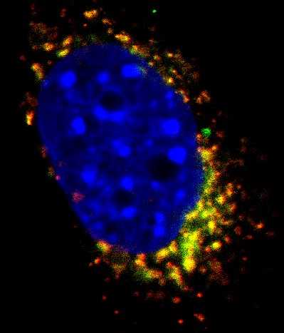
Figure 1: Immunofluorescence images of mouse embryonic fibroblasts (MEFs) expressing GFP-LRRK2 and TMEM192-3xHA. MEFs VPS35 wildtype and D620N mutants stably expressing TMEM192-3xHA and transiently expressing GFP-LRRK2 were co-immunostained with anti-HA and anti-GFP antibodies. Scale bar is 2 µm.