Xing D, Tan L, Chang CH, Li H, Xie XS (2021). Accurate SNV detection in single cells by transposon-based whole-genome amplification of complementary strands.
License: This is an open access protocol distributed under the terms of the Creative Commons Attribution License, which permits unrestricted use, distribution, and reproduction in any medium, provided the original author and source are credited
Protocol status: Working
We use this protocol and it's working
Created: January 30, 2024
Last Modified: February 01, 2024
Protocol Integer ID: 94434
Keywords: Duplex-sequencing technology, Tn5-duplex-sequencing, Somatic mutations, single nucleotide variants (SNVs), variation calling, copy number, variant allele frequency (VAF) analysis, somatic mutation, variant detection dna mutation, genome of somatic cell, robust genetic maps of somatic evolution, molecule variant detection dna mutation, identifying single nucleotide variant, characterization of dna mutation, abundance mutation, dna mutation, mutation, mutations in dna, single nucleotide variant, application to comprehensive somatic variant characterization, comprehensive somatic variant characterization, uncovering pathogenic variant, generation dna sequencing, variant allele frequency, somatic evolution, pathogenic variant, duplex consensus sequencing, recent advent of duplex consensus sequencing, genome, somatic cell, genetic diversity, comprehensive variant detection, efficient sequencing, cell whole genome amplification, variant detection, cost per variant detection, whole genome amplification, read sequenc
Funders Acknowledgements:
NIH
Grant ID: 5UG3NS132144-02
Abstract
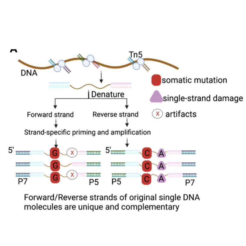
DNA mutations are the inevitable consequences of errors that arise during replication-repair of DNA damage as well as aging and disease progression. Because of their random and infrequent occurrence, quantification, and characterization of DNA mutations in the genome of somatic cells have been difficult. These mutations in DNA drive genetic diversity, alter gene function, define evolutionary trajectories, and provide targets for precision medicine and diagnostics. It is crucial to detect mutations across a wide range of abundance, i.e., variant allele frequency (VAF). Detecting low-abundance mutations (e.g., <0.1–1% VAF or in individual cells) is important for understanding human embryonic development, somatic mosaicism, and clonal hematopoiesis and uncovering pathogenic variants. Altogether somatic mutations provide important and unique insights into the biology of complex diseases. To decipher the causal inference, we must build robust genetic maps of somatic evolution in health and disease. The recent advent of duplex consensus sequencing has heralded a new generation of accuracy. However, multiple techniques focus on targeted areas of the genome (Twin Strand Biosciences) or are limited to restriction sites (Nanoseq), limiting their application to comprehensive somatic variant characterization. Furthermore, fragmentation of the genome and standard A-tailing and ligation creates errors (BotseqS, CODEC). Ligation of duplex strands for efficient sequencing has proven promising, though in practice requires complex molecular structures (Pro-Seq, CODEC) which have been observed to frequently result in incorrectly paired duplexes (CODEC). To enable comprehensive variant detection by next-generation DNA sequencing, we propose an innovative, accessible, and highly accurate Tn5 transposase-based duplex-sequencing technology (Tn5-duplex-seq) where complementary strands of DNA could be labeled at the molecular level in a single-tube reaction; thus, identifying single nucleotide variants (SNVs) from single-molecules
of DNAregardless of starting from single cells or pooled cell/DNA input. The conceptual basis of the protocol comes from META-CS (Xing et al.2021), a Tn5 based aproach optimized for single-cell whole genome amplification. We find that modifications of this approach to include flexible input and the sequencing strategy to optimize cost per variant detection enables great flexibility for all low-input applications.
Tn5-duplex-seq approach offers several benefits over other duplex approaches including.
(1) preservation of original template molecules by utilizing 16 unique sequences (Compared to the loss of 50% of
molecules due to intramolecular symmetry during TN5-based Nextera library preparation)
(2) accuracy by eliminating the requirement for A-tailing
(3) efficiency of duplex capture through specifying input
(4) accessibility by using standard reagents and oligonucleotide preparations
(5) distinction between double-stranded SNVs and single-stranded lesions.
Our method enables library preparation for short-read sequencing. Downstream analysis enables accurate and high-throughput SNV/indel and copy number analysis.
Guidelines
Optimization of proteinase K concentration for Step 1:
As different cell types may vary in the degree of chromatin condensation and material, we recommend titration of proteinase K at 0.5X, 1X, 5X, 10X our recommended concentration. The final library yield will indicate the optimal degree of digestion. In particular, this current protocol is optimized for nuclei or extracted DNA, and whole cells will likely require a higher concentration.
Expected yield and curve prior to selection for 50 cells (Note: can skip this visualization step for low yields):
ZymoClean 200ul binding buffer + 50ul reaction. Elute in 15ul TE. Run on HS Bioanalyzer.
Yield 4-8ng/ul.
Sequencing suggestion:
Ideally, part A, and B should all be sequenced separately to avoid the fragment length bias of the Illumina sequencer and to recover the most from the single-cell genome.
For cost consideration, part A alone can be sequenced on NovaSeq X Plus 10B (2x150bp), with a 20% PhiX spike-in, which
should be sufficient for determining the single-cell mutation rate.
Q5 Reaction Buffer New England BiolabsCatalog #M0491S , #9, #11Q5 polymerase New England BiolabsCatalog # M0491S , #9, #11PBS Invitrogen - Thermo FisherCatalog #2610807TL Proteinase K New England BiolabsCatalog # P8111S , #5Q5 High GC Enhancer New England BiolabsCatalog #M0491S , #9, #11
*Before use prepare 6X Stop Mix for use for 20 reactions below:
Dilute 1 μL of TL proteinase K + 19 μL PBS
Add 20 μL 12X stop solution for the final 6X Stop solution.
ADP1 and ADP2 Mix
Reconstitute the 16 ADP1 and 16 ADP2 primers separately in low TE and store in aliquots at -80 °C until ready for use.
Make an equimolar mix of the 16 ADP1 and ADP2 primers to make the ADP1 and ADP2 mix respectively.
Note
6.25 micromolar (µM)each primer x 16 primers for total 100 micromolar (µM) solution
TRANSPOSOME LOADING
Transposon Annealing
Reconstitute 16 META-CS oligos and 1 reverse oligo to 100 micromolar (µM) in Annealing Buffer (40 millimolar (mM)Tris-HCl (8), 50 millimolar (mM) NaCl)
Combine 1:1 of a singular META-CS oligo with the reverse oligo (there should be 16 separate reactions to put on the thermocycler). Mix up the reaction, spin it down briefly, and run the
thermocycler using the conditions below:
Transposon Assembly.
Combine all 16 reactions into one tube and aliquot for storage at -80 °C.
Take 10 μLof this aliquot and combine it with 10μL of unloaded Transposome
Incubate at 23 °C for00:30:00
Add 10 μL of 100% glycerol.
Aliquot and store at -80 °C.
Note
Estimated final concentration including glycerol storage (~16.7μM dimerized Tn5)
*Prior to use, dilute Tn5 in Diagenode Tn5 dilution buffer depending on the desired concentration
Optimization of Tn5 concentration:
Check on 50 cells using dilutions of 1:500, 1:750, 1:1000, and 1:1500, and check the tagmentation curve. Appendix-2
30m
TN5-DUPLEX LIBRARY PROCEDURE
3h 5m 30s
Sorting and lysing cells2 μL
Prepare nuclei for sorting.
Sort cells directly into 2 μL of 1X cell lysis buffer
Run the thermocycler using the conditions below 65 °C Lid Temp
Incubate in thermocycler using the conditions below 105 °C Lid Temp.
98 °Cfor 00:00:30
62 °C for 00:05:00
72 °C for 00:01:00
4 °C hold
6m 30s
Stop reaction1 μL
Add 1 μL Thermolabile ExoI per tube. Try to touch the minimum of the solution surface. Spin down first, then plate mix, and spin down again.
37 °C for 00:15:00
65 °C for00:05:0000:00:00
4 °Chold 75 °C Lid Temp
20m
Library prep14 μL
Make PCR Mix (per cell):
5 μL NEB Universal Primer (NEB E7335S, E7500S, E7710S, E7730S)
4 μL Q5 Reaction Buffer
4 μLQ5 High GC Enhancer
0.4 μL10 millimolar (mM) each dNTP mix
0.4 μL water (H2O)
0.2 μL Q5 polymerase *add last
2.Add 5 μL NEB Index Primer per tube, avoiding touching the liquid.
3.Add 14 μL PCR Mix per tube, avoiding touching the liquid. Vortex and spin down.
4. Incubate in thermocycler using the conditions below
98 °C for 00:00:20
12 cycles of 98 °C for 00:00:10, 72 °C for 00:02:00
72 °C for 00:05:00
4 °C hold
7m 30s
PURIFICATION AND ZYMO CLEAN
Zymo clean
Utilize the Zymo DNA Clean & Concentrator Kit with associated protocol (abbreviated version below).
For microbulk samples, use 4:1 DNA binding buffer to sample (200 μL buffer to 50 μLreaction). For single cell samples, pool desired samples first, then measure the total pooled volume and use 4:1 DNA binding buffer to sample volume. For single cell samples, pool 5 cells per spin column. For 50 cell samples, use 1 spin column per sample.
Add it to the spin column. The maximum volume that the spin column can hold is 800 μL so pooled samples should have to be run through the same column sequentially until all of the liquid has been run through, discarding flowthrough each time.
Spin for 00:00:30 at maximum speed on the tabletop centrifuge >10,000xg at RT
Add 42 μL x0.1 TE to elute and wait 00:04:00 at room temperature
Spin for 00:00:30 at maximum speed on the tabletop centrifuge >10,000xg at RT
2. Run 2 μL on High Sensitivity D5000 TapeStation chip.
5m
Size Selection (AMPure) 40 μL DNA library
Add 22 μL(0.55X) resuspended AMPure XP beads to 40 μL DNA library.
Vortex and spin down. Label the tube as “A”. Incubate for 00:05:00 at RT.
Place tube A on a magnetic stand for 00:05:00. Carefully transfer the supernatant to a
new tube. Label the new tube as “B”.
Size select tube “A” (0.55x AMPure XP beads):
a. Add 200 μL of 80% freshly prepared ethanol to all tubes while in the magnetic stand, then carefully remove and discard the supernatant.
b. repeat the ethanol wash step one more time.
c. Let air dry on magnetic stand for 00:01:00 at RT.
d. Remove the tubes from the magnetic stand. Elute DNA from beads with 12 μL 0.1X TE (for
single cell pools) or 18 μL 0.1X TE (for 50 cell pools). Vortex and gently spin down, incubate for 3 min at RT.
e. Place the tubes on the magnetic stand for 00:03:00. Transfer 18 μL of supernatant to a clean tube.
Size select tube "B" (0.8x AMPure XP beads):
a. Add 8 μL AMPure XP beads to DNA solution in tube B (0.15X). Vortex
and gently spin down. Incubate for 00:05:00 at RT.
b. Place tube B on the magnetic stand for 00:05:00. Remove supernatant from tube B.
i. IMPORTANT: save the supernatant in case AmPure did not work! (the DNA will still be in the supernatant)
c. Add 200 μL of 80% freshly prepared ethanol to all tubes while in the magnetic stand, then carefully remove and discard the supernatant.
d. repeat the ethanol wash step one more time.
e. Let air dry on magnetic stand for 1 min at RT.
f. Remove the tubes from the magnetic stand. Elute DNA from beads with 12 μL 0.1X TE (for
single cell pools) or 18 μL 0.1X TE (for 50 cell pools). Vortex and gently spin down, incubate for 3 min at RT.
g. Place the tubes on the magnetic stand for 00:03:00. Transfer 18 μL of supernatant to a clean tube.
Run 2 μLon High Sensitivity D5000 TapeStation chip. (Run1 μL of sample and 1 of 0.1x TE instead of 2 μL of sample if you need to preserve sample). (Expected concentration for 5 single cell pools is: 300-1000 pg/μL. Expected concentration for 50 cell pools is: 2000-15000 pg/μL .
Proceed to DNA quantification with final product and dilute accordingly for sequencing.
Run TapeStation, need at least 5 nanomolar (nM) concentration for sequencing. *Fraction B
yields best sequencing results.
-
Tube "A" (0.55x) contains DNA fragments with an average of 1000bp.
Tube "B" (0.15x) contains DNA fragments with an average of 400-500bp (fragment size may range from 300bp-600bp).
27m
Appendix-1
SI Appendix Table S1: Oligonucleotide sequences of META-CS transposon DNA and primers.