Dec 22, 2025
Tissue processing and RNA extraction for multi-omic molecular analyses of human surgical samples
- Matthew Sapio1,
- Evelyn Li1,
- Anthony F. Domenichiello1,
- Taichi Goto2,
- Tracy Williams1,
- Michael Iadarola1,
- Andrew J Mannes1
- 1National Institutes of Health, Clinical Center;
- 2National Institutes of Health, NINR
- Matthew Sapio: To whom correspondence should be addressed, [email protected];


External link: https://linktr.ee/matthew.sapio
Protocol Citation: Matthew Sapio, Evelyn Li, Anthony F. Domenichiello, Taichi Goto, Tracy Williams, Michael Iadarola, Andrew J Mannes 2025. Tissue processing and RNA extraction for multi-omic molecular analyses of human surgical samples. protocols.io https://dx.doi.org/10.17504/protocols.io.j8nlk81w1l5r/v1
Manuscript citation:
Sapio, M.R., Li, E., Domenichiello, A.F. et al. Longitudinal human transcriptomic and spatial gene profiling at the incisional edge during long surgical procedures. Commun Biol (2025). https://doi.org/10.1038/s42003-025-09366-0
License: This is an open access protocol distributed under the terms of the Creative Commons Attribution License, which permits unrestricted use, distribution, and reproduction in any medium, provided the original author and source are credited
Protocol status: Working
We use this protocol and it's working
Created: August 29, 2024
Last Modified: December 22, 2025
Protocol Integer ID: 106697
Keywords: RNA extraction, Human tissue processing, RNA-Seq, Dermis, epidermis, lipidomics, multi-omics, Patient reported outcomes of pain, Skin, in situ hybridization, FFPE, human tissue rna extraction, rna extraction procedures for human tissue rna extraction, rna extraction procedure, rna extraction, human surgical sample, human surgical samples this protocol, tissue processing, tissue collection, perioperative medicine, scalable processing pipeline
Funders Acknowledgements:
Intramural Research Program of the National Institutes of Health, Clinical Center
Grant ID: ZIACL090034-09, ZIACL090035-08, ZIACL0033-09
Intramural Research Program of the National Institutes of Health, NINR
Grant ID: 1ZIANR000019
Office of Behavioral and Social Sciences Research
Grant ID: Interagency Agreement
Bench to Bedside Award
Grant ID: 995123
Disclaimer
This research was supported by the Intramural Research Program of the National Institutes of Health (NIH). The contributions of the NIH author(s) are considered Works of the United States Government. The findings and conclusions presented in this paper are those of the author(s) and do not necessarily reflect the views of the NIH or the U.S. Department of Health and Human Services.
Abstract
This protocol outlines the tissue collection and RNA extraction procedures for human tissue RNA extraction developed by Sapio and colleagues at the Department of Perioperative Medicine, Clinical Center. These procedures aim to describe a reproducible, scalable processing pipeline for multi-omic human tissue collection efforts.
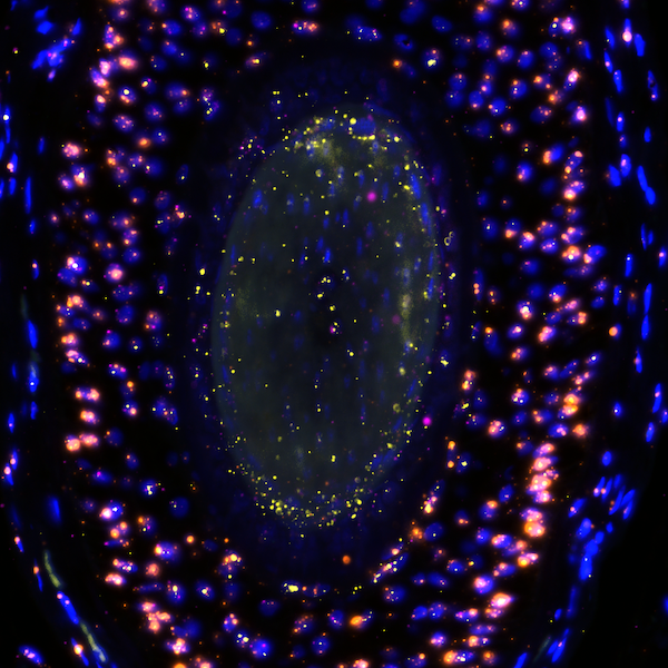
Image Attribution
Microscopic images are part of the peer-reviewed accompanying study: Sapio, et al. 2025 Communications Biology. Diagram was provided by Alison P. Manalo and created in BioRender.
Materials
Equipment:
Refrigerated mini centrifuge (such as 5425/5425 R, Eppendorf)
Bioanalyzer 2100 (Agilent)
Rotor-stator homogenizer
Bio-Gen PRO200 Homogenizer (ProScientific)
(alternatively): Bead-beating homogenizer
FastPrep-24‱ Classic bead beating grinder and lysis system (MP Biomedicals)
Reagents:
10% neutral buffered formalin
Kimwipe‱ (Kimberly-Clark, Product Code: 34120)
Invitrogen PureLink‱ RNA mini kit (Thermo Fisher Scientific, Cat. No. 12183018A)
RNAlater‱ (Thermo Fisher Scientific, Cat. No. AM7021)
Invitrogen RNAseZap‱ (Thermo Fisher Scientific, Cat. No. AM9780)
QIAzol (Qiagen, Cat. No. 79306)
alternatively: Invitrogen TRIzol‱ (Thermo Fisher Scientific, Cat. No. 15596026)
RNase-Free DNase Set (Qiagen, Cat. No. 79254)
Chloroform
RNA 6000 Nano Kit (Agilent, Cat. No. 5067-1511)
Safety warnings
Safety warning: This protocol involves human tissues and other potentially hazardous reagents. All procedures must be approved by the Institutional Biosafety Committee (IBC) and performed in compliance with institutional and regulatory safety guidelines.
Ethics statement
This study was based on an approved IRB protocol at the National Institutes of Health (20CC0031), and was registered at Clinicaltrials.gov (A single cohort study collecting interval timed
incisional epidermal and dermal tissue samples during surgical procedures to
profile temporal response of tissue after noxious stimuli, NCT04224870).
Project overview
Graphical abstract for study protocol.
Graphical overview of the study protocol. Figure was generated by Allison P. Manalo in BioRender.
Overview of study timepoints (Adapted for Manual of Procedures for Protocol # 20CC0031 at the National Institutes of Health Clinical Center)
5d
Gather participant demographics.
2h
Review Medical History and Physical Exam – performed by surgical research team and anesthesia pre-operative team.
4h
Administer screen Pain Assessment questionnaires –Brief Pain Inventory (BPI) & McGill-short form (sf-MPQ) prior to surgery.
2h
Surgery Day – document times of initial surgical incision and organize subsequent time point samples.
1d
Surgery day – obtain tissue samples for prespecified time points (0 hours, 1 hours, 2 hours, 4 hours, 6 hours, and a closure time point if the surgery lasts ≥8 hours).
1d
Post-Operative Day 1 – Pain Assessments questionnaires – Brief Pain Inventory (BPI) & short form-McGill Pain Questionnaire (sf-MPQ).
1d
Post-Operative Day 2 – Pain Assessments questionnaires – BPI & sf-MPQ.
1d
Schedule of activities for study procedures.
Intraoperative tissue sampling and tissue processing procedures.
Tissue collection staff arrive early to the operating suite, coordinate with the circulating nurse, and are present at the surgical "huddle" to make their presence known.
Note
It is important to cause minimal disruption to the procedures of the operating room, and follow the guidelines and protocols established by the clinical team.
A sample cart with instruments is prepared ahead of time to perform surgical sample processing so that samples can be divided into labeled tubes and stored for future use. Cart contains several items including sterile instrument pack (decontaminated at the end of surgery by OR staff), prepared tubes and labels, sample collection forms, writing implements, paper towels, etc. The cart serves as a workspace for tissue collection staff.
The surgical kit should contain scissors, scalpels and forceps suitable for subdivision of the tissue sample by tissue collection staff.
Tissue Sampling / Surgical Guidelines
-All specimens will be obtained by surgeon with surgical scissors or with scalpel.
-Sterile ruler (ST Ruler- 6 in) will be used for measurement of each sample.
-Tissue samples will be obtained from the lateral aspects of the incision line.
-Samples will be obtained from incision line tissue that is NOT covered by the self-retaining retractors in
use.
-DPM lab personnel responsible for monitoring protocol sample timepoints by alerting the surgeon of time for protocol sample 15 minutes prior to the intended time point.
Graphic overview of timepoints for skin collection.
Location of intraoperative tissue sampling. Samples were collected adjacent to one another to maintain a bordering edge with the initial incision. Care was taken to avoid areas of retractor contact, or other unrelated trauma, when possible. All surgical excisions were made at the surgeon's discretion.
Upon excision of tissue, tissue will be handled by designated protocol team scientist and undergo immediate specimen sectioning into the three prescribed tissue samples and the above procedures followed for each sectioned specimen.
Tissue processing in the operating room. After the sample is handed off from surgeon to tissue processing staff, the sample (24 mm) is subdivided into three major pieces of approximately equal length (8 mm each).
Pathology Tissue Identification. An initial sample is required (approximately 1 x 2 x 2 mm) at 0 hour time point for initial tissue identification and histological diagnosis per institute-specific policy and procedures. Pathology to examine a sample of the harvested tissue to confirm absence of any skin disease for protocol inclusion.
This sample will be placed in a sterile container in sterile water.
RNA-Seq (transcriptomics)
One piece of tissue (8 mm in length) is required for RNA extraction to prepare the sample for RNA-Seq analyses. The sample is entirely consumed in the RNA extraction procedure in order to perform the analysis. The sample will be stored in RNA later at the time of collection, stored overnight (10-18 hours) at 2-8°C in RNA later solution, and then frozen and stored at -80 °C until tissue processing. RNA extraction and sequencing will be performed as described previously.
Mass spectrometry
One piece of tissue (8mm in length) is required for Liquid Chromatography-Tandem Mass Spectrometry (LC-MS/MS. Using LC-MS/MS we will quantify concentrations of lipid and peptide mediators of pain and
inflammation. LC-MS/MS is the best way to quantify an array of mediators from a single sample. The sample size of 2 mm X 2 mm X 8 mm will weigh approximately 32 mg. In order to analyze a sample by LC-MS/MS a tissue homogenate must be made, essentially pulverizing the tissue in preparation for mediator extraction. Further, the lipid extraction procedures are incompatible with those used to perform RNA extraction. Therefore, a separate piece of tissue will be required for LC-MS/MS analysis.
Tissue samples will be collected and placed in 1.5 mL micro tube and frozen on dry ice and stored at -80°C until tissue processing.
Histological analysis.
One piece of tissue (8mm in length) will be used for histological analysis. This requires anatomical preservation, and therefore is incompatible with the above two methodologies which require pulverization and extraction.
In order to preserve the tissue, the sample will be collected in 10% formalin and stored at 2-8°C overnight (10-18 hours). Subsequently, the samples will be switched to 1% paraformaldehyde or 2.5% formalin and embedded in paraffin blocks following standard procedures.
Subsequently, sections can be sectioned and analyzed using either in situ hybridization, histological stains or immunohistochemistry to examine localization of proteins and/or mRNAs.
Note
During the initial tissue processing procedures, care should be taken to avoid large agglomerations of subcutaneous fat. When subdividing the tissue sample, if one area contains particular amounts of fat, that area can be used here for histology, as histological analysis is not affected by presence of adjacent regions, whereas analyses of whole tissue homogenate (such as those in RNA-Seq or mass spectrometry) are negatively impacted by adherent fat.
RNA extraction procedures for human skin
5m
These steps are adapted from the PureLink‱ RNA Mini Kit (Catalog numbers: 12183018A, 12183025, Thermo Fisher Scientific).
Set centrifuge to 4 °C .
Get dry ice and wet ice, set up in two temperature stable buckets. Place frozen samples on dry ice.
Establish an RNAse free area on the bench. Put down alumimum foil taped to a bench top with lab tape, and clean this area using decontamination procedures (spray RNAse Zap), and wipe clean with Milliq purified RNAse free water and a Kimwipe. Set up instruments with RNAse precautions: spray gloved hands, bench, and tube holders with RNAse Zap, arrange items in an RNAse free setup on the bench. This establishes the working area as "RNAse free".
Note that after establishment of an RNAse free working enviroment, care should be taken about what items travel in and out of this area. Generally, items should be decontaminated with RNAse Zap before returning to this area.
Remove Bioanalyzer reagents and DNase buffer from 4 °C fridge and DNase from -20 °C freezer.
Place tissues on Kimwipe paper and carefully remove RNAlater solution.
Place tissue into homogenization tube (round bottom, 14 ml, PCR clean), and add 1 ml of TRIzol to the tube.
Add 1 mL Qiazol to the tubes
Homogenize the tissue by rotor stator homogenizer using a 10 mm saw-toothed probe until minimal pieces of tissue remain. Fully homogenized tissue is liquid with few small visible chunks of tissue remaining.
Note
It is important to use a homogenization procedure that minimizes exposure to high temperature and does not use excessive force or homogenization time. Sample may increase in temperature during prolonged (non-pulsed) homogenization. Moving the sample tube up and down gently can lightly agitate the solution, allowing chunks to find their way into the blades. Excessive homogenization time will reduce the quality. Another key factor in obtaining high quality RNA extraction is adding enough phenol-containing buffer (QIAzol) for your sample size, and not overloading the anion exchange (RNA-binding) column.
Rest the tube for 00:05:00 .
5m
Transfer the solution to 15 mL conical centrifuge tube. Alternatively, this step can be carried out in a 2 ml tube if volume allows.
In a chemical fume hood, carefully add 200 µL of chloroform, manually mix for 00:00:15 and let sit for 2–3 min. Note: Sample should start to separate into 2 layers.
Note
Chloroform forms an immiscible organic layer (heavier than water), and can be added to excess if needed. This can be adjusted if QC (see later steps) require additional cleanup without adverse effects on yield.
15s
Centrifuge at 12,000 g for 00:15:00 at 4 °C Care should be taken to set to g and not RPM. Care should be taken to balance the centrifuge. At the end of this step set the centrifuge to 22°C.
15m
Transport tube back to "RNAse free" bench.
Carefully pipette 400 µL of clear supernatant into new microcentrifuge tube. Note that if the interface is interrupted during this step, a brief centrifugation can be introduced to re-establish the layers.
Add an approximately equal volume of 70% ethanol to microcentrifuge tube, and vortex for 5
sec.
5s
Transfer ≤ 700 µL to RNA extraction mini spin column (pink). Note: This is the holding capacity of this tube. If the amount exceeds this volume, it can be added as a second flowthrough after the first column binding centrifugation step. Care should be take not to exceed the binding capacity of the tube during this step by limiting the input.
Place the spin column in centrifuge, balance, and set to 8,000 g for 00:00:22 , decant flow-through into waste.
22s
Perform on-column DNase digest.
Add 350 µL Wash Buffer 1 to the spin column, close lid, and centrifuge for 22 sec at 12,000g, and then discard flow-through.
22s
In an extra microcentrifuge tube, add 10 µL of DNase solution to 8 μL 10xDNase buffer and 62 µL RNase free water (Per sample - ex. for 5 samples, 50 µL DNase in 350 µL buffer).
Add 80 µL of incubated mix directly to the RNA extraction mini spin column. Take care to not touch the membrane of the column with pipette and to cover membrane with liquid.
Add 350 µL Wash Buffer 1 to the spin column. Centrifuge at 12,000 g for 00:00:20 with the lid of tube open to dry the membrane.
Note
QC metrics can sometimes be improved by additional Wash Buffer 1 volume (up to 700 µl), or a 30-60 second incubation of Wash Buffer 1 with the resin at this stage. Additionally, a second Wash Buffer 1 wash step can be introduced which has a mild impact on yield and can remove additional contaminants.
20s
Add 500 µL Wash Buffer 2 (with ethanol) to the spin column. Centrifuge at 12,000g for 00:00:20 and discard flow through.
20s
Repeat the previous step.
Centrifuge your spin column at 12,000g for 00:02:00 with the lid of tube open to dry the membrane.
2m
Place the column in a new 1.5 mL collection tube (supplied, RNAse free).
To elute the final product, add 50 µL RNase-free water directly to the spin column, close lid and centrifuge for00:02:00 at 12,000 g.
2m
Label and store your samples at -80 °C .
Protocol references
This protocol was published alongside the peer-reviewed article:
Sapio MR, Li E, Domenichiello AF, et al. Longitudinal human transcriptomic and spatial gene profiling at the incisional edge during long surgical procedures. Commun Biol (2025). https://doi.org/10.1038/s42003-025-09366-0
Other references for protocol development:
1. Goto T, Sapio MR, et al. Longitudinal peripheral tissue RNA-Seq transcriptomic profiling, hyperalgesia, and wound healing in the rat plantar surgical incision model. FASEB J 35, e21852 (2021). https://doi.org/10.1096/fj.202100347R
2. Goto T, Sapio MR, et al. Longitudinal Transcriptomic Profiling in Carrageenan-Induced Rat Hind Paw Peripheral Inflammation and Hyperalgesia Reveals Progressive Recruitment of Innate Immune System Components. J Pain 22, 322-343 (2021). https://doi.org/10.1016/j.jpain.2020.11.001
3. Kim JJ, Sapio MR, et al. Transcriptional Activation, Deactivation and Rebound Patterns in Cortex, Hippocampus and Amygdala in Response to Ketamine Infusion in Rats. Front Mol Neurosci 15,
892345 (2022). https://doi.org/10.3389/fnmol.2022.892345
4. Sapio MR, Vazquez FA, et al. Comparative Analysis of Dorsal Root, Nodose and Sympathetic Ganglia for the Development of New Analgesics. Front Neurosci 14, 615362 (2020). https://doi.org/10.3389/fnins.2020.615362
5. Sapio MR, King DM, et al. Efficient removal of naturally-occurring lipofuscin autofluorescence in human nervous tissue using high-intensity white light. J Pain 30, 105359 (2025). https://doi.org/10.1016/j.jpain.2025.105359
Acknowledgements
This protocol was written by Dr. Matthew R. Sapio (to whom correspondence should be addressed) with assistance from other authors, especially Evelyn Li and Taichi Goto. It was partially based on the RNA extraction protocols developed by Matthew R. Sapio, Taichi Goto, Fernando A. Vazquez, Jenny J. Kim, and others in the Department of Perioperative Medicine at the National Institutes of Health, Clinical Center (see references to earlier publications using these methods). All authors approved the final version of this protocol.