Jun 13, 2025
Version 2
Thick 3D Tetraspeck Beads sample preparation V.2
- 1Fred Hutchinson Cancer Center



Protocol Citation: Elena Carlson 2025. Thick 3D Tetraspeck Beads sample preparation. protocols.io https://dx.doi.org/10.17504/protocols.io.n92ldnkkov5b/v2Version created by Elena Carlson
License: This is an open access protocol distributed under the terms of the Creative Commons Attribution License, which permits unrestricted use, distribution, and reproduction in any medium, provided the original author and source are credited
Protocol status: Working
We use this protocol and it's working
Created: June 13, 2025
Last Modified: June 13, 2025
Protocol Integer ID: 220162
Keywords: tetraspeck, fluoresence, microscopy, point spread function, agarose, beads, fluorescent beads, thick 3d tetraspeck beads sample preparation, fluorescent bead, sample preparation, microscopy troubleshooting, thick sample, thick sample of agar
Funders Acknowledgements:
National Institutes of Health
Grant ID: P30CA015704 (RRID:SCR_022616)
National Institutes of Health
Grant ID: P30CA015704 (RRID:SCR_022609)
National Institutes of Health
Grant ID: P30CA015704 (RRID:SCR_022610)
Abstract
Create a thick sample of agar and fluorescent beads in a well plate, to use for microscopy troubleshooting (e.g. to determine the point spread function of your system).
Materials
- Tetraspeck beads, 0.5 um, ThermoFisher Scientific cat. no: T7281. TetraSpeck‱ Microspheres, 0.5 μm, fluorescent blue/green/orange/dark red
- Agarose HOEFER | SKU: SKU:GR140-25
- P1000 and P200 micropipettes + tips
- vortexer (any)
- rice cooker or microwave
- Thermometer
- precision scale
- 15 mL falcon tubes
- glass bottom well plate, e.g. CLS-1811 CLS-1811 - GLASS BOTTOM CULTURE DISHES, TC TREATED, NEST- Chemglass Life Sciences
Prep 1% [w/v] Agarose solution
Heat DPBS to approximately 100 °C , using a rice cooker, pressure cooker, or microwave
use a thermometer to verify temperature
Measure out104 mg agar to 10.4 mL hot DPBS and mix vigorously with vortexer. If using a larger well plate, you can adjust these amounts accordingly. Verify concentration made using this website Percent (%) Solutions Calculator - PhysiologyWeb .
Mix in the beads
Let Agar solution cool to 40-50 °C
Meanwhile, vortex the stock tetraspeck vial
Equipment
Vortex mixer
NAME
Any
BRAND
xx
SKU
When DPBS-Agar mix has cooled to 40-50 °C , use a P200 micropipette to add60 µL of the stock beads to the10 mL of hot Agar-DPBS. For a sparser population of beads, add 30 microliters instead.
Vortex the resulting mixture vigorously.
Well plate
Use a simple transfer pipette to transfer the beads-agarose solution to a glassbottom well plate. Fill the plate to the very top, avoiding the formation of bubbles. Before completely filling the plate, tap the bottom of the plate gently to release any bubbles.
Carefully place a coverslip on top of the well plate, and let the agarose cool and harden. Shield the sample from light.
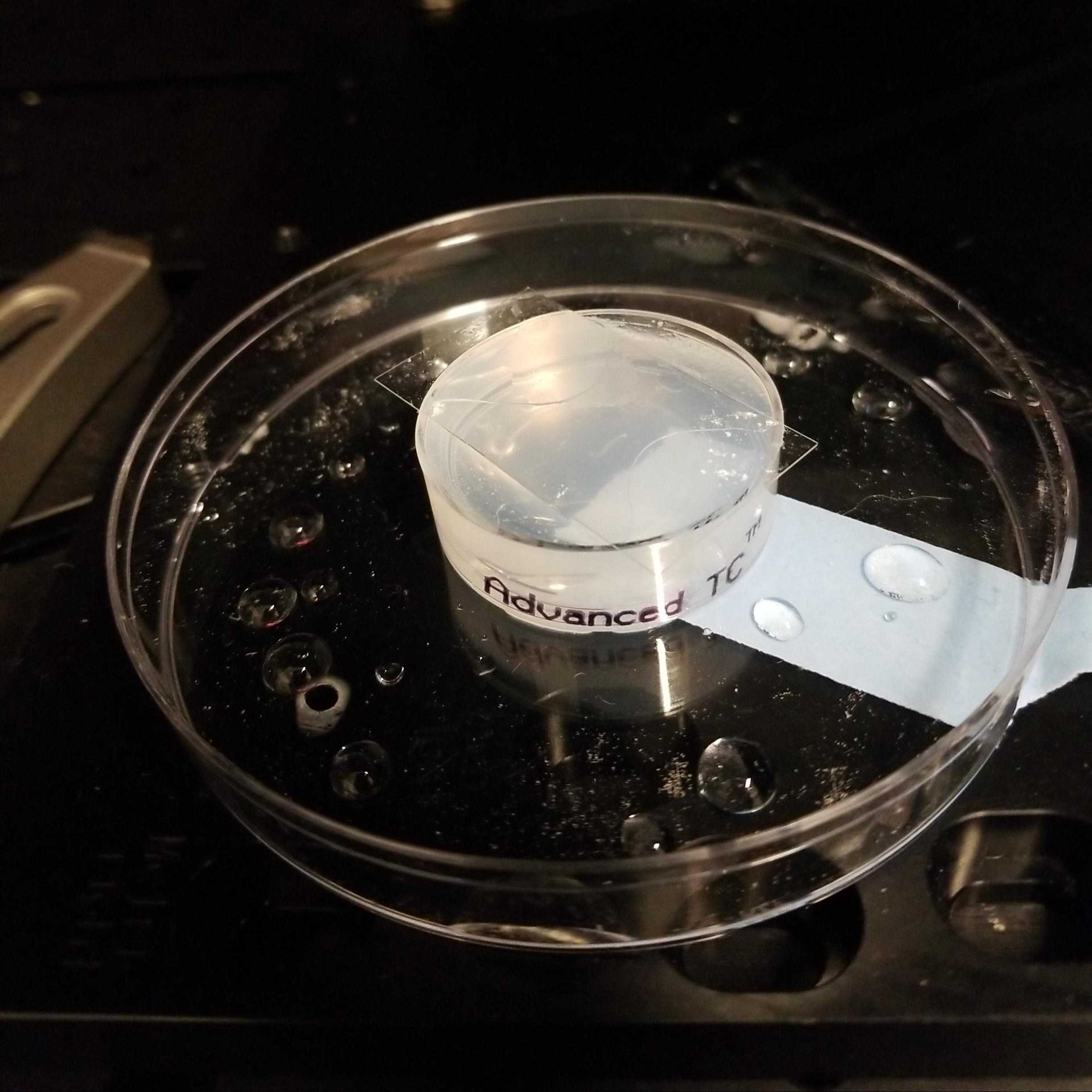
Agarose-Beads suspension in a thick well plate, with a glass coverslip on top. This is then stored in a larger well plate dish.
Store the sample in a larger well plate or similar box to keep the glass from being knocked off. Add tape.
Protocol references
Above was adapted from:
Yiming Li, Yu-Le Wu, Philipp Hoess, Markus Mund, and Jonas Ries, "Depth-dependent PSF calibration and aberration correction for 3D single-molecule localization," Biomed. Opt. Express 10, 2708-2718 (2019)
Acknowledgements
This research was supported by the Preclinical Imaging, Comparative Medicine, and Cellular Imaging Shared Resources (RRID:SCR_022616 , RRID:SCR_022609, RRID:SCR_022610 ) of the Fred Hutch/University of Washington/Seattle Children’s Cancer Consortium (P30 CA015704).