Feb 28, 2024
α-Synuclein sedimentation assay
- 1Duke University
- West lab protocols


External link: https://doi.org/10.1126/sciadv.adq3539
Protocol Citation: arpine.sokratian 2024. α-Synuclein sedimentation assay . protocols.io https://dx.doi.org/10.17504/protocols.io.6qpvr3nwpvmk/v1
Manuscript citation:
Sokratian A, Zhou Y, Tatli M, Burbidge KJ, Xu E, Viverette E, Donzelli S, Duda AM, Yuan Y, Li H, Strader S, Patel N, Shiell L, Malankhanova T, Chen O, Mazzulli JR, Perera L, Stahlberg H, Borgnia M, Bartesaghi A, Lashuel HA, West AB Mouse α-synuclein fibrils are structurally and functionally distinct from human fibrils associated with Lewy body diseases. Science Advances 10(44). doi: 10.1126/sciadv.adq3539
License: This is an open access protocol distributed under the terms of the Creative Commons Attribution License, which permits unrestricted use, distribution, and reproduction in any medium, provided the original author and source are credited
Protocol status: Working
We use this protocol and it's working
Created: January 29, 2024
Last Modified: May 31, 2024
Protocol Integer ID: 94282
Keywords: ASAPCRN, synuclein aggregation, synuclein sedimentation, synuclein over time, synuclein, precise quantification of fibril formation kinetic, fibril formation kinetic, protein
Funders Acknowledgements:
ASAP
Grant ID: 020527
Abstract
The protocol describes step-by-step instructions on how to set a sedimentation assay to monitor the aggregation of alpha-synuclein over time. The protocol allows for precise quantification of fibril formation kinetics using SDS-PAGE. This methodology enhances our understanding of the underlying mechanisms involved in protein misfolding and offers a reliable tool for investigating potential therapeutic interventions targeting alpha-synuclein aggregation.
Protocol materials
Amicon Ultra-0.5 Centrifugal Filter Unit 24 packMerck Millipore (EMD Millipore)Catalog #UFC505024
Corning® 384-well Black/Clear Bottom Low Flange Ultra-Low Attachment Microplate Bulk PackedCorningCatalog #4588
BioSpec Products 2.3 mm Zirconia/Silica Beads 1 lb bottleFisher ScientificCatalog #NC0451999
PCR Plate Heat Seal foil piercableBio-Rad LaboratoriesCatalog #1814040
4–20% Mini-PROTEAN® TGX Stain-Free™ Protein Gels, 15 well, 15 µlBio-Rad LaboratoriesCatalog #4568096
10g Coomassie Brilliant Blue R-250 DyeG-BiosciencesCatalog #786-495
Safety warnings
Hazard Identification and Risk of Exposure to the
Hazards:
Inhalation or spread through food or drink that contain fibrils aerosols or fibrils.
Protective gloves, safety glasses and lab coat must always be used when handling anything that possibly could contain α-synuclein fibrils. Food or drink is strictly prohibited in any environment where α-syn fibrils are used.
Routes of Transmission: Prior to assigning containment requirements, it is imperative to
understand the routes of transmission.
Some issues to address:
- What are the exposure routes/risks of most concern:
Inhalation or spread through food or drink that contain fibril aerosols or fibirls accordingly. Fibrils possibly might reach the brain regions through the olfactory epithelium; Risk of accidental needlestick/droplet splash while handling fibrils for in vitro or in vivo work.
- What are the consequences of exposure (potential illness, etc)
Fibrils may be considered as infectious material. Minimum to no hazard is expected from α-syn protein. There is no evidence that transmission of fibrils can lead to development Parkinson’s disease. However, taking into account prion-like properties of α-syn fibrils should therefore be handled cautiously and wisely. Strictly recommended using disposable materials and Personal Protective Equipment (PPE) such as gloves, face mask, etc.
PRECAUTIONS:
Laboratory work where high concentration of fibrils (more than 300 uM) is needed must comply with biosafety level 2 (BSL2) containment as described in the current edition of the CDC/NIH’s
Biosafety in the Microbiological and Biomedical Laboratories: http://www.cdc.gov/od/ohs/biosfty/bmbl5/bmbl5toc.htm
Sharps safety precautions:
The use of sharps (glass pipettes, glass slides and cover slips, scalpels and
lancets) should be eliminated, when possible. Appropriate precautions should be
taken to avoid percutaneous injuries. These items should be disposed of
immediately in a puncture-resistant sharps container. Bending, recapping or
clipping of needles is prohibited. As described in CDC’s sharps safety website:
https://www.cdc.gov/sharpssafety/index.html
Procedural Methods
and Materials:
- Laboratory work where high concentration of fibrils (more than 300 uM) is needed must comply with biosafety level 2 (BSL2) containment. This means all aerosol generating procedures must be performed within the biosafety cabinet.
- All the fibrils work involves using PPE, aerosol-tight centrifuges, water bath sonicator in a closed cabinet, homogenization of frozen brain samples using probe-tip sonicator under the hood (collection of protein fractions in BSL2 cabinets), chromatography equipment in a closed-door fridge, sealed plates, safe lock microfuge tubes (or tubes wrapped/sealed with parafilm), and use of filtered tips for pipettes. All personnel must strictly adhere to these procedures.
- Use of proper PPE as stated in the section below. Use of available N95 respirators is voluntary (same for the use of available sleeve protectors). Follow safety precautions for sharps (for e.g., to avoid accidental needle sticks) while working with PFFs in the lab and for doing in vivo work.
Personal Protective
Equipment (PPE): Appropriate PPE includes gloves, lab coat and safety glasses,
face mask (voluntary N95 respirator use and sleeve protectors), face / bench
top splash shield for specific procedures as stated above.
Methods to minimize personal exposure: Strictly adhere to sharps safety precautions using needles or any material that can potentially cause wounds. Use disposable supplies where possible. Use the minimal amounts of α-fibrils needed for an experiment. Keep fibrils in closed tubes. 10% of SDS solution in water must be used for decontaminating work areas. Do not use NaOH or Sodium Hypochlorite or ethanol. Do not leave samples containing fibrils unattended at the bench.
Methods to prevent the release of fibrils/protect workers from aerosols,
splashes, splatters: protective gloves and clothing always be always be worn when handling frozen vials. High concentration of fibrils(>1mg/mL) always be handled under Biosafety cabinet and containment caps will be used while centrifugation. Centrifuge cups will be opened inside a biosafety cabinet. Face shield or benchtop splash shield will be used when working at the open bench.
Specimen transport
and removal of material(s) from the laboratory: Transported in secondary
container (plastic/Styrofoam) in a closed box. The closed box is carried in a
bag.
Standard
microbiological methods: hand washing after removal of gloves and before
leaving the work area, no mouth pipetting, strictly no food or drink in
refrigerators where material is stored, no eating in work area.
Cleaning &
Disinfection: Work area must be
cleaned with 10% SDS in water. Wipes used must be immediately disposed into
biohazard waste container. Any piece of equipment or supplies that possibly
have been exposed to fibrils must be wiped with 10% of SDS.
Waste Generation and Disposal Methods: The solutions that contain α-syn fibrils must be
decontaminated with 10% of SDS in water for 30 minutes and be thrown as a
biohazard waste in a sealed container/bag (use a minimal volume of fibrils needed
for an experiment, do not generate large volumes of fibril-containing liquids).
Use small biohazard bags to collect tips and consumables of experiment
performed, appropriately tie neck of bag in single knot and place in into
secondary biohazard waste container.
Spill and Accident Response Procedure: Describe all emergency procedures including spill clean-up. Describedisinfectants and
environmental decontamination. (ex., Outside of a BSC: If spill is a
respiratory hazard, evacuate 30 minutes to allow aerosols to settle. Place absorbent towels over the spill, apply freshly
prepared 10% SDS solution to entire area of spill starting on the outer edges
and working inward, pick up sharp items with mechanical device (not hands),
place all clean-up materials in a biohazard bag)
Amplification set-up
Thaw down a-syn monomer and/or sonicated fibril aliquots. Use water-bath.
Do not generate bubbles by pipetting or shaking
Measure monomer concentration via Nanodrop
Add Amount of 5x, 10x or 20x diluted aliquots in PBS onto nanodrop piedestal;
Parameters: other proteins; coefficient extinction:
5.98; MW: 14.4 kDA for human a-synuclein
7.45; MW: 14.4 kDA for mouse a-synuclein
Perform two measurements for each dilution and confirm <10% standard error between two measurements
For fibril preparations, dilute in 6M guanidine and follow the aforementioned instructions derscribed for monomeric protein
Calculate the volume of monomer need following: 0.3 mg/mL of monomer in a reaction (30 ul reaction/well).
Dilute the monomer preparation in PBS to 2,5 mg/mL in order to filter the protein preparation to avoid any high mol. weight contaminants
Use Amicon Ultra-0.5 Centrifugal Filter Unit 24 packMerck Millipore (EMD Millipore)Catalog #UFC505024
to filter the diluted monomer concentration. Prior to adding the protein, wash the filter by adding PBS and centrifuging at high-speed.
Measure the size of proteins using DLS
Pour one zirconia bead into each well of the Corning® 384-well Black/Clear Bottom Low Flange Ultra-Low Attachment Microplate Bulk PackedCorningCatalog #4588
BioSpec Products 2.3 mm Zirconia/Silica Beads 1 lb bottleFisher ScientificCatalog #NC0451999
Prepare serial dilutions of sonicated fibrils if needed, make sure to use protein-low-binding tubes and shake the preparation each time using vortex
Prepare reaction mix containing the monomer mix, PBS and if needed, sonicated fibrils.
Transfer reaction to the plate: start filling the plate with a standard curve samples (seal the portion with a sealing foil)PCR Plate Heat Seal foil piercableBio-Rad LaboratoriesCatalog #1814040
Proceed the experimental samples and cover the whole plate with a sealing foil. Spin down the plate at 500 g for 30 sec.
Set up the program on a plate reader
Equipment
Omega - RT-QuIC / PRION Version - Fluorescence Base
NAME
BMG Fluostar plate reader
TYPE
BMG
BRAND
0415-102P
SKU
Change cycle time for each time point
Sample collection and analysis
Collect replicates at the determined time-points (combine 3 replicates for 1 technical replicate, repeat three times)
Centrifuge at 20,000g for 10 min at 10C, collect 90 uL (the pellet should be visible).
Transfer the 50 ul of supernatant into a new epperdorf tube, remove soluble fraction from the centrifuged pellets and add 90 ul of fresh PBS
Add 4x Laemmli sample buffer to the collected reactions and freeze the samples untill further application
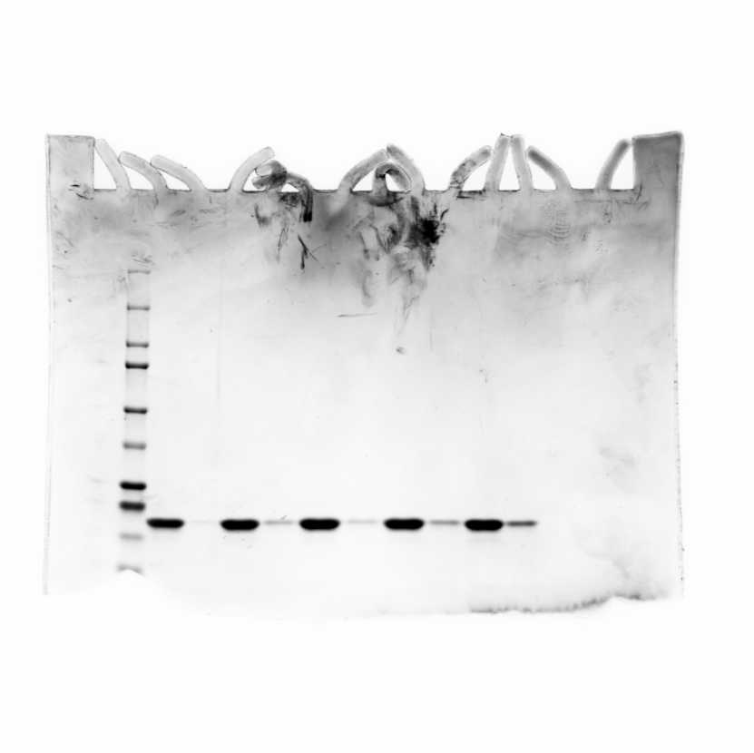
Run SDS-PAGE according the protocol described here, use 4–20% Mini-PROTEAN® TGX Stain-Free™ Protein Gels, 15 well, 15 µlBio-Rad LaboratoriesCatalog #4568096
Stop the gel and cover it with 100mL of the fixing solution (50% Methanol, 10% Acetic acid) for 30 min
Stain the gel for 1 hr at room temperature with 100 mL of Coomassie stain (0.4g of Coomassie blue 10g Coomassie Brilliant Blue R-250 DyeG-BiosciencesCatalog #786-495 in 200 mL of 40% Methanol)
Destain the gel using washing solution consisting of 10% Acetic acid
Image the resulting gels using
Equipment
ChemiDoc™ MP Imaging System
NAME
Imaging System
TYPE
Bio-rad
BRAND
12003154
SKU