Feb 06, 2024
Structural Analysis of 20S CPs and Assembly Intermediates by Electron Cryo-Microscopy
- 1Department of Molecular Machines and Signaling, Max Planck Institute of Biochemistry, 82152 Martinsried, Germany


Protocol Citation: Frank Adolf 2024. Structural Analysis of 20S CPs and Assembly Intermediates by Electron Cryo-Microscopy. protocols.io https://dx.doi.org/10.17504/protocols.io.x54v9px14g3e/v1
Manuscript citation:
License: This is an open access protocol distributed under the terms of the Creative Commons Attribution License, which permits unrestricted use, distribution, and reproduction in any medium, provided the original author and source are credited
Protocol status: Working
We use this protocol and it's working
Created: February 05, 2024
Last Modified: May 31, 2024
Protocol Integer ID: 94722
Keywords: ASAPCRN, proteasome, core particle, 20S proteasome, chaperone, molecular machine, multiprotein complex, POMP, PAC1, PAC2, PAC3, PAC4, propeptide, protease, assembly intermediates by electron cryo, structural determination by transmission electron cryo, electron cryo, transmission electron cryo, microscopy this protocol details method, assembly intermediate, structural determination, microscopy, electron
Funders Acknowledgements:
Aligning Science Across Parkinson's (ASAP)
Grant ID: ASAP‐000282
Abstract
This protocol details methods for structural determination by transmission electron cryo-microscopy of 20S CPs and assembly intermediates.
Guidelines
Please familiarise yourself with the laboratory safety rules and guidelines and follow these while performing the experiment. Please wear appropriate PE while performing the experiment.
Materials
QUANTIFOIL‱ R 1.2/1.3 Cu 200 - Quantifoil
Safety warnings
Liquid nitrogen (LN2) and other cryogens can cause severe damage to the skin and eyes. Always wear personal protective equipment when handling these cryogens.
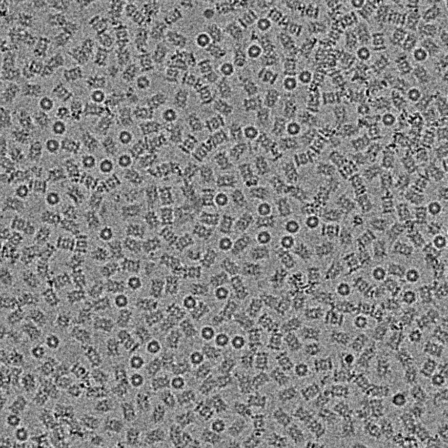
Plung freezing of 20S CPs amd 20S CP assembly intermediates
1h
Prepare Vitrobot and grids for plunging
- Set up Vitrobot as follows: blot force = 3, blot time = 00:00:03 sec, humidity 100%, temperature 4 °C
- Plasma clean Quantifoil R1.2/1.3 Cu 200 grids for 00:00:45 sec , just before plunging
48s
Apply 3.5 µL of purified 20S CPs at a concentration of 0.5-0.6 mg/mL or purified and concentrated 20S CP assembly intermediates at a concentration of 4.0-5.0 mg/mL with Fos8-cholin at a finale concentration of 0.25 millimolar (mM) on grids, automatically blot and plunge in ethane/propane mix at -180 °C with a Vitrobot Mark IV
1h
Clipp and store grids in LN2 until screening/data collection
cryo-EM screening and data acquisition
1d
Screen cryoEM grids for particle density and ice quality on a Glacios cryo-TEM (Thermo Fisher
Scientific) or cryo-TEM of your choise
1d
Data collection was carried out either on a Glacios cryo-TEM (Thermo Fisher Scientific) operated at 200 kV equipped with a K2 Summit direct electron detector (DED) camera (Gatan) or Titan Krios G2 cryo-TEM (Thermo Fisher Scientific) operated at 300 kV equipped with a Bio Quantum post-column energy filter (Gatan, 10eV) and K3 direct electron detector (DED) camera (Gatan)
Data collection on both on the Glacios and Titan Krios G2 cryo-TEM and was set up with SerialEM version 4.1 utilizing coma-corrected beam-image shift
Citation
LINK
Glacios datasets where recorded with one movie per hole in counting mode with a 3x3 or 5x5 multi hole record acquisition scheme at a pixel size of 1.181 Å/pixel with a nominal magnification of 36000x, or at a pixel size of 1.885 Å/pixel with a nominal magnification of 22000x
A total dose of 60 e-/Å2 was fractionated over 40 frames, with a target defocus range of -1.0 μm to -2.6 μm
Krios datasets where recorded with three movie per hole in counting mode with a 5x5 multi hole record acquisition scheme at a pixel size of 0.8512 Å/pixel with a nominal magnification of 105000x
A total dose of 68 e-/Å2 was fractionated over 30 frames, with a target defocus range of -1.0 μm to -2.6 μm
2d
Processing
All data processing steps were performed with cryoSPARC version 4.266
Citation
LINK
Raw movies from Glacios datasets were patch motion corrected in cryoSPARC, and raw movies of the Titan Krios K3 dataset were on the fly motion corrected with FOCUS and subsequently imported into cryoSPARC
Citation
LINK
All subsequent processing steps were preformed in cryoSPARC, for detaied processing shemes see the Extended Data Figures in https://www.biorxiv.org/content/10.1101/2024.01.27.577538v1.full
Final post-processing was preformed with DeepEMhancer
Citation
LINK
2w
Model building and refinement
1w
AlphaFold2 models of PAC1-4 and POMP along with corresponding chains from a published
model of the 20S CP (PDB 5LE5) were manually docked with ChimeraX version 1.5
Citation
LINK
Citation
LINK
Citation
LINK
Atomic models build in Coot version 0.9.8.7
Citation
LINK
Refinment was carried out in Phenix version 1.19.2 and ISOLDE
Citation
LINK
Citation
LINK
1w
Citations
Step 3
Mastronarde DN. Automated electron microscope tomography using robust prediction of specimen movements.
https://doi.org/10.1016/j.jsb.2005.07.007Step 5
Jumper J, Evans R, Pritzel A, Green T, Figurnov M, Ronneberger O, Tunyasuvunakool K, Bates R, Žídek A, Potapenko A, Bridgland A, Meyer C, Kohl SAA, Ballard AJ, Cowie A, Romera-Paredes B, Nikolov S, Jain R, Adler J, Back T, Petersen S, Reiman D, Clancy E, Zielinski M, Steinegger M, Pacholska M, Berghammer T, Bodenstein S, Silver D, Vinyals O, Senior AW, Kavukcuoglu K, Kohli P, Hassabis D. Highly accurate protein structure prediction with AlphaFold.
https://doi.org/10.1038/s41586-021-03819-2Step 5
Jumper J, Hassabis D. Protein structure predictions to atomic accuracy with AlphaFold.
https://doi.org/10.1038/s41592-021-01362-6Step 5
Goddard TD, Huang CC, Meng EC, Pettersen EF, Couch GS, Morris JH, Ferrin TE. UCSF ChimeraX: Meeting modern challenges in visualization and analysis.
https://doi.org/10.1002/pro.3235Step 5
Emsley P, Lohkamp B, Scott WG, Cowtan K. Features and development of Coot.
https://doi.org/10.1107/S0907444910007493Step 5
Liebschner D, Afonine PV, Baker ML, Bunkóczi G, Chen VB, Croll TI, Hintze B, Hung LW, Jain S, McCoy AJ, Moriarty NW, Oeffner RD, Poon BK, Prisant MG, Read RJ, Richardson JS, Richardson DC, Sammito MD, Sobolev OV, Stockwell DH, Terwilliger TC, Urzhumtsev AG, Videau LL, Williams CJ, Adams PD. Macromolecular structure determination using X-rays, neutrons and electrons: recent developments in Phenix.
https://doi.org/10.1107/S2059798319011471Step 5
Croll TI. ISOLDE: a physically realistic environment for model building into low-resolution electron-density maps.
https://doi.org/10.1107/S2059798318002425