Jun 13, 2025
Stony Coral Single Cell Isolation
- 1Boston University



Protocol Citation: Maria Ingersoll, Sarah Davies 2025. Stony Coral Single Cell Isolation. protocols.io https://dx.doi.org/10.17504/protocols.io.rm7vzkx72vx1/v1
License: This is an open access protocol distributed under the terms of the Creative Commons Attribution License, which permits unrestricted use, distribution, and reproduction in any medium, provided the original author and source are credited
Protocol status: Working
We use this protocol and it's working
Created: December 05, 2024
Last Modified: June 13, 2025
Protocol Integer ID: 114326
Keywords: Coral, Single Cell Isolation, Cnidaria, symbiotic stony coral oculina, cell rna, live cell, live cells for downstream use, cell, rna
Funders Acknowledgements:
National Science Foundation
Grant ID: IOS-1937650
National Science Foundation
Grant ID: Graduate Research Fellowship
National Science Foundation Research Traineeship
Grant ID: DGE 1735087
Boston University Marine Program
Grant ID: Warren McLeod Marine Fellowship
Abstract
This protocol was optimized in the Davies lab at Boston University for use with the facultatively symbiotic stony coral Oculina arbuscula. The protocol is designed to isolate live cells for downstream use in 10X single-cell RNA-sequencing.
Materials
Reagents
Sterile DI Water
0.2 micron filter-sterilized Calcium-Free Seawater (CfFSW)
Fetal Bovine Serum (FBS)
10X PBS
1X PBS
1M HEPES
Protector RNase Inibitor (Sigma Aldrich 3335399001)
0.4% Trypan Blue Solution (filtered through 0.2 micron filter to remove aggregates)
RNase Zap or equivalent
Materials
Heat-bath or hot-plate capable of 56°C
6-well plate
Sterile 200μl pipette tips
Forceps/tweezers
Sterile 1.5 ml tubes
Sterile 15 ml conical tubes
Sterile 70 micron cell strainers (e.g., Fisher Scientific 08-771-2)
Sterile 40 micron cell strainers (e.g., Fisher Scientific 08-771-1)
Microcentrifuge capable of 4°C
Hemocytometer
Light Microscope
Safety warnings
- As much as possible, all steps should be performed on ice
- Do not vortex cell suspension at any point during the protocol
- Limit bubbles in cell suspension
- Cells should be processed promptly following dissociation to maintain viability (viability decreases within an hour following isolation)
Heat inactivate FBS for 30 minutes at 56°C
Sterilize tools (i.e., forceps, pipettes, etc.) and bench space with RNAse Zap
Prepare cell media1 and keep on ice
To make 20 ml of cell media:
- 6.6 ml 10X PBS (final concentration of 3.3X)
- 0.4 ml heat-inactivated FBS (final concentration of 2% by volume)
- 0.4 ml 1M HEPES (final concentration of 20mM)
- Fill to 20 ml with sterile DI H2O
Add 10 mls of CafFSW2 to two wells of a 6-well plate, and add 10 mls of cell media to a third well. Keep plate on ice.
Obtain live coral frag (this protocol has been optimized for frags of symbiotic or aposymbiotic branching coral ~1.5 cm in length and ~1 cm in width) and place in first well of CafFSW
Incubate for 1 minute (on ice)
With sterilized forceps, move frag to second well of CafFSW, and incubate for 1 minute (on ice)
With sterilized forceps, move frag to well containing cell media. Using forceps and sterile 200 μl pipette tips, carefully scrape all tissue from the coral skeleton and from within the polyps3. This step should last no more than 10 minutes. If necessary, this step can be performed on the bench, not on ice.
TIP: Dig the pipette tip or forcep tip into a polyp and make small circles to extract cells from within the polyp
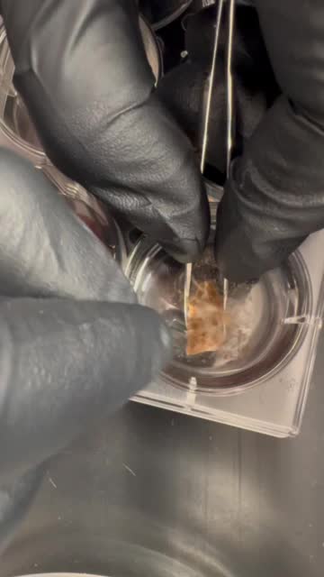
Filter full cell suspension through one 70 micron cell strainer into a 15 ml conical tube.
NOTE: for filtration steps, pipette 1 ml aliquots through strainer by holding tip against mesh, depressing the plunger slowly and assuring all liquid passes through the strainer. Keep plunger depressed while removing tip from mesh. Cell suspension may be mucus-y at this point, so some volume (and cells) will inevitably be lost.
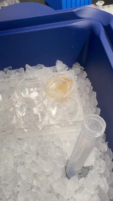
Filter 70-micron-filtered suspension through one 40 micron cell strainer into a new 15 ml conical tube
Filter suspension through a second 40 micron cell strainer into a third 15 ml conical tube
Centrifuge 1 ml aliquot(s) of filtered cell suspension at 300xg for 10 minutes at 4°C
Discard supernatent and resuspend pellet in 100 μl of cell media with 0.2 U/L Protector RNase Inhibitor
Right before use, add 0.5 μl of stock RNase Inhibitor (at 40 U/μl) per 100 μl cell media
Check cell viability and perform cell counts (using hemocytometer)
Combine 2.5 μl of 0.4% Trypan Blue with 12.5 μl of 1X PBS and 10 μl of final cell resuspension (for final concentration of 0.04% Trypan)
For 10X, cell viability should be over 90% (dead cells are completely permeated with blue dye)
Symbiotic cells (algal cells) should be counted in cell count and viability calculations
Example of cell suspension from symbiotic Oculina arbuscula with 0.04% Trypan Blue visualized at 40X magnification
Dilute cells to desired concentration in cell media with RNase Inhibitor (see step 13.1 for concentration)
For 10X at the Boston University Single Cell Core, cells were provided at concentrations of 700-1,200 cells/μl
Protocol references
1. Rosental B, Kozhekbaeva Z, Fernhoff N, Tsai JM, Traylor-Knowles N. Coral cell separation and isolation by fluorescence-activated cell sorting (FACS). BMC Cell Biology. 2017 Aug 29;18(1):30.
2. Roger LM, Reich HG, Lawrence E, Li S, Vizgaudis W, Brenner N, et al. Applying model approaches in non-model systems: A review and case study on coral cell culture. PLoS ONE 2021 Apr 8;16(4):e0248953.
3. Levy S, Elek A, Grau-Bové X, Menéndez-Bravo S, Iglesias M, Tanay A, et al. A stony coral cell atlas illuminates the molecular and cellular basis of coral symbiosis, calcification, and immunity. Cell. 2021 May 27;184(11):2973–2987.e18.