Feb 09, 2026
Standardized Protocol for Measuring Wet Weight, Volume, and Body Dimensions of Sea Anemone Polyps
- Finn Hamilton1,
- Rowan McLachlan2,
- Nathan Kirk2
- 1University of Oregon;
- 2Oregon State University
- Anemones at War Protocols


Protocol Citation: Finn Hamilton, Rowan McLachlan, Nathan Kirk 2026. Standardized Protocol for Measuring Wet Weight, Volume, and Body Dimensions of Sea Anemone Polyps . protocols.io https://dx.doi.org/10.17504/protocols.io.4r3l21o73g1y/v1
License: This is an open access protocol distributed under the terms of the Creative Commons Attribution License, which permits unrestricted use, distribution, and reproduction in any medium, provided the original author and source are credited
Protocol status: Working
We use this protocol and it's working.
Created: July 29, 2025
Last Modified: February 09, 2026
Protocol Integer ID: 223619
Keywords: wet weight, seawater displacement volume, body column dimensions, anemone, Anthopleura, elegantissima, polyp, cnidarian, pedal disk, morphometrics, preparation of anthopleura elegantissima polyps measurement, anthopleura elegantissima polyps measurement, calipers determination of polyp volume, body dimensions of sea anemone polyp, standardized protocol for measuring wet weight, measuring wet weight, polyp wet weight in air, polyp volume, sea anemone polyp, preparation of polyp, size of polyp, small anthopleura elegantissima, aggregating anemone anthopleura elegantissima, using calipers determination, wet weight, anemone anthopleura elegantissima, balance measurement, such as anthopleura xanthogrammica, body volume, anthopleura xanthogrammica, polyp, body column diameter, size of the aluminum weigh pan
Abstract
This protocol outlines four non-destructive methods for measuring the size of polyps of the aggregating anemone Anthopleura elegantissima: wet weight, volume, and body column diameter and body column height.
Although designed for the relatively small Anthopleura elegantissima, the methods can be adapted for smaller or larger species, such as Anthopleura xanthogrammica, by adjusting the size of the aluminum weigh pans, graduated cylinders, and stainless-steel sieves used.
The protocol consists of four sections:
- Preparation of Anthopleura elegantissima polyps
- Measurement of polyp wet weight in air using a balance
- Measurement of polyp contracted body column dimensions using calipers
- Determination of polyp volume via seawater displacement
Step 1 (preparation of polyps) is required before proceeding with any of the other steps, while steps 2–3 may be performed in any order. It is recommended that body volume be measured last, since this step may reintroduce water into the coelenteron that was expelled during preparation.
This protocol is adapted from the methods described in Ford (1964), Francis (1976); Francis (1979); and Sebens (1980).
Materials
Disposable Materials:
- Powder-free nitrile gloves
- 43 mm diameter aluminum weighing dishes [1 per polyp]
- A4 paper data sheet (template provided in protocol)
- Filtered seawater (natural or artificial) [~20 ml per polyp]
Reusable Materials:
- 1 x 150 mm length Vernier calipers (analog used, but digital is recommended)
- 1 x 25 ml volume borosilicate glass graduated cylinder with fine (0.2 ml) graduations
- 1 x 1000 ml volume borosilicate glass Erlenmeyer flask
- 1 x 600 ml volume borosilicate glass beaker
- 1 x Nylon filtration fabric NITEX (mesh opening 20 μm) glued to 2-inch PCV pipe
- Clean lint-free cloth towel/rag [1 per polyp]
- 1 x round-tipped precision forceps or tweezers
- Mechanical pencil
- >10 cm diameter stainless-steel sieve (pore size between 1–5 mm)
Equipment:
- Scientific scale or top-loading balance (ideally with an accuracy of≥0.01 g and resolution of 0.001 g)
- Computer (PC or Mac)
Software:
- Microsoft Excel(R)
Safety warnings
Electrical Hazards Warnings: Scientific scales and other laboratory equipment are powered by high-voltage outlets. Keep all liquids away from cords, plugs, and outlets to prevent electrical shock or short circuits. Always ensure hands and work surfaces are dry before handling electrical equipment.
Broken Glass and Sharps Warning: Glassware is fragile and may break easily. Handle with care to avoid cuts. Never dispose of broken glass in regular trash — always place it in a designated sharps or glass disposal container.
Biological Hazard Warning: While nematocysts of Anthopleura spp. are typically unable to penetrate human skin, repeated or prolonged handling of live specimens has been reported to cause sensitization and subsequent allergic responses in some individuals.
Ethics statement
All Anthopleura elegantissima specimens used in this protocol were collected in compliance with state regulations under the Oregon Department of Fish and Wildlife’s Scientific Taking Permit (Permit number 28795, Permit Applicant: Maya Watts, Organization: Oregon Institute of Marine Biology). Collection and handling procedures followed institutional guidelines for the ethical use of invertebrates in research and teaching. All efforts were made to minimize stress and potential harm to the animals, and only the number of specimens necessary to achieve the educational and methodological objectives was collected.
Before start
Preparation of Datasheet
- Create hardcopy datasheet. Create and print a hardcopy datasheet to record data (download template datasheet here
 Protocol Example Spreadsheet.xlsx10.4KB ). Record all collected data on the printed datasheet in pencil. After completing the protocol, digitize the records by entering the data into a computer using Microsoft Excel(R).
Preparation of Anthopleura elegantissima polyps
Remove any undigested food particles from the sea anemone's gastrovascular cavity. If the polyp has been recently collected from the wild or fed large prey items (e.g., mussel, squid) in the laboratory, this step in necessary to ensure an accurate wet weight measurement. Otherwise, this step can be skipped. To remove food, the mouth can be gently encouraged to open using the approach described in Bedgood et al (2020). Briefly, use tweezers or forceps to touch a food item to the tentacles gently. This typically triggers a feeding response and will cause the mouth to open, allowing for the removal of any undigested material, using blunted or round-tipped forceps.
Dry the sea anemone polyp. Gently blot dry the polyp with a clean, lint-free towel, applying light but firm pressure to expel water from the coelenteron (Fig. 1). Use the towel to remove excess mucus. Avoid rubbing the tissues during blotting, as this can cause damage to the polyp.
Fig. 1. Drying procedure for an Anthopleura elegantissima polyp. (A) Initial appearance of the polyp before drying. (B) Polyp wrapped in a clean towel and gently blotted. (C) Final appearance after removal of excess water.
Remove debris from the external surface of the sea anemone. Polyps of A. elegantissmia often adhere particles such as sand and shell fragments to tubercules/verrucae on their external body wall. Similarly, the pedal disk may adhere to encrusting algae. To obtain accurate size, weight, and volume measurements, all debris must be removed before proceeding. Using tweezers or forceps, carefully remove debris from the body column (Fig. 2A). Clear away shell fragments, loose particles, and any lint remaining from the drying step from the body column (Fig. 2B). Any encrusting algae on the pedal disk can be gently removed by carefully scraping it away with a fingernail (Fig. 2C–D). Take care throughout this process to avoid damaging the underlying tissue.
Note
Fig. 2. Cleaning procedures for an Anthopleura elegantissima polyp. (A) Gentle removal of shell fragments and debris from the body wall using forceps. (B) Polyp after removal of external debris. (C) Encrusting algae on the pedal disk being carefully scraped away with a fingernail. (D) Final appearance of the cleaned pedal disk.
Fig. 2. Cleaning procedures for an Anthopleura elegantissima polyp. (A) Gentle removal of shell fragments and debris from the body wall using forceps. (B) Polyp after removal of external
debris. (C) Encrusting algae on the pedal disk being carefully scraped away with a fingernail. (D) Final appearance of the cleaned pedal disk.
Measurement of polyp wet weight in air using a balance
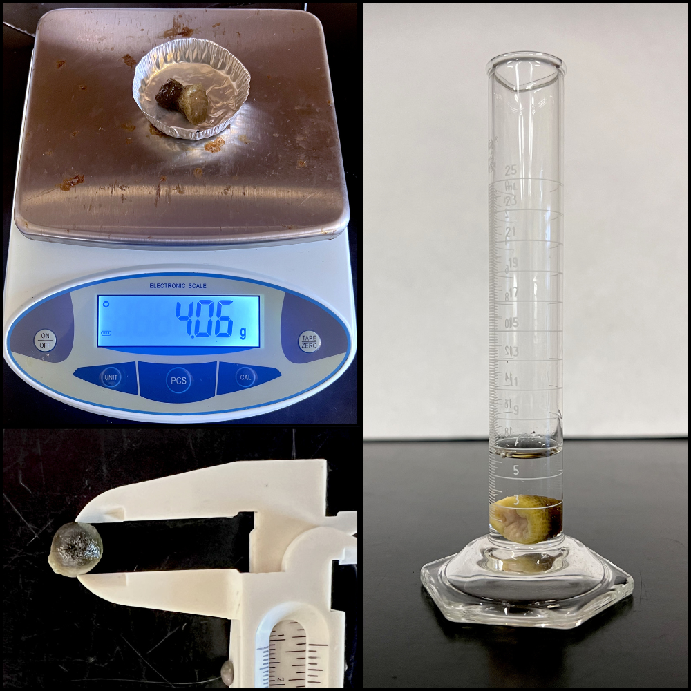
Measure the sea anemone wet weight. Turn on the balance, place a clean, empty aluminum weigh dish on the pan, and tare the balance to zero (Fig. 3A–C). Carefully transfer the dried and cleaned Anthopleura elegantissima polyp into the dish and record the wet weight in grams on the hardcopy data sheet (Fig. 3D).
Fig. 3. Procedure for measuring the wet weight of an Anthopleura elegantissima polyp. (A) Balance zeroed without an aluminum weigh dish. (B) Aluminum weigh dish placed on the balance. (C) Balance tared with the dish in place. (D) Final wet weight of the polyp measured in the aluminum dish.
Measurement of polyp contracted body column dimensions using calipers
Step 5: Prepare the anemone for caliper measurements. The anemone polyp should be fully contracted into a compact, dome-like (hemispherical or short-cylindrical) shape with tentacles completely withdrawn before measuring body column height and width. If necessary, place the polyp on a clean, flat surface and gently shape it into a compact hemishere, taking care to avoid extension or distortion into an irregular form (Fig. 4A–C).
Fig 4. Proper and improper polyp shapes of measurement in Anthopleura elegantissima . (A–B) Examples of incorrect body shapes. (C) Correct, fully retracted, compact polyp shape prior to measurement.
Step 6: Measure body width. Place the pedal disk of the Anthopleura elegantissima polyp flat on a clean surface. Using calipers, measure the body column diameter by positioning the caliper tips on opposite sides of the column at its widest point, ensuring they just contact the tissue without compressing or distorting the fully contracted polyp (Fig. 5). Keep the calipers parallel to the pedal disk (not angled) to obtain an accurate measurement. Measure pedal disk diameter in the same manner, spanning from one edge of the disk to the other. Record all measurements to the nearest millimeter on the hardcopy data sheet.
Fig. 5. Measurement of body column diameter in an Anthopleura elegantissima polyp using analog or digital calipers.
Step 7: Measure body height. Place the Anthopleura elegantissima polyp on its side on a clean, flat surface so that the pedal disk is perpendicular to the table. Using calipers, measure the distance from the center of the pedal disk to the center of the oral disk, taking care not to compress or alter the shape of the fully contracted polyp (Fig. 6). Keep the calipers parallel to the table and perpendicular to the pedal disk. Record the measurement to the nearest millimeter on the data sheet.
Fig. 6. Measurement of the body column diameter in an Anthopleura elegantissima polyp using analog or digital calipers.
Determination of polyp volume via seawater displacement
Filter seawater. Collect unfiltered seawater in a clean beaker or Erlenmeyer flask large enough to hold the total volume required for subsequent polyp volume measurements and comparable in size to the receiving beaker to prevent overfilling (a 1000 mL flask was used in this protocol). Place a 20 µm NITEX filter over a second clean beaker, ensuring it fits securely just inside the rim without shifting. Slowly pour the unfiltered seawater through the filter into the collection beaker (Fig. 7A), taking care not to overfill either the filter or the beaker (Fig. 7B). Use the filtered seawater for the following steps.
Fig. 7. Demonstration of filtering seawater through a 20 μm filter, (A) pouring the unfiltered water through the filter into a beaker, (B) pausing to allow the seawater to drain through the filter, without overflowing.
Fill the graduated cylinder with an exact volume. Select the smallest graduated cylinder (10–50 mL) that can accommodate the Anthopleura elegantissima polyp without risk of lodging. Fill the cylinder with a measured volume of prepared 20 µm–filtered seawater sufficient to fully submerge the polyp (e.g., 5 mL in a 25 mL cylinder for a medium-sized polyp; Fig. 8A). Use a cylinder with appropriately fine graduations (e.g., 0.2 mL for a 25 mL cylinder or up to 1 mL for a 50 mL cylinder). Read the water level at eye level from the bottom of the meniscus (Fig. 8B) and record the initial volume to the nearest milliliter on the data sheet.
Fig. 8. Volume measurement of an Anthopleura elegantissima polyp using seawater displacement. (A) Initial seawater volume (5 mL) in a 25 mL graduated cylinder. (B) Correct technique for reading liquid volume in a graduated cylinder; the arrow indicates the bottom of the meniscus, which should be read at eye level for accuracy. (C) Final volume (6.4 mL) after the polyp is placed in the graduated cylinder.
Measure polyp volume. Gently lower the polyp into the graduated cylinder, taking care to avoid splashing or wetting the cylinder walls, as this would alter the final volume measurement. Once the polyp is fully submerged (Fig. 8C), record the new water volume to the nearest milliliter on the hardcopy data sheet.
Return the sea anemone to the water table. Gently pour the contents of the graduated cylinder into a stainless-steel sieve, allowing the seawater to drain while retaining the polyp. Carefully lift the polyp from the sieve and return it to the water table or to a laboratory holding aquarium.
Calculate the volume of the polyp. Enter the data from the paper hardcopy datasheet into Microsoft Excel(R). To determine the volume of the Anthopleura elegantissima polyp, use the displacement equation:
Polyp Volume (mL) = Final Water Volume (mL) - Initial Water Volume (mL)
Protocol references
Bedgood, S.A., Mastroni S.E., Bracken, M.E.S. (2020). Flexibility of nutritional strategies within a mutualism: food availability affects algal symbiont productivity in two congeneric sea anemone species. Proc. R. Soc. B 287: 20201860.
Ford Jr, C. E. (1964). Reproduction in the aggregating sea anemone, Anthopleura elegantissima. Pacific Science, 18(2):138–145.
Francis, L. (1976). Social organization within clones of the sea anemone Anthopleura elegantissima. Biological Bulletin. 150(3):361-376.
Francis, L. (1979). Contrast between solitary and clonal lifestyles in the sea anemone Anthopleura elegantissima. American Zoology. 19:669-681.
Sebens, K. P. (1980). The regulation of asexual reproduction and indeterminate body size in the sea anemone Anthopleura elegantissima (Brandt). The Biological Bulletin. 158(3):370-382.
Acknowledgements
We gratefully acknowledge the University of Oregon’s Oregon Institute of Marine Biology for providing access to teaching facilities, equipment, and support from faculty and staff. We thank the administrative staff, Laura Screen and Shawna Johnston, for their assistance in ordering supplies and materials. Special thanks are extended to Ian Washington, Education Support Specialist, for his support in setting up the teaching laboratory space and equipment, as well as for his help in locating additional supplies. Finally, we thank all students enrolled in BI457 Anemones at War for their contributions in refining and troubleshooting the methods described in this protocol.
Funders Acknowledgements: All supplies and materials not available through the Oregon Institute of Marine Biology’s teaching inventory were purchased using student class fees.