Jan 06, 2026
Standardized Protocol for Blow Fly (Phormia regina and Lucilia sericata) Development on Control Burned Vertebrate Remains
- Hayden McKee-Zech1,
- Dillon G. Gaines1,
- Linnea N. Briley1,
- Chantel M. Gurak1,
- Samantha S. Richards1,
- Andrew Tease1,
- Cameron Koonter1,
- Miah Beck1
- 1Department of Biology, Northern Michgian


Protocol Citation: Hayden McKee-Zech, Dillon G. Gaines, Linnea N. Briley, Chantel M. Gurak, Samantha S. Richards, Andrew Tease, Cameron Koonter, Miah Beck 2026. Standardized Protocol for Blow Fly (Phormia regina and Lucilia sericata) Development on Control Burned Vertebrate Remains. protocols.io https://dx.doi.org/10.17504/protocols.io.8epv5kyrjv1b/v1
License: This is an open access protocol distributed under the terms of the Creative Commons Attribution License, which permits unrestricted use, distribution, and reproduction in any medium, provided the original author and source are credited
Protocol status: Working
We use this protocol and it's working
Created: December 02, 2025
Last Modified: January 06, 2026
Protocol Integer ID: 234029
Keywords: Phormia regina, entomotoxicology, development, blow fly, growth rate, morphine, lorazepam, drugs, pharmaceuticals, protocols for entomotoxicological develo..., larval blow fly, entomotoxicological developmental study, estimating larval age, larval age, laboratory colony of the black blow fly, specific developmental dataset, death investigation, developmental duration, black blow fly, abundant blow fly, phormia regina, minimum postmortem interval, important phenotype, generation of drug, Burn, Burned , burned decomposition , burned ecology , burned vertebrate remain, burned vertebrate, developmental data for the forensic blow, mammalian carcass, vertebrate remain, examining blow fly development, insect development, larval development, reproducible developmental datasets relevant to forensic casework, blow fly development, burned remain, substitution of alternative vertebrate, alternative vertebrate, standardized mammalian analog, standardized protocol for blow fly, blow fly, reproducible developmental dataset, forensic blow, altered rem
Disclaimer
DISCLAIMER – FOR INFORMATIONAL PURPOSES ONLY; USE AT YOUR OWN RISK
The protocol content here is for informational purposes only and does not constitute legal, medical, clinical, or safety advice, or otherwise; content added to protocols.io is not peer reviewed and may not have undergone a formal approval of any kind. Information presented in this protocol should not substitute for independent professional judgment, advice, diagnosis, or treatment. Any action you take or refrain from taking using or relying upon the information presented here is strictly at your own risk. You agree that neither the Company nor any of the authors, contributors, administrators, or anyone else associated with protocols.io, can be held responsible for your use of the information contained in or linked to this protocol or any of our Sites/Apps and Services.
Abstract
This protocol describes a standardized laboratory method for generating developmental data for the forensic blow flies Phormia regina (Meigen) and Lucilia sericata (Meigen) using experimentally burned vertebrate remains. Mammalian carcasses (Mus musculus) are burned to defined Crow–Glassman Scale (CGS) levels (Glassman & Crow 1996) and used as rearing substrates under controlled environmental conditions. Larval development, survivorship, and post-feeding metrics are documented at regular intervals through adult emergence.
This protocol is designed to support reproducible developmental datasets relevant to forensic casework involving burned remains, where insect development may be altered by thermal damage and tissue modification. Although mice are used here as a standardized mammalian analog, the protocol is intentionally structured to allow substitution of alternative vertebrate or human analog substrates, provided appropriate ethical approvals and safety considerations are met. The methods described here aim to improve consistency and comparability across studies examining blow fly development on thermally altered remains, thereby strengthening PMImin estimation in thermally altered contexts.

Image Attribution
Image depicting FADE Lab experimental housing created by Graycen C. McKee
Experimental image included in this publication were taken by Hayden S. McKee-Zech, Chantel M. Gurak, Dillion G. Gains and Samatha S. Richards.
Guidelines
Recorded data include all observation timestamps, larval counts, developmental milestones, pupal and adult masses, emergence times, and mortality events. Time measurements are reported relative to larval inoculation. Any missing data points or excluded replicates are documented with explanatory notes.
This protocol is intended to support reproducible developmental datasets for forensic entomology applications and to facilitate comparison across studies involving burned remains.
Materials
| Equipment/Materials | Amount | Purpose/Description | |
| Biohazard bag/containers | - | For disposal | |
| Bucket | - | To hold water | |
| Bunsen burner | 1 | To burn mice | |
| Calipers | 3 | To measure larvae length | |
| Camera (Digital/Phone) | 1 | For field documentation | |
| Collection jars | 15 | For temporary storage | |
| Cultured butter milk ad libitum | - | To give flies nutrients | |
| Disinfectant wipes | 4 containers | To clean | |
| Equipment/Material | Amount | Purpose/Description | |
| Ethanol (70-90%) | 1 bottle | For preserving larvae | |
| Face maks | 1 box | In case people want to wear while burning | |
| Field notebook | 7 | To take notes/data during the experiment | |
| Fire extinguisher | 1 | For fire safety | |
| First aid kit | 1 | For safety | |
| FLIR thermal imaging camera | 1 | For larval mass detection on burned tissue | |
| Forceps/tongs | 3 | To handle burned and unburned pork | |
| Gloves (Large) | 4 boxes | PPE | |
| Gloves (Medium) | 4 boxes | PPE | |
| Goggles/safety glasses | 8 | PPE (In case anyone wants to wear) | |
| Grosse probes | 3 | To move larvae to mice | |
| Humidity tray | - | In case we need to increase the humidity | |
| Hygrometer | 1 | To measure humidity | |
| Incubator | 1 | For temperature control | |
| Kettle | 1 | For boiling water for parboiling larvae before storage | |
| Kimwipes | - | For laying eggs | |
| Laptops | - | For data, photos, writing our report, etc. | |
| Light cycle control/timer | - | To control the light cycle | |
| Lucilia sericata | 1200 larvae | One of the two species of flies we are using | |
| Magnifying glass | 2 | To magnify and see better if needed | |
| Markers (black Sharpie) | 4 | For external labeling | |
| Metal grate/screen | 2 | To put meat on | |
| Mice (burn) 21g-35g | 10 | Meat for experiment | |
| Mice (control) 25g-32g | 10 | Meat for experiment | |
| Micro probe | 3 | For inoculation | |
| Microscope slides | - | To put specimens on | |
| Microscopes | 8 | To examine specimens | |
| Napkins (paper) | - | To act as a lid on the deli cups | |
| O-ring | 1 | To hold the mice above the flame | |
| Pencils | 4 | For writing on the labels inside the ethanol | |
| Petri dishes | - | For eggs and larvae | |
| Plastic cups (mini) | - | For rearing substrate | |
| Plastic tubs | 3 | To sift the sand over | |
| Rearing containers / Fly dorms | 2 | To house our flies | |
| Rearing substrate (Liver) | - | To get flies to lay eggs | |
| RStudio or Excel | - | For data collection | |
| Rubber bands | - | To hold a secure napkin covering over deli cup | |
| Sawdust or sand | - | For rearing cages | |
| Scales | 2 | To weigh larval and adult flies | |
| Sifters | 3 | To sift out sand from larvae and pupa | |
| Soft, synthetic paintbrushes | 8 | For gentle examination and inoculation | |
| Specimen jars | - | For preserving specimens and long-term storage | |
| Striker | 1 | To light Bunsen burner | |
| Sugar | - | To feed flies | |
| Tall cylindrical continer | 2 | To hold water for flies | |
| Tape (colored) | 2 rolls | To label | |
| Thermometer | 1 | To take the temperature of the meat | |
| Tweezers | 8 | To handle eggs, larvae, pupae, and adult flies | |
| UTK Phormia Regina | 1,200 Larvae | One of the two species of flies we are using | |
| Water | - | To maintain humidity and to hydrate flies |
Basic materials used in experiment.
Safety warnings
Refer to your facilities guidelines for working with potentially hazardous material and Blood Borne Pathogen (BBP) protocol. This work was performed in a Biohazard Safety Level (BSL)-2 laboratory.
Refer to your facilities fire safety protocols and standards.
Ethics statement
All procedures involving vertebrate animals were conducted in accordance with internationally accepted standards for the ethical use of animals in research and were reviewed and approved by the Northern Michigan University Institutional Animal Care and Use Committee (NMU IACUC). The mouse breeding colony from which specimens were obtained was maintained under an approved NMU IACUC protocol (Application No. 465), approved on April 29, 2024, and valid through May 29, 2027. For the purposes of the experiments reported here, no live animals were subjected to experimental manipulation. All mice used in this study were received already deceased, having been euthanized under approved IACUC protocols by the colony managers prior to transfer. No additional euthanasia, invasive procedures, or live-animal experimentation was performed as part of this study.
Before start
Each individual mouse carcass represents one biological replicate. Larvae placed onto a single mouse are treated as subsamples within that replicate and are not considered independent experimental units. All developmental metrics (e.g., time to wandering, pupation, adult emergence, mass) are summarized at the replicate level for statistical analyses unless otherwise specified.
Five replicate mice were used per treatment group (control and CGS-3 burned). This level of replication is consistent with previously published developmental datasets for Phormia regina and Lucilia sericata and was selected to balance logistical constraints with the ability to characterize treatment-level variation.
1) Environmental Conditions and Monitoring
All experiments were conducted in a controlled growth chamber maintained at 25 °C with 70% relative humidity and a 12:12 (L:D) photoperiod. Ambient temperature and humidity were monitored using a calibrated hygrometer placed at the container level within the chamber.Because larval aggregations can generate metabolic heat, surface temperatures of the substrate and larval masses were documented at each observation period using a FLIR thermal imaging camera. Thermal images were collected with consistent camera distance and angle to allow qualitative comparison among replicates. These measurements are intended to document relative thermal patterns rather than replace ambient chamber conditions.
2) Printing labels for each step of the experiment that can be filled in prior to starting each step with speed the process along immensely.
3) Ensure mice are room temperature and dry.
4) Ensure all materials used with the Bunsen burner are not flammable and heat resistance.
5) This project was conducted in a fume hood, ensure proper ventialiation before starting.
Colony Establishment
Fly colonies should be established using local populations of blow flies. Adult flies can be baited with aged organ or muscle tissue, or they can be collected from a vertebrate carcass, refuse, or feces. Flies should be collected with an aerial sweep net, sorted to the desired species, and placed in a BugDorm or other enclosure specifically for insects. Flies can be maintained using bug dorms inside Percival growth chamber (model I-36VL Environmental Chamber), and given sugar, water, and cultured buttermilk ad libitum. With both species having a simulated consistent 12 hour Light:Dark cycle, while in the Percival. To ensure sufficient genetic diversity and to minimize the risk for genetic drift within the colony, wild fly collections should span multiple timepoints and the total founding generation (G0) should be ~200 individuals.
Note
This set of experiments utilized two species (Lucilia seracata and Phormia regina), the L. seracata flies where wild-caught on-location in Marquette, MI, while the P. regina fly eggs where sourced from the University of Tennessee from wild-caught specimens.
Egg Collection
Fly colonies should be presented with a fresh protein source daily for ~1 week prior to the start of experiments. For this experiment, we exposed colonies to a kimwipe soaked in chicken blood in a 3 oz bath cup each day.
24 - 48 hours prior to the start of experiments, provide the colony with ~5g chicken liver and a kimwipe soaked with chicken blood in a 3 oz cup.
Check for eggs every ~3 hours.
Once eggs are observed, remove them from the cage and record the time and prepare to setup the rearing substrate section.
Rearing Substrate Set-up
Weigh out ~5g of lean pork into a 3 oz bath cup.
Place a pencil-eraser sized clump of eggs onto the pork.
L. sericata eggs placed on Pork Liver.
Place the 3 oz bath cup with pork and eggs into a 32-oz deli cup filled with 3 inches of sterile sand, then transfer into the Percival, checking every ~8 hours for hatching larvae.
Standard experimental housing in the FADE lab.
When larvae have hatched, they can now be used for the inoculation of treatments once ready to begin. This can be found in the Inoculation of Treatments section.
Substrate cup containing Larvae, from our eggs.
Treatment Preparation
For Control treatment - Source 5 mice that have been cervically dislocated for the control, with each being roughly 25-32 grams.
Note
See ethics statement under the Guidelines and Warning tab
Ensure your mice are at room temperature before inoculation. Transfer your mice to individually labeled petri dishes. Make sure to take the weight of the mice, so there is comparable initial weight data to our pre-burn treatment replicates.
The eggs from the Egg Collection section will be used for inoculation. Proceed to the Inoculation of Treatments section for the next steps.
For Burn Treatment - Source 5 mice that have been cervically dislocated for the burning process, with each being roughly 21-35 grams.
Note
See ethics statement under the Guidelines and Warnings tab
Ensure your mice are at room temperature, from freezing. Before beginning the burn process.
Before burning your mice, place them in individually labeled weigh boats. Take individual weights of the mice in order to gain a pre-burn data point and add this to your metadata.
Note
While in the process of weighing your mice for pre-burn data, take individual pictures of the five being weighed. (This gives us a before-and-after comparison of our burn outcome)
Weighing of mice for a pre-burn data point.
Creating your burning setup.
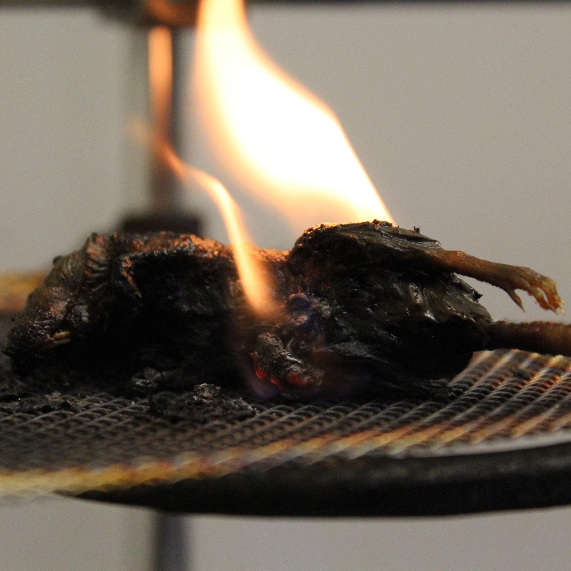
You'll want to create your setup directly in the fume hood. Once in the fume hood, you can attach the O-ring to the metal frame, placing it above the Bunsen burner. With this done, put a metal screen on the O-ring. This will be used to hold the mice while burning.
Note
Make sure when placing the O-ring above the Bunsen burner that it's at an appropriate height, so that the mice can get an even burn.
Burning setup created and used for our experiment.
The burn treatment was designed to approximate Crow–Glassman Scale (CGS) level 3. Mice were positioned supine on a metal screen suspended above a Bunsen burner flame to promote even heat exposure. During the burn process, each carcass was continuously observed for visible indicators of CGS progression, including epidermal blistering, charring of soft tissue, exposure of underlying musculature, and partial limb involvement.
Thermal images were periodically collected during burning to document surface temperature patterns. Burn duration was adjusted on an individual basis to achieve CGS-3 characteristics rather than a fixed time endpoint.
Once CGS-3 was achieved, carcasses were immediately removed from heat and allowed to cool to room temperature prior to larval inoculation.
Note
As you are conducting your burn, take note of the time when burning/charring is first seen, any calcined bone, limbs falling odd, and the impact of fire on the abdomen (creates leakage and internal organs can fall out).
Crow-Glassman Scale (CGS) in accordance with our own burning images.
Once CGS level 3 is reached, take the mouse off the heat and let it cool to room temperature. Once the mouse has cooled down, make sure to transfer it to a labeled petri dish before beginning the inoculation of treatments.
Inoculation of Treatments
Each replicate was inoculated with 100 first-instar larvae.
Use a synthetic plastic paintbrush or precision probe to hand count 100 individual larvae onto each replicate. Larvae were placed primarily within the cervical wound resulting from decapitation and secondarily along the body surface to mimic natural colonization patterns observed in burned remains.
Note
This density was selected to ensure consistent colonization while limiting excessive larval mass heating that could confound developmental timing. Larvae were hand-counted using a synthetic paintbrush or precision probe to maintain consistent density across replicates. This may change if larger animal models are used.
Counting of Individual larvae to reach 100 per replicate.
Placing of larvae onto a burn replicate mouse.
Place the petri dish in 3" of sterile sand within a 32oz deli cup and cover with a paper napkin and 2 rubber bands, making sure there are no creases in the napkin.
Note
See image of experimental housing under description
Updated experimental housing for our inoculated mice.
Data collection - Larvae
Larval development was observed every 12 hours until the onset of larval wandering. Observations occurred at approximately 11:00 AM and 11:00 PM. At each observation period, photographs and FLIR thermal images were collected prior to any handling.
Before starting the inspection of larvae, take a picture of the mice along with a FLIR image of the mice (including their label in the pictures).
A FLIR thermal imaging camera being used to take a thermal image of larvae on a mouse.
Larval activity was recorded descriptively, including visible aggregation, feeding activity, dispersal, and mortality. Replicates were not disturbed during routine observations.
Note
Do not disturb the mice, only record what you can visibly see. Record any visible larval masses, dead or presumed dead larvae observed in the cup and movement/behavior of larvae. See attached data spreadsheet.
Once larval wandering was observed, sand was gently sieved using a 40 mm mesh sieve to recover wandering third-instar larvae. The number of wandering larvae was recorded, along with the date and time of observation. After counting, larvae, sand, and feeding substrate were returned to the replicate container to minimize disruption of the remaining cohort.
Note
All handling was conducted as efficiently as possible to minimize disturbance to larvae and substrate. Repeated observation and sieving may influence microclimate conditions and larval behavior; however, all replicates were subjected to identical handling schedules to maintain consistency across treatments.
Sieving of sand to find the hidden 3rd wandering instar.
Record the number of wandering larvae present. If there are dead larvae, make sure to record location, date, and time for those larvae as well.
Return 3rd wandering instar larvae, sieved sand, and the feeding cup back to the replicant container.
Replace paper napkin and rubber bands as needed.
Note
If any rips are beginning to appear or the napkin is being saturated from larval movement up to the top of the container, replace the napkin. This will prevent larvae from escaping and creating mishaps in the experiment.
Wash plastic 12"x9" bin used for sand cast off with distilled water and dry before sampling the next replicate/treatment
Data collection - Diet
After all larvae have dispersed away from the mice, take a final mass of the mice using an analytical balance and record the date and time.
Data collection - Pupae
Once wandering larvae are observed, continue with 8-hour observations
We Sampled at:
8:00 AM
4:00 PM
12:00 AM
Pupae were collected individually as they were encountered during scheduled observations. Each pupa was placed into a labeled 1-oz container.
First pupa found.
Labeled example for pupa container.
Note
Label each lid with:
Treatment name:
Date:
Sampling collection time:
Sample #:
After collecting all pupae from all treatments during the sampling period, immediately weigh each individual pupa to the nearest microgram (0 µg ).
Pupae were monitored every 8 hours for adult emergence, and emergence time was recorded for each individual.
We Sampled at:
8:00 AM
4:00 PM
12:00 AM
Record the emergence date and time for each individual.
First emergence of blowfly from pupa.
Example of label adjusted to blowfly emergence. Labels included treatment, replicate ID, date, time of collection, and sample number. Following emergence the date and time was added.
Data collection - Adults
2h
Adults were allowed to fully sclerotize prior to processing.
Note
This may take a few hours.
Following sclerotization, adults were frozen at −20 °C.
Record mortality of pupae.
Sex was determined based on eye separation using external morphology.
Note
Blow flies exhibit sexual dimorphism that can be easily seen with the naked eye or a dissecting microscope. The eyes of males touch, or are spaced very close together. Females typically have a large gap between their eyes.
Record any abnormalities in fly morphology (e.g., wings not formed).
Adults were dried at 60 °C 02:00:00 prior to final mass measurement to the nearest microgram.
2h
Weigh each individual adult to the nearest microgram (0 µg )with a microbalance and record.
Protocol references
This protocol is forked from:
Hayden S. McKee‑Zech, Charity G. Owings (2024) Protocols for entomotoxicological experiment of Phormia regina (Megnin) development using morphine and lorazepam in the rearing substrate. Protocols.io. DOI:10.17504/protocols.io.n2bvjnrpngk5/v1
This protocol is loosely based on the methods and materials outlined in:
Byrd, J. H., & Allen, J. C., (2001). The development of the black blow fly, Phormia regina (Meigen). Forensic Science International, 120 (1-2), 79-88.
Glassman, D.M., Crow, R. M., (1996) Standardization model for describing the extent of burn injury to human remains. J Forensic Science: 41(1):152-4.
This project was derived from:
Owings, C. G., McKee-Zech, H. S., Orebaugh, J. A., Devlin, J. L., & Vidoli, G. M., (2024). The utility of blow fly (Diptera: Calliphoridae) evidence from burned human remains. Forensic Science International, 356, 111962.
Acknowledgements
We are grateful to Dr Charity Owings for her thoughtful guidance and constructive insight, which meaningfully shaped the development of this project and Dr Jill Leonard for her enthusiasm and support.