Feb 24, 2022
Staining Protocols for Safety Study of Wireless Fecobionics Device
- Yanmin Wang1,
- Ghassan Kassab1,
- Hans Gregersen1,
- Bhavesh Patel1
- 1California Medical Innovations Institute
- SPARCTech. support email: [email protected]


Protocol Citation: Yanmin Wang, Ghassan Kassab, Hans Gregersen, Bhavesh Patel 2022. Staining Protocols for Safety Study of Wireless Fecobionics Device. protocols.io https://dx.doi.org/10.17504/protocols.io.b4u8qwzw
License: This is an open access protocol distributed under the terms of the Creative Commons Attribution License, which permits unrestricted use, distribution, and reproduction in any medium, provided the original author and source are credited
Protocol status: Working
We use this protocol and it’s working
Created: February 09, 2022
Last Modified: February 24, 2022
Protocol Integer ID: 57984
Keywords: wireless fecobionics device protocols for hematoxylin, safety study of wireless fecobionics device, wireless fecobionics device, fecobionics device group, wireless fecobionics device protocol, tissue sample, dogs in the chronic group, calmi2 institutional animal care, dogs in the acute subgroup, fecobionic, staining protocol, animals in each group, immunofluorescence, animal, mongrel dog, dog, protocols for safety study, hematoxylin, procedure, weeks after the device removal
Abstract
Protocols for Hematoxylin and Eosin staining and Immunofluorescence for two SPARC datasets: Safety Study of Wireless Fecobionics Device and Safety testing of predicate device for Fecobionics. The procedures were approved by Calmi2 Institutional Animal Care and Use Committee (IACUC)
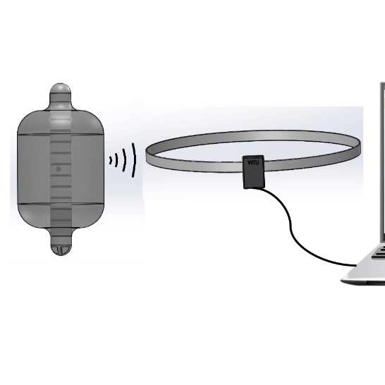
Mongrel dogs were randomly assigned into two groups: the Fecobionics device group, and the THD SensyProbe control group. All animals were anesthetized, the devices were transanally inserted into the rectum, and then inflated at 20 ml. Six minutes later the devices were removed either naturally or manually. The animals in each group were randomly assigned into two subgroups, acute and chronic. The dogs in the acute subgroups were euthanized immediately after the removal of the devices, whereas the dogs in the chronic groups were euthanized two weeks after the device removal. The tissue samples were harvested upon sacrifice.
Materials
Animals
Device:
H&E Staining
Immunofluorescence
Macrophages
- Primary Antibody:
- Secondary Antibody:
- For mucosa staining:
- For nuclear counterstain:
Caspase-3
- Primary Antibody:
- Secondary Antibody:
- For mucosa staining:
- For nuclear counterstain:
Equipment
Cryostat - Leica CM1850
Camera - pco.panda 4.2
Troubleshooting
Hematoxylin and Eosin Staining
Fix the tissues with 4% paraformaldehyde (pH 7.4) overnight at4 °C
Embed the tissues with Optimal Cutting Temperature (OCT) compound and freeze with liquid nitrogen.
Cut 7 µm sections with cryostat (Leica, CM1850).
Rinse slides in 1X PBS for 3 minutes.
Rinse in a gentle stream of tap water until OCT is washed off for 3 minutes.
Stain in Hematoxylin for 20 minutes.
Wash in a gentle stream of tap water for 1 minute, then dip in saturated lithium carbonate solution for 45 seconds.
Blue nuclei in 1X PBS for 20 seconds.
Wash in a gentle stream of tap water for 1 minute.
Dip in 70% EtOH for 30 seconds.
Dip in 95% EtOH for 30 seconds.
Counterstain in Eosin Y for 10 seconds.
Dehydrate through 2 changes of 95% EtOH for 15 seconds.
Dehydrate through 3 changes of 100% EtOH for 15 seconds.
Clear through 3 changes of Xylene for 1 minute each and coverslip.
Immunofluorescence
Put the frozen slides at room temperature for 30min.
Wash slides with PBS 3 times. Each time 5 min.
1% SDS for 5min, then wash 3 times.
0.25% triton in 1xPBS for 15min.
10% Donkey serum in 1xPBS for 1hour.
Incubate slide with primary antibody diluted in 0.25% triton 2.5% Donkey serum 1xPBS at room temperature for 3 hours.
Wash slides with PBS 3 times. Each time 5 minutes.
Incubate slide with secondary antibody (1:100 dilution) for 1 hour.
Wash in PBS 3 times. Each time 5 minutes.
Stain with Wheat Germ Agglutinin (1:100 dilution) for 30 minutes.
Wash slides with PBS 2 times. Each time 5min.
Stain with Hoechst 33442 (1:100 dilution) for 30 minutes.
Dry and mount coverslip.