Jun 05, 2025
Version 2
Solcia‘s lead hematoxylin stain (diffuse endocrinocytes) V.2
- 1Sechenov University



Protocol Citation: Egor Kuzmin 2025. Solcia‘s lead hematoxylin stain (diffuse endocrinocytes). protocols.io https://dx.doi.org/10.17504/protocols.io.6qpvrq5xolmk/v2Version created by Egor Kuzmin
Manuscript citation:
License: This is an open access protocol distributed under the terms of the Creative Commons Attribution License, which permits unrestricted use, distribution, and reproduction in any medium, provided the original author and source are credited
Protocol status: Working
We use this protocol and it's working
Created: June 03, 2025
Last Modified: June 05, 2025
Protocol Integer ID: 219460
Keywords: lead hematoxylin, enteroendocrinocytes, endocrine cell granules, diffuse endocrinocytes, Solcia‘s hematoxylin, Hematoxylin, identifying diffuse endocrine cell, diffuse endocrinocyte, diffuse endocrine cell, prominent modification of the macconaill procedure, macconaill procedure, toluidine blue method, classical histochemical approach, secretory granule, staining without additional section processing, hematoxylin, solcia method, microscopic monitoring, stain
Abstract
Classical histochemical approaches demonstrate considerable diversity in techniques for identifying diffuse endocrine cells, which is directly attributable to the distinct composition of their secretory granules. Among these, the Solcia method stands out as a prominent modification of the MacConaill procedure. Notably, this protocol offers three key advantages: it enables staining without additional section processing, exhibits alcohol resistance (unlike the HCl-toluidine blue method; Solcia, 1968), and critically, functions as a single-step technique – in sharp contrast to multi-step аrgyrophilic methods (Grimelius 1968) and argentaffin methods (Masson, 1914). Furthermore, the approach facilitates controlled progressive staining through real-time microscopic monitoring, thereby ensuring highly reproducible and morphologically optimal outcomes.
Guidelines
- Reagents must be prepared immediately prior to use. The working solution undergoes rapid oxidation and becomes unsuitable for high-quality staining (maximum stability: 4 hours at room temperature).
- While staining may be performed at elevated temperatures to accelerate reaction kinetics, this markedly increases the risk of precipitate formation. Such particulate matter deposits on specimens, compromising histological integrity. Agitating the solution during staining or repeated immersion/withdrawal of slides is strongly discouraged.
- Post-staining, thorough rinsing in multiple changes of distilled water constitutes a critical step. Due to rapid contaminant accumulation, frequent water replacement is essential.
- Although the original protocol omits acidified water (HCl) and ammoniacal water treatments, empirical evidence indicates their controlled application significantly reduces background staining, eliminates dye-derived precipitates, and enhances chromatic contrast.
Materials
- Lead nitrate; Pb(NO3)
- Ammonium acetate; NH4CH3CO2
- Formalin 10%
- Hematoxylin
- Hydrochloric acid; HCL
- Ortho-xylene; C6H4(CH3)2
- Ethanol 100% or isopropanol 100%
- Distilled water
Safety warnings
- Formalin is a volatile compound classified as carcinogenic.
- Xylene, isopropyl alcohol (IPA), and ethyl alcohol are volatile substances hazardous to human health, especially following long-term exposure. Procedures utilizing these solvents require a ventilated fume hood and the use of medical gloves (e.g., nitrile).
- Lead compounds are toxic substances possessing potent oxidizing activity. Lead(II) nitrate is recognized as a carcinogen. All procedures involving these compounds must be conducted within a fume hood with appropriate safety precautions to prevent skin and mucous membrane exposure.
Before start
Autopsy specimens were fixed by immersion in neutral buffered formalin for 48 hours. Processing involved a standard protocol utilizing a graded series of isopropyl alcohols, followed by paraffin embedding. Histological blocks were cut on a microtome to yield 3-μm sections.Sections were placed on silane-coated adhesive glass slides for secure adhesion. All staining reagents were freshly prepared immediately before staining commenced.
Introduction
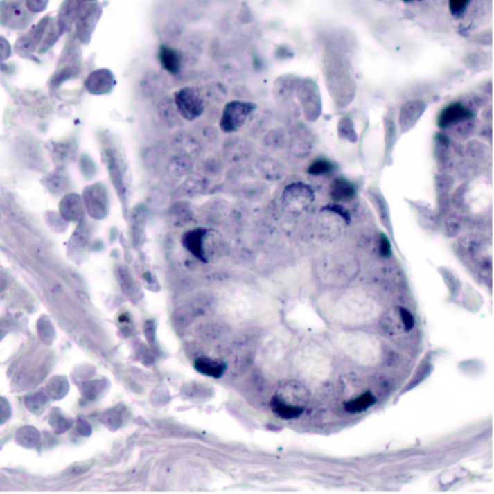
Fig. 1. Micrographs of cow duodenum stained with lead hematoxylin. A positive reaction in the endocrinocytes of the crypt epithelium is shown on the right.
Preparation of solutions
1h
Stable lead solution100 mL
Mix 1:1
- 40ml lead nitrate 5% (Pb(NO3)2 5g/100ml) solution in distilled water [40 ml]
- saturated ammonium acetate (NH4CH3CO2 60-65%) solution in distilled water [40 ml]
Add
10% formalin (4% formaldehyde) 20 ml per 100 ml of solution [16 ml]
Note
The original protocol uses 40% formaldehyde, which has a concentration 10 times higher, but is less common.
40% formaldehyde 2 ml per 100 ml of solution [1,6 ml]
Stable for weeks at room temperature.
10m
Preparation of solutions
1h
Working staining solution100 µL
Mix 1:1
- 10 mL stable lead solution with
- 10 mL distilled water
Add
0.2 g hematoxylin (not hematein).
Stir, let stand 30 minutes, filter.
Dilute to 100 mL
Prepare fresh before use; discard if darkened or precipitated. Stand 30 minutes, filter.
Note
The working solution is unstable—oxidizes within hours. Perform staining at 37-45 °C . Avoid precipitate artifacts.
40m
Reducing solution 1100 mL
HCL-water
5m
Reducing solution 2100 mL
Scott’s tap water buffer
5m
Protocol
2h 48m 10s
Dewaxing, rehydration, rinsing in distilled water.
- ortho-xylene 5 min.
- ortho-xylene 5 min.
- absolute ethanol or isopropanol 5 min
- 50% ethanol or isopropanol 5 min
- distilled water 5 min
20m
Stain in lead hematoxylin working solution for 2 hours at37 °C
Produce without mixing at a constant temperature
2h
Rinse in distilled water
Rinse well from sediment
5m
Differentiate carefully (Not obligatory)
Be carried out with caution, staining is very unstable to acidic pH.
10s
Rinse in ammonia water for 3 minutes (Not obligatory)
In order for the hematoxylin to acquire a bluer hue
3m
Rinse, dehydrate, mount.
- distilled water 5 min
- 50% ethanol or isopropanol 5 min
- absolute ethanol or isopropanol 5 min
- ortho-xylene 5 min.
- ortho-xylene 5 min.
- mount
20m
Result
Granules of endocrine cells: dark blue
(from black to blue depending on the time of exposure in the dye and differentiation)
EC cells of the gastrointestinal tract
AL-cells (X-cell, glucagon) and G-cells (gastrin) of the stomach
A-cells (glucagon) and D-cells (somatostatin) of the pancreatic islets of Langerhans
Chromophilic cells of the pituitary gland
Cells of the adrenal medulla
Chemoreceptor cells of the carotid body
And other representatives of the diffuse neuroendocrine system
Expected result
Fig. 2. Microphotographs of bovine duodenum stained with lead hematoxylin. A positive reaction of EC cells is observed at the base of the crypts. Nucleoli are intensely stained, and background nuclear staining is evident.
Fig. 3. Microphotographs of sections from various organs stained with lead hematoxylin.
The right panel shows a region of Layer V (internal pyramidal lamina) of the primary somatosensory cortex in the mouse brain. The staining highlights the cytoplasm of pyramidal neurons within this layer. Intense staining is observed in specific cortical laminae and in select neurons within subcortical structures. The center panel displays the anterior horns of the mouse spinal cord. Intense cytoplasmic staining is evident in the somata of motor neurons, while other neurons remain unstained. The right panel features a gastric section from a rat, where scattered endocrine cells (endocrinocytes) are visualized in the mucosa, specifically within the gastric (fundic) glands.
Note
Pre-oxidation with 1% HCl for 3–17 hours (as in HCl-TB method) improves staining quality.
Protocol references
1. Solcia E, Capella C, Vassallo G. Lead-haematoxylin as a stain for endocrine cells. Significance of staining and comparison with other selective methods. Histochemie. 1969;20(2):116-26. doi: 10.1007/BF00268705
2. Solcia E, Vassallo G, Capella C. Selective staining of endocrine cells by basic dyes after acid hydrolysis. Stain Technol. 1968 Sep;43(5):257-63. doi: 10.3109/10520296809115078