May 19, 2026
siRNA Delivery in Retinal Explant using Reverse Magnetofection with XPMag
- 1OZ Biosciences


External link: https://ozbiosciences.com/explants/125-140-xpmag-explant-transfection.html#/24-capacity-250_l
Protocol Citation: Cédric Sapet 2026. siRNA Delivery in Retinal Explant using Reverse Magnetofection with XPMag. protocols.io https://dx.doi.org/10.17504/protocols.io.x54v9qqjql3e/v1
License: This is an open access protocol distributed under the terms of the Creative Commons Attribution License, which permits unrestricted use, distribution, and reproduction in any medium, provided the original author and source are credited
Protocol status: Working
We use this protocol and it's working
Created: May 12, 2026
Last Modified: May 19, 2026
Protocol Integer ID: 316894
Keywords: Reverse Magnetofection, XPMag, magnetic nanoparticles, siRNA delivery, RNA interference, retina explants, organotypic culture, adult retina, gene silencing, RPE, non-viral delivery, transfection, ocular gene therapy, organ explant transfection, sirna delivery in retinal explant, reverse magnetofection with xpmag nucleic acid delivery, xpmag nucleic acid delivery, retina explant, adult retina explant, surface of adult retina explant, retinal explant, retinal tissue, adult retinal tissue, magnetic nanoparticle, retinal layer, reverse magnetofection with xpmag, sirna delivery, challenging problems in ophthalmic research, transferable to other organ explant, using reverse magnetofection, reverse magnetofection, ophthalmic research, classical transfection method, full transfection step, sirna across the entire thickness, posterior segment of the eye, viral vector, external supermagnetic plate, magnetic complexes from the rpe side
Funders Acknowledgements:
OCUTHER
Grant ID: 722717
Disclaimer
For Research Use Only. Not for use in humans. Not for use in diagnostic or therapeutics purposes.
Abstract
Nucleic acid delivery to the posterior segment of the eye remains one of the most challenging problems in ophthalmic research: adult retinal tissue is thick, multilayered, post-mitotic and protected by an intact limiting membrane that restricts diffusion of conventional vectors. Classical transfection methods (lipofection, electroporation, viral vectors...) all reach mainly the surface of adult retina explant and reach mainly the surface of adult retina explants and frequently induce toxicity in deeper layers.
This protocol describes Reverse Magnetofection with XPMag, a magnetic nanoparticle reagent specifically designed to deliver siRNA across the entire thickness of adult retina explants. siRNA/XPMag complexes are added to the culture medium below the explant, and an external Supermagnetic plate is placed above the culture lid to pull the magnetic complexes from the RPE side up to the ganglion layer. The full transfection step takes only 30 minutes and reproducibily achieves 60-70% gene silencing in all retinal layers without inducing photorecpetor death, microglial activation or gliosis. The procedure is also transferable to other organ explants such as cornea, brain slice, skin patch and lung.

Image Attribution
All images in this protocol are the property of OZ Biosciences. (c) OZ Biosciences - All right reserved.
Guidelines
- This protocol is a standard starting point: optimal siRNA concentration and XPMag volume may need to be optimized depending on target gene expression level, animal age and explant type.
- siRNA/XPMag complexes must be added to the culture medium within 1 hour of formation.
- Use medium without any supplement (serum, antibiotics, supplement) for complex formation.
- For co-silencing with several siRNAs, calculate XPMag volume against total siRNA amount.
- Prefer to use chemically modified siRNA (≤21 bp) to avoid triggering innate immune response in the retina.
Materials
Magnetofection Reagents (OZ Biosciences)
- XPMag transfection reagent (OZ Biosciences, Ref #XP00500). Store at -20°C; thaw at Room Temperature before use.
- Supermagnetic plate (OZ Biosciences, Ref #MF14000).
Nucleic Acids
- siRNA targeting gene of interest (modified 21-mer siRNAs ). Recommanded: use a pool of at least 3 sequences targeting the same mRNA for enhanced specificity.
- Scrambled siRNA (negative control, e.g. Ambion Ref. 4390843).
- GAPDH siRNA (positive control).
Culture medium and Reagents
- For eye dissection: R16 serum-free culture medium (Invitrogen Life Technologies, Ref. 07490743A).
- For complex formation and explant culture: Neurobasal A medium (Gibco, Ref. 10888022).
- Supplement for complete culture medium: B-27 (2%, Gibco, Ref. 17504044), N2 (1%, Gibco, Ref. 17502048), penicillin (1%, ThermoFisher, Ref. 15140-122), Glutamax (0.4%, Gibco, Ref. 35050061).
- For enzyme inactivation only: Fetal Bovine Serum (FBS, Sigma-Aldrich, Ref. F7524).
Consumables
- 0.4 µm polycarbonate Transwell membrane (Corning Life Sciences, Ref. CLS3412).
- 6-well tissue culture plate.
- Sterile plastic tubes and pipette tips.
- Sterile dissection tools (fine forceps, scalpel, micro-scissors).
- Glass tube (internal diameter 0.5 cm) with rubber bulb to transfer flattened retinas.
Equipment
- Laminar airflow hood.
- Humidified incubator (37°C, 5% CO2).
- Stereomicroscope for retinal dissection.
Troubleshooting
Problem
Poor or inhomogeneous silencing, especially in the GCL
Solution
Possible cause: complexes are not being actively pulled trough the tissue; the magnet has en placed underneath the plate (classical Magnetofection).
Solutions:
• Place the Supermagnetic plate above the culture lid directly over the explants.
• Check explant orientation (RPE/photoreceptors facing the membrane, GCL, facing up). Increase reverse Magnetofection incubation time with magnetic plate above the lid.
• Check the orientation of the Supermagentic plate; OZBiosciences' logo facing the explant.
Problem
Complex aggregation in the well
Solution
Possible cause: complexes were done in medium containing suppléments (serum, antibiotics, growth factors…)
Solution: Use serum-free medium only for complex formation, do not vortex.
Problem
Increased cell death in the explant after transfection
Solution
Possible cause: non-optimized transfection conditions, long culture duration past 72H or biomechanical stress during dissection
Solution:
• As a starting point do not exceed 100nM siRNA and refer to standard procedure for optimization procedure. Alternatively contact [email protected]
• Evaluate silencing at 72h post transfection ; for long term culture, adopt a replacement plan for culture medium.
Problem
Microglial activation or gliosis after transfection
Solution
Possible cause: non-modified siRNA longer than 21 bp triggering innate immune system or off-target effects.
Solution: use chemically modified siRNA of ≤21 bp and include scrambled siRNA and XPMag-only controls.
XPMag alone is non-toxic at the recommended doses.
Problem
No silencing despite correct procedure
Solution
Possible cause: siRNA does not match the species mRNA or target protein has a very long half-life
Solution: Verify sequence mathc against the animal model used; extend evaluation to 72-96 h post-transfection for long-lived proteins.
Safety warnings
- Reverse Magnetofection requires the appropriate magnetic field delivered by OZ Biosciences Supermagnetic plates.
- Handle and dispose siRNA-containing reagents according to local biosafety regulations
- Animal procedure must follow institutional and national guidelines.
Ethics statement
This protocol involves the use of laboratory animals for the harvesting of ocular tissue. Users must obtain prior approval from their Institutional Animal Care and Use Comittee (IACUC) or equivalent ethics committee before performing any procedure described here and must comply with all applicable national and institutional regulations.
The protocol has been validated in adult Sprague-Dawley rats and C57BL/6J mice with original procedure approved by the Tübingen University committee on animal protection.
The method is transferable to other rodent strains and to other organ explants (cornea, brain slice, skin, lung), provided that corresponding ethical approvals are in place.
Before start
- Allow reagents to reach room temperature before starting.
- Replace culture medium 24 H after transfection, perform assay 48 to 96 h after Reverse Magnetofection.
Rationale
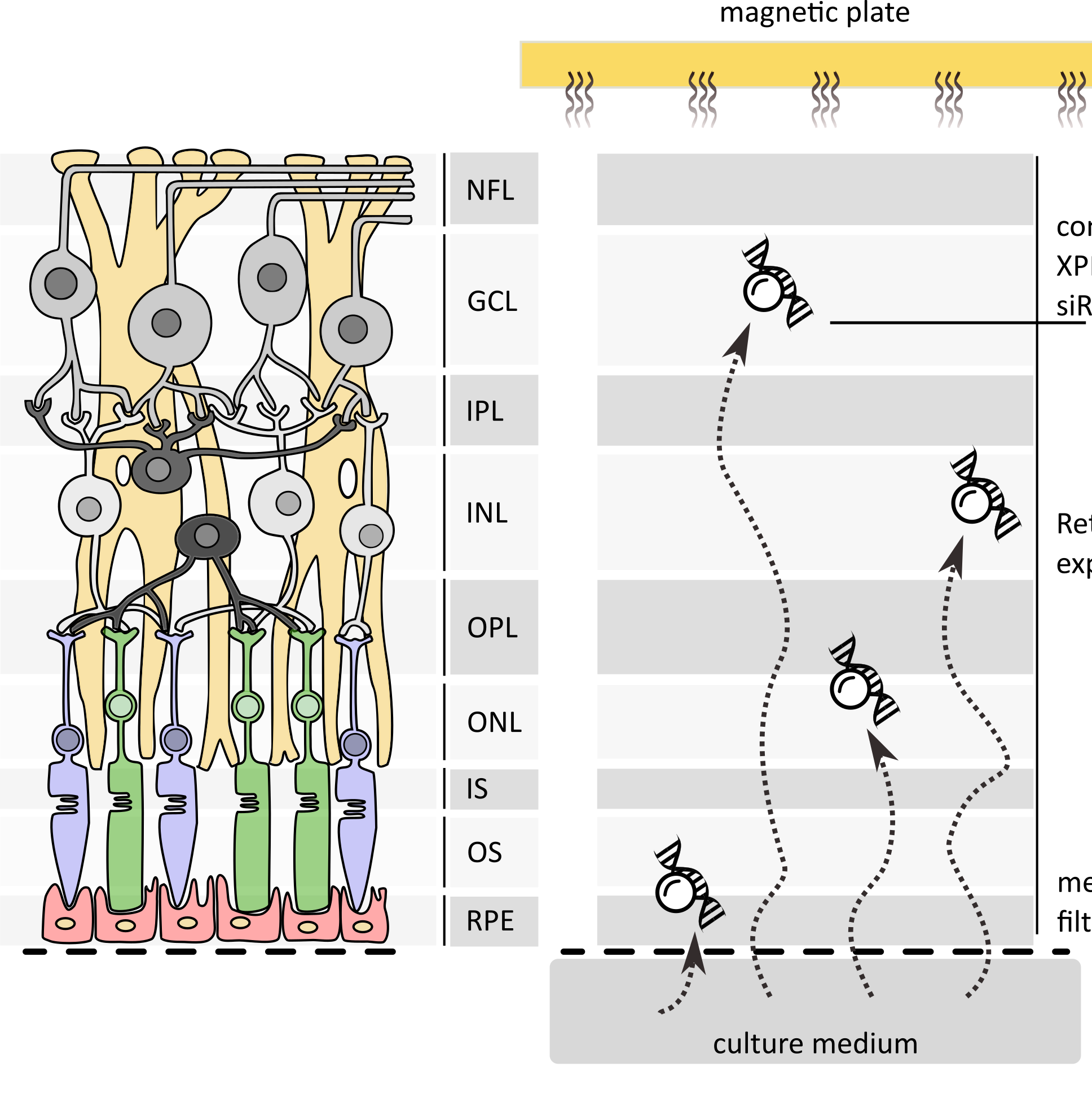
Reverse Magnetofection consists in attracting magnetic complexes of siRNA / XPMag upward from the culture medium to the inside layers of the retinal explant.
Principle of reverse Magnetofection with XPMag. magnetic complexes are pulled across all retinal layers (RPE to GCL) by Supermagnetic plate positioned above the explant ensuring homogeneous siRNA delivery through the full thickness of the adult retina (REP, Retinal Pigment Epithelium; OS, Outer Segment; IS, Inner Segment; ONL, Outer Nuclear Layer; OPL, Outer Plexiform Layer; INL Inner Nuclear Layer; IPL, Inner Plexiform Layer; GCL, Ganglion Cell Layer; NFL, Nerve Fiber Layer).
The flat-mounted retina is cultured on a polycarbonate membrane with the RPE side facing the culture medium and the GCL side facing up.
1. Preparation of adult rat retina explants.
Sacrifice the animal according to approved-ethical guidelines.
Clean the head with 70% ethanol.
Enucleate the eyes and wash them in serum-free R16 medium for 5 min at room temperature.
Transfer eyes into 0.12% Proteinase K in R16 medium pre-warmed at 37°C and incubate for 15 min at 37°C.
Stop enzymatic activity by transferring the eyes into R16 medium supplemented with 20% FBS for 5 min at room temperature.
Wash 5 min in fresh R16 medium.
In a sterile Petri dish filled with R16, remove the sclera while keeping the RPE attached to the neural retina.
Open the eyeball at the ora serrata with a scalpel, remove cornea, lens and vitreous body.
Make 4 equidistant radial incisions on the retina to flatten it.
Transfer the flattened retina onto a 0.4 µm polycarbonate Transwell membrane with RPE/photoreceptors facing the membrane and the GCL facing up.
Note
Using a 0.5 cm-diameter glass tube with a rubber bulb makes the transfer easier and protects the tissue.
Place the membrane carrying the explant into a 6-well plate and add complete culture medium.
Maintain the explants at 37°C in a humidified 5% CO2 atmosphere until transfection.
2. Preparation of siRNA / XPMag complexes
30m
Bring XPMag, siRNA and Neurobasal A medium to room temperature.
Note
Use supplement-free medium only for complex formation.
Gently resuspend XPMag by pipetting up and down.
In an empty tube, transfer 2-3 µL XPMag per explant.
In a second tube, prepare 200 µL of serum-free Neurobasal A per well and add the appropriate amount of siRNA to reach final concentration of 50-100 nM in 1 mL (final transfection volume per well).
Note
50-100 nM are usually sufficient. Concentrations above 100 nM may reduce efficacy
Transfer the siRNA-containing solution onto XPMag and mix gently by pipetting
Safety information
Critical: do not vortex once siRNA and XPMag are combined.
Incubate the mixture for 20-30 min at room temperature to allow proper complex formation.
3. Reverse Magnetofection
1h
In a sterile 6-well plate, dispense 800 µL of complete culture medium per well
Add the 200 µL of siRNA/XPMag complexes to each well (final transfection volume: 1 mL/well).
Incubate 30 min at 37°C in a humidified 5% CO2 atmosphere with the magnet in place.
Reverse Magnetofection in a 4-steps procedure: complex formation (1), incubation (2), Addition to culture medium (3) and upward Magnetic attraction (4).
Remove the Supermagnetic plate and incubate under standard culture conditions until evaluation of the experiment.
| Parameters | Value | Parameters | Value | |
| XPMag | 2-3 µL / explan | Complex incubation | 20-30 min, RT | |
| Final siRNA | 50-100 nM | Magnet incubation | 30 min, 37°C, 5% CO2 | |
| Complex volume | 200 µL (Neurobasal A) | Magnet position | Above the lid | |
| pre-loaded medium | 800 µL complete medium | First medium change | 24 h post-transfection | |
| Total volume | 1 mL / well | Evaluation | 72 h post-transfection |
Recommended quantities per explant (one well of a 6-well plate)
4. Post-transfection culture
24 h after transfection, replace the culture medium with fresh complete supplemented Neurobasal A.
Incubate for an additional 48 h before evaluating gene silencing.
Note
Always include matched untreated and scrambled-siRNA controls at the same time point.
Note
Cultures can be extended beyond 72 h, but cell death due to axotomy and biomechanical insult will increase progressively.
5. Downstream analyses
Suggestions of downstream analysis
Cryosectioning (14 µm vertical sections) followed by fluorescent immunohistochemistry of the target protein in GCL, INL and ONL.
TUNEL assay on cryosections to verify the absence of induced cell death.
Iba1 and GFAP immunostaining to confirm absence of microglial activation or reactive gliosis.
Western blot on retina lysate (RIPA + protease/phosphatase inhibitors, 12000 x g, 15 min, 4°C)
Expected result
• 60-70 % reduction of target protein expression in ONL, INL and GCL of adult rat retina explants (validated for VCP and GAPDH - see papers in reference).
• No increase in Iba1-positive activated microglia of GFAP-positive reactive gliosis.
• in RHO P23H rat and Rho∆I255/+ mouse retina explant, VCP silencing via Reverse Magnetofection restores rhodopsin trafficking to the outer segments, increases OZ length and preserves photoreceptors cell rows.
Protocol references
Bassetto M, Sen M, Poulhes F, Arango-Gonzalez B, Bonvin E, Sapet C, Ueffing M, Zelphati O. New Method for Efficient siRNA Delivery in Retina Explants: Reverse Magnetofection. Bioconjug Chem. 2021 Jun 16;32(6):1078-1093. doi: 10.1021/acs.bioconjchem.1c00132. Epub 2021 Jun 3. PMID: 34081855.
Sen M, Bassetto M, Poulhes F, Zelphati O, Ueffing M, Arango-Gonzalez B. Efficient Ocular Delivery of VCP siRNA via Reverse Magnetofection in RHO P23H Rodent Retina Explants. Pharmaceutics. 2021 Feb 6;13(2):225. doi: 10.3390/pharmaceutics13020225. PMID: 33562020; PMCID: PMC7914601.
Cao B, Almansa-Garcia AC, Sen M, Mühlfriedel R, Seeliger MW, Petremann-Dumé AS, Kilger E, Vollert A, Bolz S, Henes C, Caliceti P, Salmaso S, Ueffing M, Arango-Gonzalez B. Ocular delivery of different valosin-containing protein (VCP) inhibitory formulations prevents retinal degeneration in rho∆I255 mice. J Control Release. 2026 Apr 10;392:114670. doi: 10.1016/j.jconrel.2026.114670. Epub 2026 Jan 28. PMID: 41617017.
Acknowledgements
The development and validation of Reverse Magnetofection with XPMag was supported by the European Union's Horizon 2020 Research and Innovation programme under the Marie Skłodowska-Curie grant agreement No. 722717—project OCUTHER.