Apr 11, 2026
Single Cell Isolation from Murine Kidney for Single-Cell RNA Sequencing
- Rachel Njeim1,
- Antonio Fontanella1,
- Alla Mitrofanova1
- 1Peggy and Harold Katz Family Drug Discovery Center, University of Miami, Miller School of Medicine, Miami, Florida, USA
- Mitrofanova's Lab


Protocol Citation: Rachel Njeim, Antonio Fontanella, Alla Mitrofanova 2026. Single Cell Isolation from Murine Kidney for Single-Cell RNA Sequencing. protocols.io https://dx.doi.org/10.17504/protocols.io.q26g79oy8vwz/v1
License: This is an open access protocol distributed under the terms of the Creative Commons Attribution License, which permits unrestricted use, distribution, and reproduction in any medium, provided the original author and source are credited
Protocol status: Working
We use this protocol and it's working
Created: June 14, 2025
Last Modified: April 11, 2026
Protocol Integer ID: 220155
Keywords: kidney, single cell, Parse Bioscience, mouse, single cell isolation from murine kidney, single cell isolation, adaptations for murine kidney, murine kidney, cell rna, rnase inhibitor, rna, physiol genomic, sequencing, sequence of strainer
Funders Acknowledgements:
Chernowitz Medical Research Foundation
Grant ID: GR021608
Carl W. Gottschalk Research Scholar Grant, American Society of Nephrology
Grant ID: GR018262
Career Development Award, American Heart Association
Grant ID: 24CDA1267060
Miami Clinical and Translational Science Institute
Grant ID: UL1TR002736
Disclaimer
DISCLAIMER – FOR INFORMATIONAL PURPOSES ONLY; USE AT YOUR OWN RISK
The protocol content here is for informational purposes only and does not constitute legal, medical, clinical, or safety advice, or otherwise; content added to protocols.io is not peer reviewed and may not have undergone a formal approval of any kind. Information presented in this protocol should not substitute for independent professional judgment, advice, diagnosis, or treatment. Any action you take or refrain from taking using or relying upon the information presented here is strictly at your own risk. You agree that neither the Company nor any of the authors, contributors, administrators, or anyone else associated with protocols.io, can be held responsible for your use of the information contained in or linked to this protocol or any of our Sites/Apps and Services.
Abstract
This protocol is based upon Miao Z., et al., Nature Communications, 12: 2277, 2021 and Robertson J.N., et al., Physiol Genomics, 56(7):469-482, 2024 with adaptations for murine kidney including: mincing, homogenization strokes, digestion enzymes optimization, addition of protease inhibitor and SUPERase-In RNAse inhibitor and adjustments to strainer size and sequence of strainers used.
Materials
TrypLE Select Enzyme (1x), no phenol redThermo Fisher ScientificCatalog #12563011
Dispase (5 U/mL)STEMCELL Technologies Inc.Catalog # 07913
Collagenase DMerck MilliporeSigma (Sigma-Aldrich)Catalog #11088882001
HBSS, no calcium, no magnesium, no phenol redSartoriusCatalog #02-018-1A
SUPERase• In™ RNase Inhibitor (20 U/μL)Thermo Fisher ScientificCatalog #cat# AM2694
DNase I, RNase-free (1 U/µL)Thermo FisherCatalog #EN0521
Gibco™ Bovine Albumin Fraction V (7.5% solution)Thermo Fisher ScientificCatalog #15260037
Trypan Blue Solution 0.4%Thermo Fisher ScientificCatalog #15250061
RNase AWAY™ Surface Decontaminant, BottleThermo FisherCatalog #7000TS1
LO-bind protein and genomic microcentrifuge tubesThermo Fisher ScientificCatalog #AM12400
LO-bind tips 10 µlCorningCatalog #4150
LO-bind tips 200 ulCorningCatalog #4148
LO-bind tips 1000 ulCorningCatalog #4140
100 m cell strainersFalconCatalog #352360
70 m cell strainersFalconCatalog #352350
40 m cell strainersFalconCatalog #352340
30 m cell strainerspluriSelect Life ScienceCatalog #43-50030-01
20 m cell strainers EASYStrainer smallgreiner bio-oneCatalog #542120
Mr. Frosty freezing containerThermo Fisher ScientificCatalog #5100-0001
Equipment
Swinging bucket rotor centrifuge
NAME
Centrifuge
TYPE
Beckman Coulter Allegra X-30
BRAND
B05794
SKU
LINK
Equipment
Thermal Shake lite
NAME
Thermal shaker
TYPE
VWR
BRAND
WRI460-0249
SKU
LINK
Safety warnings
Note
All calculations and volumes are given for 1 sample with 2 kidneys.
Note
Use RNase Away frequently to clean hands, pipettes, and surfaces to avoid RNA degradation.
Ethics statement
All animal studies complied with the relevant ethical regulations and were performed in accordance with the
National Institutes of Health Guidelines. The study protocol was approved by the Institutional Animal Care and Use Committee of the University of Miami, Miller School of Medicine (#22-104).
Before start
Pre-coat tubes with 1% BSA in nuclease-free water: prepare 4 of 1.5 mL tubes per sample.
Incubate at room temperature for 30 min. Discard the 1% BSA solution, remove any
residual liquid using a pipette, and leave the tubes uncapped for another 30 minutes to air dry.
!! Make this step 1-2 days before tissue processing.
♦ Use sterile cell culture hood and close the sash when “air drying” the tubes.
♦ 1% BSA stock solution in water can be stored at 4°C for up to 1 week.
Harvest the kidneys
30m
Follow the lab protocol to anesthetize and perfuse the animal using HBSS buffer to harvest kidneys.
Harvest the kidneys
30m
Weigh and collect kidneys in ice-cold HBSS buffer.
Prepare glomerular and tubule fractions
30m
Separate glomerular portion from tubules: using a syringe plunger, mince the kidney on a 100 mm cell strainer placed over a 50 mL Falcon tube and pass the minced tissue through the strainer using up to 20 mL of ice-cold HBSS.
5m
Repeat this step using a fresh clean 100 mm strainer and a new 50 mL Falcon tube. Wash the strainer with an additional 20 mL of ice-cold HBSS.
5m
Pass the liquid collected from step 4 through a 70 mm cell strainer placed over a clean 50 mL Falcon tube. Collect 5 mL of liquid containing mostly tubular fraction into a new 50 mL Falcon tube and add 5 mL of ice-cold HBSS.
Set on ice. This is tubular fraction.
5m
Invert the 70 mm strainer containing the glomerular fraction over a clean 50 mL Falcon tube and rinse the contents into the tube using 10 mL of ice-cold HBSS. This is glomerular fraction.
5m
Check how the suspension looks under a microscope.
5m
Spin down at 500xg for 5 min at 4°C. Use swinging bucket rotor!
4 °C Centrifugation 00:05:00
5m
Discard supernatant carefully.
Digestion
38m
Prepare 3 mL of digestion solution for each of the glomerular and tubular fractions, following the recipe in the table. Prepare the non-enzymatic and enzyme-containing solutions in two separate tubes*.
*CRITICAL NOTE: This approach is necessary because the enzymes begin acting immediately, and
resuspending the pellet directly in an enzyme-containing solution may lead to tissue clumping.
| Reagent/Conc | Stock conc | To get 3 mL | |
| TrypLE/0.5% | 1X | 15 μl | |
| BSA/1% | 7.5% | 400 μl | |
| HBSS | 1X | 746 μl |
Non-enzyme solution
| Reagent/Conc | Stock conc | To get 3 mL | |
| Collagenase D, 2U/mL | 10U/mL | 600 μl | |
| Dispase II, 2U/mL | 5U/mL | 1,200 μl | |
| DNase 10U/mL | 1U/uL | 30 μl | |
| SUPERase-In RNase inhibitor 60U/mL | 20U/uL | 9 μl |
Solution with enzymes
5m
Divide each mixture into 3 LO-bind tubes with 1 mL each and incubate on a thermomixer for 20 min at 37°C, 300xRPM.
37 °C on a thermomixer
25m
Triturate samples by pipetting every 5 min. Mix gently using a cell culture glass* pipette, pipetting to the first stop.
*CRITICAL NOTE: : Shearing the tissue with a metal needle leads to cell death. Although longer incubation increases the number of isolated cells, it decreases cell viability. Therefore, it is better to reduce the volume and shorten the incubation time.
Prepare 2x15 mL Falcon tubes containing 5 mL of 7.5% BSA and 2 μl
*CRITICAL NOTE: Adding DNase at this step helps to neutralize DNA in the solution from the dead cells and to avoid forming sticky DNA-containing webs-like.
Stop the digestion reaction by adding the mixtures from 3 LO-bind tubes containing digested glomeruli or tubules into 15 mL Falcon tubes. Add 3 mL of 7.5% BSA and 2 μl
3m
Centrifuge the tubes at 500xg for 5 min at 4°C. Use swinging bucket rotor!
4 °C Centrifugation 00:05:00
5m
Discard supernatant carefully.
Sieving
50m
Make 10 mL of ice-cold HBSS and 1% BSA: 1,330 mL BSA + 8,670 mL
5m
Wash each pellet with 5 mL of ice-cold HBSS and 1% BSA.
5m
Pass the mixtures through BSA-pre-wetted 30 mm cell strainer* placed over 50 mL Falcon tubes. Add 4 μl
*CRITICAL NOTE: Do not touch the strainer mesh to avoid forcing larger particles through the sieve.
5m
Centrifuge the tubes at 500xg for 5 min at 4°C. Use swinging bucket rotor!
4 °C Centrifugation 00:05:00
5m
Make 2 mL ice-cold HBSS with 1% BSA containing SUPERase-In RNase inhibitor and DNase.
| Reagent/Conc | Stock conc | To get 2 mL | |
| BSA, 1% | 7.5% | 266 μl | |
| HBSS | 1X | 1,732 μl | |
| SUPERase-in RNA inhibitor, 60U/mL | 1U/mL | 2 μl | |
| DNase, 1U/mL | 1U/mL | 2 μl |
Remove the supernatant with a pipette and resuspend each pellet in 1 mL ice-cold HBSS with 1% BSA.
Assess the quality and cell count of the suspension: take 10 μl
15m
In a new BSA-coated tube, combine all glomerular fractions and add a matching number of tubular cells to achieve a glomeruli-to-tubule ratio of 1:2, respectively.
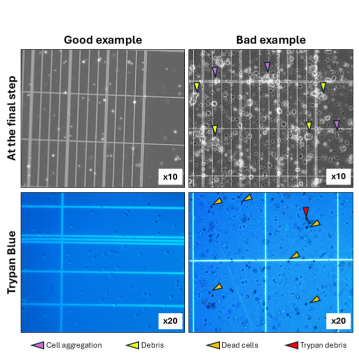
Assess cell viability: Take another 5 μL of the cell mixture and add 5 μL of Trypan blue 0.4% stock solution*. Mix well and incubate 3 min at room temperature. Capture images under a microscope (10X or 20X). Count the number of dead cells.
*CRITICAL NOTE: Remove of Trypan debris: take a 40 μl aliquot and spin it down for 30 sec; use the top fraction only.
Expected result
There should be at least 75% of viable cells (no Trypan blue staining), <5% cell aggregation and no debris. The Protocol typically generates 0.5x10⁶– 9.0x10⁶ cells per kidney with >90% viability.
Check images attached for reference.
15m