Feb 23, 2026
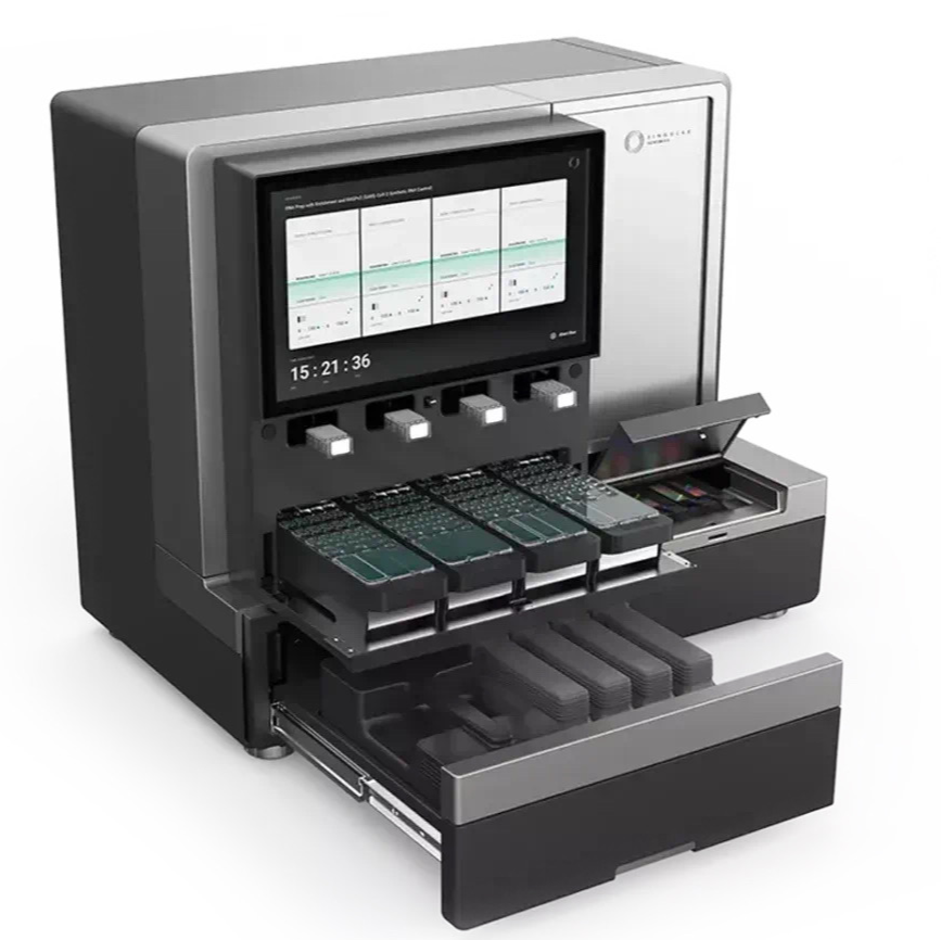
Short read sequencing (PE 2x100) on Singular Genomics G4 NGS sequencer
- Shuoshuo Wang1,2,3,
- Antonella Arruda de Amaral1,3,
- Ioannis Vlachos1,2,3
- 1Beth Israel Deaconess Medical Center;
- 2Harvard Medical School;
- 3Broad Institute of MIT and Harvard
- protocols.io Ambassadors


Protocol Citation: Shuoshuo Wang, Antonella Arruda de Amaral, Ioannis Vlachos 2026. Short read sequencing (PE 2x100) on Singular Genomics G4 NGS sequencer. protocols.io https://dx.doi.org/10.17504/protocols.io.81wgbnpq1gpk/v1
License: This is an open access protocol distributed under the terms of the Creative Commons Attribution License, which permits unrestricted use, distribution, and reproduction in any medium, provided the original author and source are credited
Protocol status: Working
We use this protocol and it's working
Created: April 01, 2025
Last Modified: February 23, 2026
Protocol Integer ID: 125881
Keywords: sequencing, SBS, G4, Singular, short read, NGS, Sequencer, singular genomics g4 ngs sequencer, g4 sequencing platform, g4 system from singular genomic, singular genomic, routine sequencing, genomic, g4 system, g4, short read, labs with unprecedented power
Disclaimer
DISCLAIMER – FOR INFORMATIONAL PURPOSES ONLY; USE AT YOUR OWN RISK
The protocol content here is for informational purposes only and does not constitute legal, medical, clinical, or safety advice, or otherwise; content added to protocols.io is not peer reviewed and may not have undergone a formal approval of any kind. Information presented in this protocol should not substitute for independent professional judgment, advice, diagnosis, or treatment. Any action you take or refrain from taking using or relying upon the information presented here is strictly at your own risk. You agree that neither the Company nor any of the authors, contributors, administrators, or anyone else associated with protocols.io, can be held responsible for your use of the information contained in or linked to this protocol or any of our Sites/Apps and Services.
Abstract
The G4 Sequencing Platform from Singular Genomics empowers labs with unprecedented power, speed, and flexibility in a benchtop instrument. By combining novel, high-performance chemistry and advanced engineering, the G4 delivers highly accurate results across 1-4 flow cells and 4-16 independent lanes in a single day. The Spatial Technologies Unit (STU) has been uninterruptedly operating the G4 system from Singular Genomics since 2022 for routine sequencing of short-read libraries. This protocol represent typical workflow in our lab.
Attachments

G4-Sequencing-Platfo...
12.7MB
Materials
| Category | Material | Vendor / Catalog # | Notes / Typical Use | |
| Instrumentation & Quantification | Qubit 4 Fluorometer or Qubit Flex | Thermo Fisher | Use with dsDNA HS Assay for library quantification | |
| Qubit dsDNA HS Assay Kit (1×) | Thermo Fisher | Measure library concentration before conversion | ||
| TapeStation (Agilent) or Fragment Analyzer (Advanced Analytical) | — | Optional; for library size distribution and QC | ||
| Library Conversion / PCR | Ice | — | Keep PCR master mix and primers cold | |
| Nuclease-free water | — | For reagent preparation and dilutions | ||
| Fresh 80% ethanol | — | For SPRI bead purification steps | ||
| PCR Master Mix (KAPA HiFi or equivalent) | Roche / KAPA | For library amplification during conversion | ||
| Dual-Indexed PCR Primers | Singular Genomics, #500007 | FWD=S1, REV=S2; used in library conversion | ||
| SPRI beads | SPRIselect or AMPure XP | DNA cleanup and size selection | ||
| Sequencing Consumables | G4 Reagent Cartridge (50–300 cycles) | Singular Genomics, PN-500006 | Contains Extension dibromo, Chase dibromo, Cleave, Quenching solution; thaw and handle per SOP | |
| Flow Cells: F2 (standard), F3 (high-density) | Singular Genomics | Up to 4 per run; each has 4 lanes | ||
| Pipette tips | Low-retention, filtered | For all liquid handling steps | ||
| Thermal Cycler | — | For PCR conversion, 3-step program | ||
| QC / Miscellaneous | Microcentrifuge tubes | Nuclease-free | For reagent preparation, intermediate storage | |
| Vortex mixer | — | Mix reagents thoroughly | ||
| Plate or tube spinner | — | Brief centrifugation of reagents or beads | ||
| Personal Protective Equipment | Gloves, lab coat, eye protection | Required when handling chemical reagents and DMSO-based quenchers |
Basics of G4 Sequencing Platform: Laboratory Implementation and Chemistry
The Spatial Technologies Unit at BIDMC has operated the Singular Genomics G4 sequencing platform since 2022 for routine sequencing of short-read libraries, including single-cell and Visium libraries generated with 10x Genomics kits (Zajac et al., 2025).
The Singular Genomics G4 Sequencing Platform utilizes a distributed system designed to balance fluidic, optical, and computational demands. The platform is comprised of three primary physical units: the G4 instrument, a dedicated High Performance Computing (HPC) station, and a set of uninterruptible power supply (UPS) units.
The G4 supports up to four independent flow cells per run, each divided into four fluidically isolated lanes, enabling 4–16 simultaneous lanes depending on configuration. Two flow-cell formats were used: F2, a standard-density format for bulk RNA-seq and single-cell RNA-seq, and F3, a higher-density format for large libraries or highly multiplexed experiments. Typical sample loading included 4–8 single-cell RNA-seq libraries per flow cell, or 8–32 libraries when all four flow cells were utilized, with throughput dependent on target read depth and recommended reads-per-cell.
The G4 uses a four-color reversible terminator SBS chemistry. Each cycle consists of discrete chemical and imaging steps:
1. Extension (Incorporation): A mixture containing DNA polymerase and all four nucleotides (A, C, G, T) is added. Each nucleotide has a 3′ reversible terminator to restrict polymerase extension to a single base per cycle, and a fluorescent dye attached via a dibromo-based chemical linker. The dibromo linker is cleavable under defined conditions to allow rapid removal of the dye and terminator in subsequent steps.
2. Chase: A second pulse of nucleotides, unlabeled but 3′-blocked, is applied immediately after extension to incorporate any bases missed during the initial step, maintaining cluster synchronization and reducing phasing errors over successive cycles.
3. Imaging: Clusters are imaged using four distinct fluorescent channels corresponding to each base. Imaging occurs during the chase step, enabling real-time signal capture without additional cycle delay.
4. Cleavage: The cleavage reagent removes both the fluorescent dye and the 3′-OH terminator, restoring a free hydroxyl group for the next incorporation.
5. Quenching: A quenching reagent dissolved in DMSO terminates the cleavage reaction and prevents residual reactive species from interfering with subsequent cycles.
This cycle of incorporation, chase, imaging, cleavage, and quenching is repeated to achieve the desired read length, producing standard SBS output suitable for base calling.
Library Architecture and Conversion PCR Dynamics
5m
Libraries intended for G4 sequencing were first confirmed for platform compatibility.
At the 5' ends of the final library fragments, Singular Genomics requires platform-specific S1 (forward) and S2 (reverse) sequences to serve as anchors for cluster formation on the flow cell. These sequences are placed upstream of traditional sequencing primer sites such as SP1 and SP2 (equivalent to Illumina TruSeq or Nextera read primers). Illumina-compatible primer sequences (TruSeq Read 1/Read 2, Nextera R2, P5/P7 adapters, or small RNA-specific sequences) were incorporated downstream of S1/S2 as required.
The PCR-based conversion protocol uses limited-cycle PCR to append the S1 and S2 sequences. The typical structure of a converted library includes the S1/S2 anchors, unique dual indices (UDI), and the SP1/SP2 sites flanking the insert of interest.
The conversion PCR protocol is highly sensitive to the initial library concentration. Input libraries should be quantified using high-sensitivity dsDNA assays (e.g., Qubit fluorometer) and diluted to 1 ng/μL. A critical insight into this process is the potential for increased duplication rates. Benchmarking the G4 against other platforms has indicated that for input amounts below 125 ng, the additional PCR cycles required for conversion can lead to a significant proportion of duplicate reads, sometimes ranging from 34% to 96% depending on the input amount (Zajac et al. 2025).
The conversion PCR is followed by a bead-based cleanup, typically utilizing a 0.9x ratio of SPRI beads (e.g., SPRIselect or AMPure XP) to the final PCR product. This ratio is chosen to exclude primer dimers and small non-specific fragments while retaining the larger library inserts. Noteworthy, the conversion process, while potentially increasing duplicates at low input, often results in lower adapter dimer abundance compared to standard Illumina runs. This is attributed to the combined effect of the specific conversion primers and the mandatory cleanup step. However, for very low input amounts below 15 ng, the duplication rate on the G4 is observed to be higher than that of native Illumina sequencing. To mitigate this, the use of unique molecular identifiers (UMIs) is strongly recommended. UMIs allow for the in silico deduplication of the data, which recovers the biological diversity of the sample by removing artifacts introduced during the conversion PCR.
Program a thermal cycler. Lid temperature at 105 °C and volume at 50 µL .
| Conversion PCR Step | Temperature (∘C) | Duration | Cycles | |
| Initialization | 98 | 2 min | 1 | |
| Denaturation | 98 | 20 sec | 7 | |
| Annealing | 57 | 30 sec | 7 | |
| Extension | 72 | 30 sec | 7 | |
| Final Extension | 72 | 1 min | 1 | |
| Hold | 4 | Infinite | N/A |
Library Conversion Workflow
Libraries intended for G4 sequencing were first verified for compatibility with the platform’s sequencing chemistry. Input libraries were quantified using a Qubit fluorometer (preferred), and when necessary diluted to 1 ng/µL prior to conversion and kept on ice. If the original concentration was below 1 ng/μL, add 2 ng of total library into the reaction and reduce water accordingly without changing the total volume of 50 μL.
A PCR master mix was prepared immediately before use and kept on ice. Library conversion PCR was performed for up to 7 cycles, followed by bead-based purification according to the manufacturer’s instructions. Bead cleanup time varied with sample number and operator experience (typically 30 min to 2 h). Purified libraries were evaluated by fragment analysis (e.g., Fragment Analyzer, TapeStation) and Qubit quantification, requiring approximately 90 min.
Prepare the master mix and place on ice. On ice, aliquot 48 μL of Master Mix to a PCR strip for each reaction.
Pre-heat the thermocycler for hot start at 98°C.
Add 2 uL of the strip tube with 48uL PCR mastermix, and gently aspirate to mix. Spin down the reaction tubes and place them in the pre-heated thermal cycler.
The PCR step takes less than 20 minutes on a BioRad C1000 Touch.
Remove samples from block when thermal cycler program is complete. Keep at room temperature for the following bead purification step.
Thoroughly resuspend the beads on a vortex.
Add 45 uL (0.9x) of resuspended beads to the final PCR samples and mix by pipetting up and down, or gentle vortexing.
Incubate for 00:05:00 at Room temperature .
5m
Place the strip tube on a magnet for 3 minutes. We use the magnet from Chromium accessory kits (1x8 or 4x8). For 96 format: https://www.alpaqua.com/product/magnum-flx/
Keep the strip tube on magnet during the ethanol wash.
Remove and discard the supernatant, and add 200 uL of fresh 80% ethanol immediately.
Remove the ethanol after 30 sec to 1 min, repeat the ethanol wash once.
Spin the strip tube quickly, and use a P10 pipette to remove the remaining ethanol without disturbing the beads. Do not dry the beads.
Remove the strip tube from the magnet and resuspend the beads in 17 uL of water.
Measure the concentration on Qubit. Typical samples give a concentration of ~ 7ng/uL.
Remark to the conversion free approach:
The elimination of PCR amplification from NGS library preparation is highly desirable for mitigating biases and artifacts, thereby enabling a more accurate representation of the original nucleic acid sample.
In principle, ligating a single closed “stem–loop” adapter to each fragment and then opening it adds all necessary sequences (cluster primers and barcodes) without PCR. This should yield very uniform coverage and high library complexity from low‐bias input DNA.
Stem-loop adapter strategies have been conceptualized as a means to achieve true PCR-free libraries by ligating a single, closed hairpin adapter containing all necessary sequencing elements, which is subsequently opened enzymatically. While conceptually elegant, this approach faces current practical challenges, primarily concerning ligation and cleavage efficiencies, which directly impact library yield and necessitate high DNA input quantities. The enzymatic basis for hairpin cleavage, typically involving Uracil-DNA Glycosylase (UDG) and an AP-lyase, is well-established and robust. However, the overall performance of stem-loop protocols has led to the exploration and adoption of alternative PCR-free methods, such as tagmentation-based approaches and advanced ligation chemistries.
In practice, eliminating PCR placed very heavy demands on every ligation and cleanup step. First, ligation efficiency becomes critical: if even a fraction of fragments fails to get the stem–loop adapter, those templates are permanently lost (no PCR will rescue them). Latest public data shows only ~70–80% conversion of 100 ng input with four PCR cycles, implying significant losses even with amplification.
Without PCR, efficiency may drop further unless input DNA is very high. Indeed, all reported uses of this protocol have required comparatively large inputs (e.g. 75–100 ng) to get usable yield. This high input demand is a barrier for many samples (e.g. clinical biopsies, cfDNA) where DNA is precious. Second, the stem–loop workflow is complex. It requires a precise two‐step adapter handling: ligation of a closed adapter and then enzymatic cleavage to open the hairpin, Incomplete cleavage or residual hairpin structures could reduce library yield or produce short oligo byproducts. In practice, users found that even increasing adapter concentration only gave ~20% more yield, indicating diminishing returns.
However, the overall performance of stem-loop protocols has led to the exploration and adoption of alternative PCR-free methods, such as tagmentation-based approaches and advanced ligation chemistries. In short, the stem–loop scheme trades off simplicity (one ligation) for the difficulty of achieving near‐perfect ligation/cleavage.
Specifically, Singular kit used a “Cleave Enzyme” to open the hairpin adapter loops after ligation. The loop contains a deoxyuridine (dU) at a cleavable site. In effect, the cleavage reagent is the standard uracil excision mix: a combination of Uracil‐DNA Glycosylase (UDG) and an AP‐lyase (Endonuclease VIII). UDG first removes the uracil base and Endonuclease VIII then nicks the DNA backbone on both sides of the abasic site, yielding a linear fragment with the SP1/SP2 ends exposed. NEB’s USER Enzyme and Qiagen’s Uracil Cleavage System use the identical two enzymes (UDG + Endo VIII) are functionally interchangeable.
Seeding Densities and Flow Cell Loading Procedures
Each flow cell contains four lanes, allowing users to run separate samples, libraries, or even different loading concentrations simultaneously.
This is particularly advantageous for optimizing new library types or for small-scale pilot studies where batching requirements would otherwise cause delays.
Currently, the platform supports two primary flow cell formats, the F2 and the F3, which differ in their cluster density and total output. The F3 flow cell is the high-density version, enabling up to 400 million reads per flow cell, or 1.6 billion reads per run when using all four flow cell slots.
| A | B | C | D | |
| Flow Cell Type | Raw Reads per Lane | Total Reads per Flow Cell | Application Use-Case | |
| F2 (Standard) | ∼40M | 150M−165M | Bulk RNA-seq; Targeted Panels | |
| F3 (High-Density) | ∼100M | 300M−450M | Whole Genome; WES; Single-Cell | |
| Max Read (discontinued) | ∼200M | 800M | High-throughput Single-Cell |
Loading a library onto the G4 requires a denaturation and neutralization step to convert dsDNA into ssDNA. This is typically performed using a freshly prepared 0.1M NaOH solution diluted from the samples cartridge kit.
The library is incubated with NaOH for 5 minutes at room temperature, followed by the addition of a neutralization buffer (Tris-HCl, pH 7.5-8) to stabilize the fragments.
Note
The final loading concentration is the most significant determinant of sequencing quality.
An useful tool is https://www.idtdna.com/Calc/library-concentration-conversion
Over-seeding the flow cell results in "cluster merging," where the imaging software cannot distinguish individual clusters, leading to a precipitous drop in the percentage of reads passing filter (%PF). Under-seeding, while producing high-quality data, results in inefficient use of the flow cell and reduced throughput.
Singular Loading Concentration Calculator helps users determining the optimal picomolar (pM) concentration for their specific library type.
For standard single-cell RNA-seq libraries, post-conversion concentrations are typically around 30nM, which must then be diluted by several orders of magnitude before loading.
Sequencer loading
Sequencing Day and Reagent Preparation
Reagent cartridges were thawed in an ambient-temperature water bath for ≥4 h, with the tray filled with deionized water to just below the cartridge chip. Cartridges were not submerged, and the upper surface was kept dry. Water was replaced hourly or gently inverted until thawing was complete. Alternatively, cartridges could be thawed overnight at 4 °C for use up to one week.
| A | B | C | D | |
| Thaw Method | Minimum Time | Maximum Stability Post-Thaw | Notes | |
| Water Bath (RT) | 4-6 hours | 18-24 hours | Water depth <10 cm; Do not submerge | |
| Refrigerator (4∘C) | 16-24 hours | 1 week | Ideal for pre-planning; Stackable | |
| Room Temperature | 12 hours | 16 hours | Requires air circulation on all sides |
Note
The reliability of a sequencing run is heavily dependent on the stability of the reagents within the G4 Reagent Cartridge. These cartridges are temperature-sensitive and must be stored at-25 °C to -15 °C .
Proper thawing is critical; if the reagents are not fully equilibrated or are overheated, the accuracy of the sequencing-by-synthesis chemistry will be compromised.
Two thawing methods are validated for the G4 Reagent Cartridge:
the room-temperature water bath method and the refrigerator method.
For the water bath method, the cartridge is placed in a deionized water bath at room temperature 20 °C to 25 °C for at least 04:00:00 . The water depth must be carefully controlled to reach just below the cartridge cover; the cartridge must not be submerged, and the upper surface should remain dry to prevent moisture from entering the fluidic ports.
It is recommended to replace the water hourly or gently invert the cartridge to ensure uniform thermal distribution.
Stability of the cartridge after thawing. If a run is delayed, the cartridge can be stored at 2 °C to 8 °C for up to 72:00:00 , but it must be brought back to room temperature for 00:15:00 before the run commences. Refreezing a thawed cartridge is strictly prohibited, as the thermal cycling can cause precipitation of the nucleotides or denaturation of the sequencing enzymes.
The flow cells must not be frozen. Before loading, these components should be equilibrated to room temperature for at least 00:30:00 . The presence of condensation on the flow cell surface can interfere with optical registration, so it is imperative that the flow cells are handled only by the edges and kept in their sealed bags until the moment of loading.
The quenching reagent was fully thawed, vortexed, and protected from light before use. Appropriate volumes were added according to the sequencing kit and cycle number: 70 µL (50 cycles), 90 µL (100 cycles), 130 µL (200 cycles), and 170 µL (300 cycles). If necessary, solutions were briefly spun down to collect contents at the bottom of the well. The quenching reagent ensures reaction specificity, minimizes background signal, and stabilizes nucleotide incorporation prior to cleavage of the 3′ blocking group.
| A | B | C | |
| Run Format | Cycle Limit (Total) | Quenching Reagent Volume (µL) | |
| 50 Cycles | 100 | 70 μL | |
| 100 Cycles | 150 | 90 μL | |
| 200 Cycles | 250 | 130 μL | |
| 300 Cycles | 350 | 170 μL |
The volume of the quenching reagent must be precisely calibrated to the total cycle count of the run.
if the incorrect volume is added for the selected cycle number, the signal-to-noise ratio will degrade prematurely.
Inadequate quenching leads to rising background fluorescence and a progressive decline in signal-to-noise ratios, particularly in runs exceeding 100 cycles.
Note
The use of DMSO in the quenching reagent (light-sensitive) necessitates specific personal protective equipment (PPE), including gloves, lab coats, and eye protection, as it can act as a carrier for chemical residues. Furthermore, the quenching reagent must be protected from light during storage and preparation to prevent photodegradation of its reactive components.
Protocol inaccuracies often stem from the failure to vortex the quenching reagent thoroughly after thawing, which can result in non-uniform termination of the cleavage reaction across the flow cell. A problematic practice is the improper handling of the DMSO-based quencher in the dark walled tube, it may remain in a solid or semi-solid state even after other cartridge components have thawed. The quenching reagent well must be inspected visually to ensure complete liquefaction, followed by vigorous vortexing. Failure to do so can lead to a gradient of quenching efficiency across the flow cell, manifesting as high background signal in specific lanes or flow cell regions.
The quenching reagent plays a critical role in ensuring reaction specificity, minimizing background signal, and stabilizing the sequencing chemistry prior to the cleavage of the 3′ blocking group and subsequent nucleotide incorporation. This is especially important for photocleavable SBS chemistries, where precise temporal control of nucleotide addition is essential for high-speed, accurate sequencing.
if the reagent cartridge is not fully thawed, specifically the DMSO-based quencher well, the fluidic system may ingest air or insufficient volumes of reagent, resulting in a "dry run" that ruins the flow cell.
Run Timing and Throughput
Single-cell RNA-seq runs typically required 12–15 hours, depending on flow-cell configuration and read length. The dibromo-based photocleavable 3′ blocking chemistry, combined with imaging during the chase step and GPU-accelerated base calling, reduces total cycle time compared to conventional chemically cleavable SBS chemistries.
Quality Control and Maintenance
Library concentrations were verified after conversion using Qubit and/or fragment analysis. Accurate quantification in nanomolar units ensures optimal cluster density, high-quality sequencing, and reproducible throughput.
Routine sequencer maintenance is essential for consistent performance. Wash and rinse cycles should be performed at manufacturer-recommended intervals to preserve fluidic and optical system integrity.
Fluidic maintenance is essential for consistent performance. The platform requires routine wash and rinse cycles using manufacturer-recommended solutions to preserve the integrity of the fluidic lines. Salt crystals from the buffers or residual nucleotides from the sequencing cycles can accumulate in the valves, leading to pressure fluctuations or cross-contamination between lanes. Following a standard maintenance schedule ensures that the system's rapid fluidic exchanges remain precise.
Troubleshooting and Critical Considerations
- Library denaturation and neutralization: Incorrect pH or omission of neutralization steps can cause sequencing failure. Freshly prepared NaOH should always be used, as atmospheric CO2 can neutralize the base over time, leading to incomplete strand separation. Undiluted NaOH must never be applied, as it will cause irreversible damage to the DNA backbone.
- Library quantification and seeding: Inaccurate library concentration or incorrect seeding density may lead to suboptimal cluster formation, low read quality, or unusable data. Verification using Qubit is strongly recommended.
- Quenching reagent handling: Protect from light, vortex thoroughly, and ensure the correct volume is added for the selected cycle number.
| A | B | C | |
| Troubleshooting Symptom | Probable Cause | Corrective Action | |
| Low % PF Reads | Over-seeding; Cluster overlapping | Reduce loading concentration by 20% | |
| Falling Q-scores (Late Cycles) | Quenching reagent exhaustion | Verify quenching volume for cycle count | |
| Registration Failure | Vibrations; Optical misalignment | Check bench stability; Avoid nearby centrifuges | |
| High Mismatch Rate | Conversion PCR errors; Low input | Reduce PCR cycles; Increase DNA input | |
| Incomplete Cleavage | Inactive Cleave Enzyme | Keep on ice; Do not vortex enzyme |
G4 Platform Architecture and Computational Workflow
The Singular Genomics G4 sequencing platform employs a distributed hardware architecture that separates sequencing chemistry, imaging, and data processing functions.
The system consists of three components:
- the G4 Sequencing Platform, containing fluidic and optical modules;
- a dual-node Computing Tower, comprising physically separate Primary and Secondary computers within a single chassis;
- and the Singular Control System (SCS), a network appliance that manages data transfer and security.
The Primary Computer operates the G4 SeqOS human-machine interface and controls all instrument operations, including run configuration, pump and valve actuation, stage positioning, and monitoring of sensor telemetry (e.g., pressure and temperature).
The Secondary Computer is dedicated to image analysis and base calling. Raw image data generated by the optical system are streamed to this node, where cluster registration, intensity extraction, and base calling are performed. This separation allows resource-intensive computation to occur independently of instrument control processes.
The Singular Control System (SCS) provides an intermediate storage layer between the instrument and external storage. Base-called data are transferred directly from the Secondary Computer to the SCS, after which the run is marked complete on the instrument. The SCS then asynchronously transfers data to user-specified storage locations (e.g., local servers, cloud storage). This configuration decouples the instrument duty cycle from network performance and enables continuous run scheduling.
The SCS also functions as a network isolation appliance. It separates the G4 and its internal computers from institutional intranets and the internet while providing controlled data transfer. The SCS executes a security suite (FortiClient with Enterprise Management Server), which is maintained and patched by the IT department to maintain secure connectivity without requiring modifications to the instrument operating system.
Sequencing Output and Bioinformatics Compatibility
This workflow can be used to demultiplex the raw output fastq files from the G4 instrument.
Sequencing produces FASTQ files with base calls and per-base quality scores. The predominant error type is single-base substitutions, with low insertion/deletion rates. This error profile is compatible with standard short-read bioinformatics pipelines (e.g., BWA, GATK, STAR) without modification.
Primary data processing utilizes the Nextflow Workflow (DSL2) for Demultiplexing G4 FASTQ Files
similar to DemuxFastqs (https://github.com/fulcrumgenomics/fqtk). For automated environments, the platform processes raw bgzipped FASTQ files and handle complex dual-index structures.
Protocol references
Zajac, N., Vlachos, I.S., Sajibu, S., Opitz, L., Wang, S., Chittur, S.V., Mason, C.E., Knudtson, K.L., Ashton, J.M., Rehrauer, H. and Aquino, C., 2025. The impact of PCR duplication on RNAseq data generated using NovaSeq 6000, NovaSeq X, AVITI, and G4 sequencers. Genome Biology, 26(1), pp.1-17.
Glezer, E., Spaventa, A. and Barker, D., Singular Genomics Systems Inc, 2023. Nucleic acid sequencing-by-synthesis (SBS) methods that combine SBS cycle steps. U.S. Patent 11,591,647.
Ju, J., Kim, D.H., Bi, L., Meng, Q., Bai, X., Li, Z., Li, X., Marma, M.S., Shi, S., Wu, J. and Edwards, J.R., 2006. Four-color DNA sequencing by synthesis using cleavable fluorescent nucleotide reversible terminators. Proceedings of the National Academy of Sciences, 103(52), pp.19635-19640.
Zajac, N., Vlachos, I.S., Sajibu, S., Opitz, L., Wang, S., Chittur, S.V., Mason, C.E., Knudtson, K.L., Ashton, J.M., Rehrauer, H. and Aquino, C., 2023. The impact of PCR duplication on RNAseq data generated using NovaSeq 6000, NovaSeq X, AVITI and G4 sequencers. bioRxiv, pp.2023-12.
Loading Concentration Calculator - https://singulargenomics.com/loading-concentration-calculator/
G4 Quick Reference Guide - https://singulargenomics.com/g4-qrc/
G4 Support Page - https://singulargenomics.com/g4/support/