Feb 15, 2022
Sequential smFISH Allen Institute
- Jennie Close1,
- Zoe Maltzer1
- 1Allen Institute for Brain Science
- Human Cell Atlas Method Development Community
- CZI Spatial Transcriptomics Protocol Repository


Protocol Citation: Jennie Close, Zoe Maltzer 2022. Sequential smFISH Allen Institute. protocols.io https://dx.doi.org/10.17504/protocols.io.mhic34e
License: This is an open access protocol distributed under the terms of the Creative Commons Attribution License, which permits unrestricted use, distribution, and reproduction in any medium, provided the original author and source are credited
Protocol status: Working
We use this protocol and it’s working
Created: January 08, 2018
Last Modified: February 15, 2022
Protocol Integer ID: 9482
Keywords: multiplexed single molecule fish protocol for use, multiplexed single molecule fish protocol, sequential smfish allen institute, mouse tissue, smfish, protocol, human tissue, nature protocol
Disclaimer
DISCLAIMER – FOR INFORMATIONAL PURPOSES ONLY; USE AT YOUR OWN RISK
The protocol content here is for informational purposes only and does not constitute legal, medical, clinical, or safety advice, or otherwise; content added to protocols.io is not peer reviewed and may not have undergone a formal approval of any kind. Information presented in this protocol should not substitute for independent professional judgment, advice, diagnosis, or treatment. Any action you take or refrain from taking using or relying upon the information presented here is strictly at your own risk. You agree that neither the Company nor any of the authors, contributors, administrators, or anyone else associated with protocols.io, can be held responsible for your use of the information contained in or linked to this protocol or any of our Sites/Apps and Services.
Abstract
We have developed a multiplexed single molecule FISH protocol for use at the Institute. This protocol was optimized on human tissue, but will work on mouse tissue as well. It was adapted from Lyubimova et. al., Nature Protocols, 2013.
Attachments

Guidelines
Ensure that all reagents are in recombinant and RNAse-free format, as we have noticed RNA degradation in solutions that contain enzymes derived from whole organisms.
We filter every solution with a 0.2um syringe filter prior to use. This reduces background spots and dust that interfere with imaging of diffraction limited spots.
For the SDS treatment after fixation and permeabilization, be gentle when dropping SDS onto the section, as well as during washes. This treatment is relatively harsh and the tissue must be treated somewhat delicately.
Materials
MATERIALS
TE buffer pH 8, 100mlMerck MilliporeSigma (Sigma-Aldrich)Catalog #93283-100ML
Deionized Formamide, 500 mlMerck MilliporeSigma (Sigma-Aldrich)Catalog #4650-500ML
DAPI (4,6-diamidino-2-phenylindole)Merck MilliporeSigma (Sigma-Aldrich)Catalog #32670-5MG-F
RBS 35 ConcentrateInvitrogen - Thermo FisherCatalog #27950
tRNA from E.Coli MRE 600Merck MilliporeSigma (Sigma-Aldrich)Catalog #10109541001
Dextran Sulfate Sodium SaltMerck MilliporeSigma (Sigma-Aldrich)Catalog #D8906-5G
Glucose OxidaseMerck MilliporeSigma (Sigma-Aldrich)Catalog #G2133-10KU
CatalaseMerck MilliporeSigma (Sigma-Aldrich)Catalog #C3515-10MG
TroloxMerck MilliporeSigma (Sigma-Aldrich)Catalog #238813-1G
GlucoseMerck MilliporeSigma (Sigma-Aldrich)Catalog #G8270 - 1KG
Buffers and Solutions:
Imaging Buffer
Imaging buffer stock (can be stored at 4°C)
0.4g glucose
48.5mL nuclease free water
1mL 1M Tris-HCl
500μL 5M NaCl
Enzymes/Trolox (per 5mL of imaging buffer, added right before use)
5μL glucose oxidase (3.7mg/mL stock)
8.78μL catalazse (7mg/mL stock
Wash buffer
5 ml 20X SSC
10 ml Formamide
35 ml deionized, nuclease free water
Hybridization buffer
7.3 ml deionized, nuclease free water
1 ml 20X SSC
1 g Dextran Sulfate
(rotate on tube rotator until Dextran dissolves)
then add:
1 ml Formamide (can substitute 10% ethylene carbonate for formamide)
500 ul tRNA stock (20 mg/ml)
100 ul RVC stock (200 mM)
40ul BSA stock (50 mg/ml)
Troubleshooting
Safety warnings
Avoid exposure to formamide, DAPI
Before start
Ensure all incubators and ovens are at the appropriate temperature prior to experiment.
Tissue and Sectioning
10-14 um cryosections are taken from fresh-frozen tissue, which are collected on poly-lysine-treated #1 coverslips at room temperature (RT). After 5-10 min at RT, sections are placed at 4°C until sectioning is complete. At that point, proceed immediately to fixation and permeabilization.
Fixation/Permeabilization
- Post-fix sections for 15 min with 4% PFA @ 4C
- Wash with PBS 3X
- Permeabilize with room temperature isopropanol 3 min
- Air dry for 30 min in fume hood (Stopping point: store coverslips at -80C)
Optional: Treat sections with 8% SDS/PBS for 10 minutes, followed by 3 – 5 rinses with PBSor 2XSSC
Add 2ml 2X SSC
Hybridization
Place sections in hyb buffer without probes for 5 min.
Add probes to 400ul hyb buffer at a final concentration of 2ng/ul* (specific to 6-well plate format – if using perfusion chamber, this volume can be reduced)
*We store a working 200ng/ul stock of probes in the dark at 4C. These are diluted 1:100 for hybridizations, but this may need to be adjusted depending on the probe.
Incubate at 37 C for 2H
Wash
Add 2 ml wash buffer to each well, incubate at 37 C for 15 min
Remove wash buffer
Add 2 ml fresh wash buffer and incubate at 37 C for 15 min
Replace wash buffer with fresh wash buffer + DAPI (final 5ug/mL) and incubate at 37 C for 15 min
(GLOX buffer step if performing antibody stain)
Mount and image or store at 4 C in 2XSSC until imaging session
Stripping
65% formamide/2X SSC, 10 min X 3, 30 C
3 washes in 2XSSC
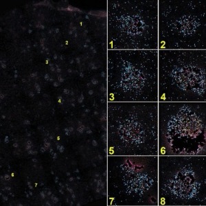
Imaging
Add enzymes to Imaging Buffer just prior to imaging.