Nov 09, 2022
Scanning electron microscopy protocol
- 1Universidade Estadual de Campinas
- Rene Flores Clavo: CENTRO DE INVESTIGACIÓN E INNOVACIÓN EN CIENCIAS ACTIVAS MULTIDISCIPLINARIAS
- Francisco Breno Teófilo: Electron Microscopy Laboratory - University of Campinas
- RENE FLORES
- Protocol Electronic Microscopic


Protocol Citation: Rene Flores Clavo, Francisco Breno Teófilo 2022. Scanning electron microscopy protocol . protocols.io https://dx.doi.org/10.17504/protocols.io.j8nlkk796l5r/v1
License: This is an open access protocol distributed under the terms of the Creative Commons Attribution License, which permits unrestricted use, distribution, and reproduction in any medium, provided the original author and source are credited
Protocol status: Working
We use this protocol and it's working
Created: August 16, 2022
Last Modified: November 09, 2022
Protocol Integer ID: 68749
Keywords: Scanning electron, microscopy protocol, bacterial identification's, LBME, CIICAM, scanning electron microscopy processing, electron microscopy protocol, electron microscopy processing, electron microscopy, scanning electron, microscopy, critical point chamber, sputter coating, acquisition of image, fixation
Abstract
This protocol briefly summarizes the basic steps of a scanning electron microscopy processing. The methods adopted for fixation, post-fixation, dehydration, drying in a critical point chamber, sputter coating, and the visualization and acquisition of images are described here.
Materials
1. Sample;
2. Glutaraldehyde, 2,5%;
3. Sodium cacodylate buffer (pH 7.3) 0.1 M;
4. Osmium tetroxide, 1.0%;
5. Acetone;
6. CO2;
7. Aluminum stubs;
8. Balzers CPD-030 Critical Point Dryer;
9. Balzers SCD 050 Sputter-Coater;
10. Scanning Electron Microscope JEOL JSM 5800LV, at 10 kV;
11. SemAfore 5.21 software.
Protocol materials
Glutaraldehyde 25% Aqueous Solution 10 x 10 ml ampoulesElectron Microscopy SciencesCatalog #16220
Sodium cacodylate trihydrateMerck MilliporeSigma (Sigma-Aldrich)Catalog #C0250
MATERIAL SELECTION
1 mm2 samples were selected in the colony.
FIXATION
4h
Samples were fixed in a solution of 2.5 % volume Glutaraldehyde 25% Aqueous Solution 10 x 10 ml ampoulesElectron Microscopy SciencesCatalog #16220 and 0.1 Mass Percent Sodium cacodylate trihydrateMerck MilliporeSigma (Sigma-Aldrich)Catalog #C0250 buffer 7.3)) , at Room temperature , for 04:00:00 .
4h
WASHING AFTER FIXATION
Samples was then rinsed three times in 0.1 Mass Percent Sodium cacodylate trihydrateMerck MilliporeSigma (Sigma-Aldrich)Catalog #C0250 (7.3 ), at Room temperature , for 00:20:00 for each rinse.
20m
POST-FIXATION
Samples was then post-fixed with 1 % volume osmium tetroxide in 0.1 Mass Percent Sodium cacodylate trihydrateMerck MilliporeSigma (Sigma-Aldrich)Catalog #C0250 buffer, 7.3 , at Room temperature , for 01:00:00 , protected from ambient light.
1h
WASHING AFTER POST-FIXATION
Samples was then rinsed three times in sodium 0.1 Mass Percent Sodium cacodylate trihydrateMerck MilliporeSigma (Sigma-Aldrich)Catalog #C0250 buffer 7.3 , at Room temperature , for 00:20:00 for each rinse.
20m
DEHYDRATION
Samples were dehydrated with ascending acetone series, for 00:20:00 minutes each step - 30 % volume ; 50 % volume , 70 % volume , 90 % volume , 100 % volume (last one step, for three times).
20m
CRITICAL POINT DRYING
Samples was then dried using the critical point method with CO2.
MOUTING ON THE STUBS
Samples was then placed on aluminum stubs.
SPUTTER-COATER
Samples was then coated with a layer of 30−40 nm gold using a Balzers SCD 050 sputter-coater.
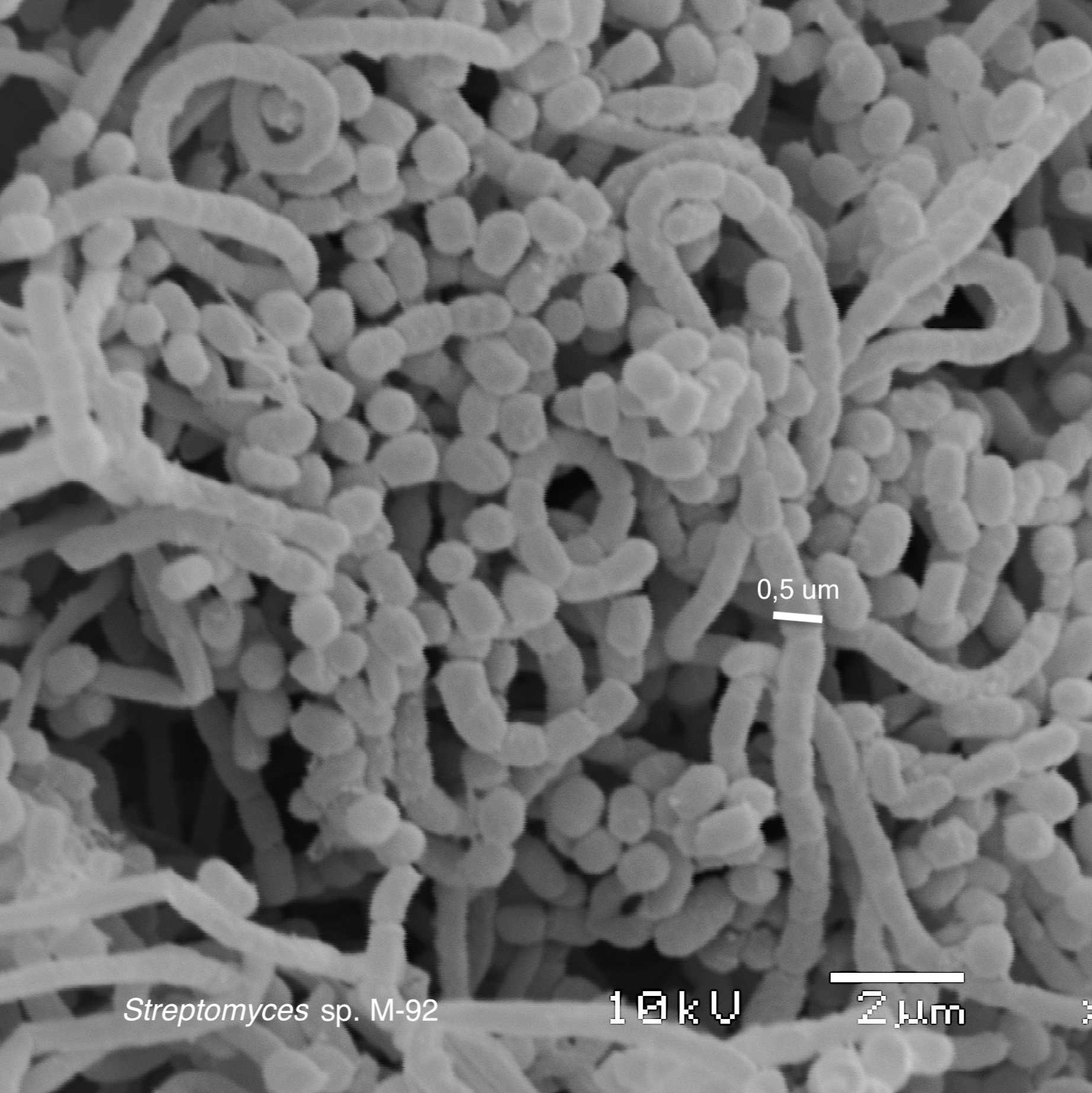
OBSERVATIONS AND IMAGE ACQUISITION
Observations and photomicrograph aquisitions were obtained using a JEOL JSM 5800LV at 10 kV with SemAfore 5.21 software.