Aug 10, 2025
Version 5
SARS-CoV-2 nsp3 macrodomain Time-Resolved FRET peptide displacement assay V.5
- Haim Barr1,2,
- Noa Lahav1,2
- 1The Weizmann Institute of Science;
- 2ASAP Discovery Consortium
- Haim Barr: General acknowledgement: The Wohl Drug Discovery institute, The Nancy and Stephen Grand Israel National Center for Personalized Medicine.;
- ASAP Discovery



Protocol Citation: Haim Barr, Noa Lahav 2025. SARS-CoV-2 nsp3 macrodomain Time-Resolved FRET peptide displacement assay. protocols.io https://dx.doi.org/10.17504/protocols.io.eq2ly7r2mlx9/v5Version created by Nick Lynch
License: This is an open access protocol distributed under the terms of the Creative Commons Attribution License, which permits unrestricted use, distribution, and reproduction in any medium, provided the original author and source are credited
Protocol status: Working
We use this protocol and it's working
Created: June 09, 2025
Last Modified: August 10, 2025
Protocol Integer ID: 219785
Keywords: Coronaviridae, Time-Resolved FRET assay, Fluorescence assay, Mac1, Nsp3, TR-FRET, HTRF, ADPr, Displacement, Screening, Assay, Inhibitor, Fragment, Binding, Macrodomain, SARS-CoV-2, resolved fret peptide displacement assay, fret peptide displacement assay this protocol detail, peptide, modified peptide, binding of adenosine, affinity of mac1, assay for sar, sars cov2 mac1183000, potential inhibitor, adenosine, inhibitor, sar
Funders Acknowledgements:
National Institutes of Health/National Institute Of Allergy and Infectious Diseases (NIH/NIAID)
Grant ID: U19AI171399
Disclaimer
The content is solely the responsibility of the authors and does not necessarily represent the official views of the National Institutes of Health.
Abstract
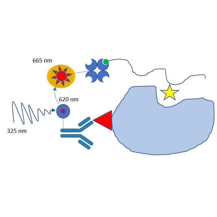
This protocol details the Time-Resolved FRET (TR-FRET) assay for SARS-CoV-2 nsp3 (non-structural protein 3) macrodomain (Mac1) binding of adenosine diphosphate (ADP)–ribosylated (ADPr) peptide. This method measures the affinity of Mac1 to its potential inhibitors by using a specific ADPr-modified peptide that allows the detection of binding. When bound, the biotinylated-peptide and the HIS-tagged Mac1 form a proximity complex that is detected by TR-FRET using Streptavidin-Eu Cryptate and anti-HIS-XL665 as a donor/acceptor pair. Excitation of the Eu Cryptate complex at 325 nm emits a resonant energy of 625 nm which in turn excites the XL665 to emit fluorescence at 665 nm. This energy transfer occurs only when ADPr modified peptide is in sufficient proximity to Mac1 while inhibitors which displace the peptide will prevent energy transfer. Binding is reported as the ratio of Acceptor/Donor (Em/Em) X 10,000.
-------------------------------------------------------
Experiment Concentrations (From Stock to Assay)
| A | B | C | D | E | |
| Reagent | Stock | Loaded into Combi | Final in assay plate | Units | |
| His-SARS CoV-2 MAC1 | 183000 | 50 | 12.5 | nM | |
| Substrate (Biotin-ADPr) | 10000000 | 1600 | 400 | nM | |
| Detection solution | |||||
| Streptavidin-XL665 (SA-XL) | 1 | 0.25 | 0.125 | % | |
| MAb Anti-6HIS-Eu cryptate Gold | 100 | 0.25 | 0.125 | % | |
| Assay buffer | |||||
| HEPES pH=7.0 | 250 | 25 | 25 | mM | |
| NaCl | 200 | 20 | 20 | mM | |
| BSA | 0.5 | 0.05 | 0.05 | mg/ml | |
| Tween 20 | 0.5 | 0.05 | 0.05 | % | |
| HTRF PPI Europium Detection Buffer | 100 | 10 | 10 | % | |
For more information, please check out the "Materials" Section
-------------------------------------------------------
Compound Plate Design for Dose Response:
Total Assay Volume: 16 µL
Compounds Top Assay Concentration: 100 µM
Dilution Factor: 3
Dose Response Points: 10
Number of Replicates: 2
Backfill with DMSO: Yes
Compounds Plate Design for 2-Point Assay:
Total Assay Volume: 16 µL
Compounds Assay Concentration: 100 µM and 50 µM
Dilution Factor: 2
Dose Response Points: 2
Number of Replicates: 2
Backfill with DMSO: Yes
Materials
Assay Buffer Reagents (Concentration listed are from Stock Solutions)
- 250 millimolar (mM) HEPES 0.5M buffer soln. pH 7.0Fisher ScientificCatalog #AAJ60064AE (or similar)
- 200 millimolar (mM) Sodium chlorideMerck MilliporeSigma (Sigma-Aldrich)Catalog #S9888 (or similar)
- 0.5 mg/mL Bovine Serum Albumin (BSA)Merck MilliporeSigma (Sigma-Aldrich)Catalog #A7030
- 0.5 % volume TWEEN® 20Merck MilliporeSigma (Sigma-Aldrich)Catalog #P9416
- 100 % volume HTRF PPI Europium Detection BufferCISBIO BIOASSAYS (PerkinElmer)Catalog #61DB9RDF
*Note: There are several forms of the Assay Buffer in this experiment. The Assay Buffer is the final, active buffer used throughout the experiment and has all of the five above reagents included. HTRF PPI Europium Detection Buffer needs to be added fresh before each experiment. Thus, there was an intermediate Buffer called Mac1 Buffer that contained HEPES, NaCl, BSA, and Tween only. Mac1 Buffer was filtered and stored at 4˚C. HTRF PPI buffer was then added to Mac1 Buffer fresh (to a final concentration of 10%) prior to performing the experiment--creating the active Assay Buffer.
---------------------------------------------------------
Detection Solution Reagents (Concentration listed are from Stock Solutions)
1 % volume Streptavidin-XL665CISBIO BIOASSAYS (PerkinElmer)Catalog #610SAXAC
- Note: Streptavidin-XL665 was dissolved in triply distilled water and diluted with HTRF PPI buffer to its stock concentration and then was aliquoted into 1.5mL sterile conical tubes
100 Mass Percent MAb Anti-6HIS-Eu cryptate GoldCISBIO BIOASSAYS (PerkinElmer)Catalog #61HI2KLA
Note: MAb Anti-6HIS-Eu cryptate Gold was dissolved in tripled distilled water and then aliquoted into 1.5mL sterile conical tubes
---------------------------------------------------------
Additional Reagents:
183000 nanomolar (nM) His-SARS COV2 MAC1 Enzyme
- The Enzyme stock was originally 183000 nanomolar (nM) and was diluted to 50 nanomolar (nM) before every experiment in freshly made Assay Buffer. The final assay concentration is 12.5 nanomolar (nM)
10000000 nanomolar (nM) Substrate (Biotin-ADPr) MAC1
- Substrate stock (ARTK(Bio)QTARK(Aoa-RADP)S) was dissolved in DMSO to the stock concentration. Before each experiment, the substrate stock was diluted to 1600 nanomolar (nM) in freshly made Assay Buffer.
Protocol materials
Sars Cov 2 Mac domain PlasmidaddgeneCatalog #204787
Safety warnings
Please be sure to wear proper Personal Protective Equipment (PPE) while performing this experiment.
Before start
Note: Inhibitor compounds stock concentration is 20 millimolar (mM) . Compounds are pre-dispensed into 384 plates and stored at -20˚C until use.
SARS-CoV-2 nsp3 expression and purification
The protein used in this assay was expressed and purified using the following protocol.
We used the following plasmid in the protein expression and purification protocol.
Sars Cov 2 Mac domain PlasmidaddgeneCatalog #204787
Protocol
CREATED BY
korvus.wang
Prepare Reagents
PREPARE all of the reagents/buffers required for this experiment.
Assay Buffer
| A | B | C | D | E | |
| Reagent | Stock | Loaded into Combi | Final in assay plate | Units | |
| HEPES pH=7.0 | 250 | 25 | 25 | mM | |
| NaCl | 200 | 20 | 20 | mM | |
| BSA | 0.5 | 0.05 | 0.05 | mg/ml | |
| Tween 20 | 0.5 | 0.05 | 0.05 | % | |
| HTRF PPI Europium detection buffer | 100 | 10 | 10 | % |
Reagents (dilute reagents in assay buffer for required volume)
| A | B | C | D | E | |
| Reagent | Stock | Loaded into Combi | Final in assay plate | Units | |
| His-SARS-CoV-2 MAC1 | 183000 | 50 | 12.5 | nM | |
| Substrate (Biotin-ADPr) | 10000000 | 1600 | 400 | nM |
Detection Solution (dilute reagents in assay buffer for required volume)
| A | B | C | D | E | |
| Reagent | Stock | Loaded into Combi | Final in assay plate | Units | |
| Streptavidin-XL665 (SA-XL) | 1 | 0.25 | 0.125 | % | |
| MAb Anti-6HIS-Eu cryptate Gold | 100 | 0.25 | 0.125 | % |
Prepare 384-well Plate
16m
PRIME Multi-Drop Combi Tube Dispensing Cassette with Assay Buffer by selecting the PRIME button on the Combi Dispenser until the tubes are filled completely.
- Note: Be sure to cycle dispensing several times on a clean plate lid (This confirms there are no bubbles in the Dispensing Cassette).
DISPENSE 4 µL Mac1 Buffer to Columns 1 and 23 of assay plate
- Note: These will represent the inhibitor control columns
EMPTY Multi-Drop Combi Tube Dispensing Cassette (by selecting the EMPTY button on the Combi Dispenser until the tubes of the cassette are emptied). Discard the Assay Buffer discharged from the cassette.
PRIME Multi-Drop Combi Tube Dispensing Cassette with His-SARS COV2 MAC1 Enzyme by selecting the PRIME button on the Combi Dispenser until the tubes are filled completely.
- Note: Be sure to cycle dispensing several times on a clean plate lid (This confirms there are no bubbles in the Dispensing Cassette).
DISPENSE 4 µL 50 nanomolar (nM) His-SARS COV2 MAC1 Enzyme to Columns 2-22 and 24 of assay plate
Note:
- 50 nanomolar (nM) His-SARS COV2 MAC1 is four times the final concentration for the assay. It will be diluted to be a final concentration of 12.5 nanomolar (nM) His-SARS COV2 MAC1 Enzyme
- Column 2 and Column 24 are neutral control columns (Contain: Enzyme, Substrate, DMSO; no experimental compounds)
EMPTY Multi-Drop Combi Tube Dispensing Cassette (by selecting the EMPTY button on the Combi Dispenser until the tubes of the cassette are emptied). Discard the 50 nanomolar (nM) His-SARS COV2 MAC1 Enzyme discharged from the cassette.
PRIME Multi-Drop Combi Tube Dispensing Cassette with Assay Buffer by selecting the PRIME button on the Combi Dispenser until the tubes are filled completely. Then, EMPTY the Multi-Drop Combi Tube Dispensing Cassette (by selecting the EMPTY button on the Combi Dispenser until the tubes of the cassette are emptied). Discard the Assay Buffer discharged from the cassette.
CENTRIFUGE plate 1500 rpm, Room temperature, 00:01:00 to remove bubbles
1m
INCUBATE plate for 00:15:00 atRoom temperature
15m
PRIME Multi-Drop Combi Tube Dispensing Cassette with1600 nanomolar (nM) MAC1 Substrate (Biotin-ADPr) by selecting the PRIME button on the Combi Dispenser until the tubes are filled completely.
- Note: Be sure to cycle dispensing several times on a clean plate lid (This confirms there are no bubbles in the Dispensing Cassette).
DISPENSE 4 µL 1600 nanomolar (nM) MAC1 Substrate (Biotin-ADPr) into Columns 1-24 (full plate)
Note:
- 1600 nanomolar (nM) MAC1 Substrate (Biotin-ADPr) is four times the final concentration for the assay. It will be diluted to be a final concentration of400 nanomolar (nM) MAC1 Substrate (Biotin-ADPr)
EMPTY Multi-Drop Combi Tube Dispensing Cassette (by selecting the EMPTY button on the Combi Dispenser until the tubes of the cassette are emptied). Discard the 1600 nanomolar (nM) MAC1 Substrate (Biotin-ADPr) discharged from the cassette.
CENTRIFUGE plate 1500 rpm, Room temperature, 00:01:00 to remove bubbles
PRIME Multi-Drop Combi Tube Dispensing Cassette with Assay Buffer by selecting the PRIME button on the Combi Dispenser until the tubes are filled completely. Then, EMPTY the Multi-Drop Combi Tube Dispensing Cassette (by selecting the EMPTY button on the Combi Dispenser until the tubes of the cassette are emptied). Discard the Assay Buffer discharged from the cassette.
PRIME Multi-Drop Combi Tube Dispensing Cassette with 0.25 % volume Detection Solution by selecting the PRIME button on the Combi Dispenser until the tubes are filled completely.
- Note: Be sure to cycle dispensing several times on a clean plate lid (This confirms there are no bubbles in the Dispensing Cassette).
DISPENSE 8 µL 0.25 % volume Detection Solution into full plate
Note:
- 0.25 % volume Detection Solution is two times the final concentration for the assay. It will be diluted in the plate to be a final concentration of 0.125 % volume Detection Solution
EMPTY Multi-Drop Combi Tube Dispensing Cassette (by selecting the EMPTY button on the Combi Dispenser until the tubes of the cassette are emptied). Discard the 1600 nanomolar (nM) MAC1 Substrate (Biotin-ADPr) discharged from the cassette.
CENTRIFUGE 1500 rpm, Room temperature, 00:01:00 plate to remove bubbles
INCUBATE plate for 01:00:00 at Room temperature
Recommended: Clean/Empty the Multi-Drop Combi Reagent Dispenser and Dispensing Cassette during this incubation step
1h
Read Plate Fluorescence
READ and RECORD the plate Relative fluorescence units (RFU) via the "Mac1 Protocol" on the PHERAstar FS Control Software.
Equipment
PHERAstar FS
NAME
Microplate reader
TYPE
BMG LABTECH
BRAND
0471B0001A
SKU
LINK
Expected result
Donor 325/620 ex/em should be ~ 5000 . Acceptor ~ 3000
Diagram of assay
Figure 1 graphical depiction of assay principal and its use in screening campaign
Principal of ADPr peptide displacement assay
A - Binding of ADPr-modified peptide to Mac1 protein B- Detection reagents added to protein+peptide complex C- TR-FRET detects binding of peptide to Mac1 if proximity of donor and acceptor detection reagents is sufficient to enable resonant energy transfer D- Inhibitor compounds are detected by reduced TR-FRET signal when inhibitor displaces/prevents binding of ADPr-peptide to Mac1
Experimental Design
384 plate layout
Protocol references