Sep 26, 2025
Version 2
Sanger Tree of Life HMW DNA Extraction: Manual Tissue Nanobind® V.2
- Pacific Biosciences1,
- Adam Bates2,
- Amy Denton3,
- Caroline Howard3
- 1Pacific Biosciences;
- 2Wellcome Sanger Institute;
- 3Tree of Life, Wellcome Sanger Institute, Hinxton, Cambridgeshire, CB10 1SA
- Tree of Life at the Wellcome Sanger Institute
- Earth BioGenome Project


Protocol Citation: Pacific Biosciences, Adam Bates, Amy Denton, Caroline Howard 2025. Sanger Tree of Life HMW DNA Extraction: Manual Tissue Nanobind®. protocols.io https://dx.doi.org/10.17504/protocols.io.14egn36nyl5d/v2Version created by Amy Denton
Manuscript citation:
License: This is an open access protocol distributed under the terms of the Creative Commons Attribution License, which permits unrestricted use, distribution, and reproduction in any medium, provided the original author and source are credited
Protocol status: Working
We use this protocol and it's working
Created: August 19, 2025
Last Modified: September 26, 2025
Protocol Integer ID: 224939
Keywords: HMW DNA extraction, mollusc extraction, Nanobind, manual DNA extraction, reference genome, long read sequencing, life hmw dna extraction, sanger tree of life hmw dna extraction, extracting hmw dna, manual extraction of hmw dna, hmw dna from black mystery snail tissue, hmw dna, hmw dna fragmentation, quantity hmw dna, mollusc sample, mollusc species, effective for mollusc species, dna extraction, acronyms hmw, black mystery snail tissue, dna, li pacbio protocol, procedure from pacbio, read sequencing, molecular weight li, high molecular weight li, manual extraction, extraction, animal tissue extraction, mammal tissue DNA extraction, fish tissue DNA extraction, bird tissue DNA extraction, amphibian tissue DNA extraction, sanger tree of life hmw dna fragmentation, life hmw dna fragmentation, extracting dna, dna from animal tissue, nanobind, animal tissue sample, pacbio li protocol, including mollusc, sanger tree, tissue sample, tree of life programme
Funders Acknowledgements:
Wellcome Trust
Grant ID: 218328
Wellcome Trust
Grant ID: 206194
Gordon and Betty Moore Foundation
Grant ID: GBMF8897
Abstract
This protocol describes the manual extraction of HMW DNA from animal tissue samples intended for long-read sequencing using the Nanobind® PanDNA kit and and following the ‘Extracting DNA from animal tissue using Nanobind® PanDNA kit’ procedure from PacBio. This process is effective for several animal species covered by the Tree of Life Programme, including molluscs, mammals, birds, fish and amphibians. The output of this protocol is high quality and quantity HMW DNA, which can be directed towards the Sanger Tree of Life HMW DNA Fragmentation: Diagenode Megaruptor® 3 for LI PacBio or the Sanger Tree of Life HMW DNA Fragmentation: Opentrons® OT-2 for PacBio LI protocols.
Acronyms
HMW: high molecular weight
LI: low input
Guidelines
- This protocol uses the ‘Extracting DNA from animal tissue using Nanobind® PanDNA kit’ procedure, with the inclusion of the sample preparations, standard sample inputs and standard elution volumes used by Sanger Tree of Life.
- For molluscs, this protocol works best with cryoprepped material of around 30–50 mg input weight. If the mollusc tissue is not cryoprepped, place in a chilled petri dish and on ice, cut the tissue into smaller chunks, then powermash the sample in CT buffer.
- For vertebrate species, tissue can be either cryoprepped/bead beaten, with 25–35 mg input weight, or powermashed, with smaller tissue input <20 mg.
- Before starting the protocol, keep samples on dry ice to maintain temperature and prevent nucleic acid degradation.
- An experienced operator can expect to comfortably process 8 samples with a start to finish period of 4 hours. This estimation excludes overnight incubation at room temperature and subsequent QC checks.
Additional Notes:
- FluidX tubes are used throughout the Tree of Life programme in order to track samples, therefore rather than the microcentrifuge tubes which have been mentioned in this protocol for DNA storage, all routine DNA extracts are stored in FluidX tubes.
Materials
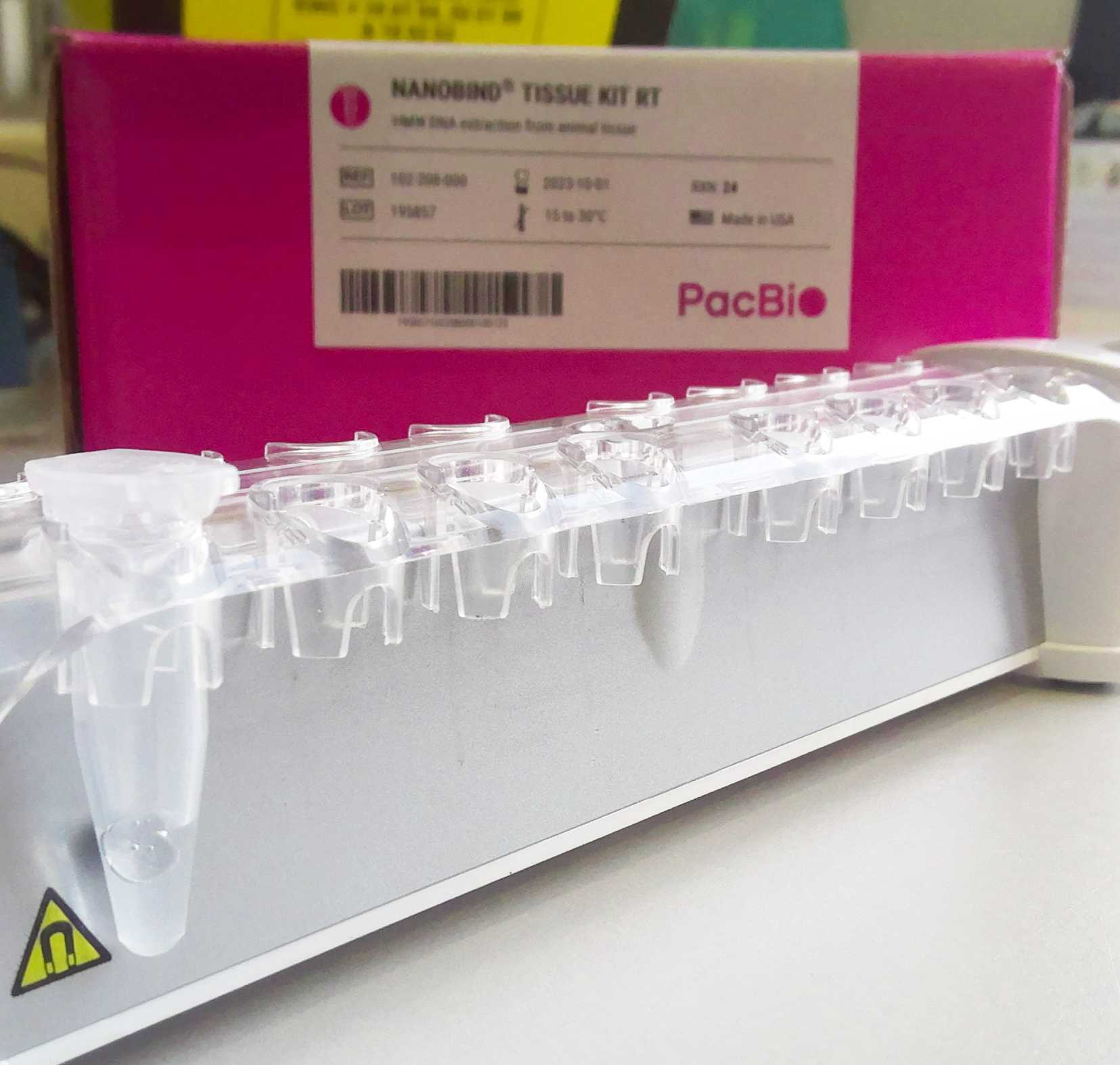
- Nanobind® PanDNA kit (Pacific Biosciences PN 103-260-000)
- 2 mL DNA LoBind microcentrifuge tubes (Eppendorf Cat. no. 0030108078)
- 1.5 mL Protein LoBind microcentrifuge tubes (Eppendorf Cat. no. 0030108116)
- 1.5 mL BioMasher II tubes and pestles (sterile) (Takara Cat. no. 9791A)
- 100% absolute ethanol
- 100% absolute isopropanol
Equipment:
- Pipettes for 0.5-1000 μL and filtered tips
- Wide-bore pipette tips (200 μL, filtered if available)
- Scalpel (Thermo Fisher Scientific Cat. no. 22-079-712)
- Diagnocine PowerMasher II tissue disruptor (Cat. no. FNK-891300)
- DynaMag™-2 magnetic rack (Cat. no. 12321D) or similar
- Eppendorf ThermoMixer C (Cat. no. 5382000031)
- Eppendorf SmartBlock 2.0 ml (Cat. no. 5362000035)
- Mini centrifuge (Cat. no. SS-6050)
- Eppendorf Centrifuge 5425/5425 R (Cat. no. 5405000263)
- HulaMixer Sample Mixer (Cat. no. 15920D)
- Vortexer (Vortex Genie™ 2 SI-0266)
- Timer
Protocol PDF:  Sanger Tree of Life HMW DNA Extraction_ Manual Tissue Nanobind.pdf128.3KB
Safety warnings
- The operator must wear a lab coat, powder-free nitrile gloves and safety specs to perform the laboratory procedures in this protocol. Cotton glove liners are strongly recommended when handling the samples on dry ice.
- Waste needs to be collected in a suitable container (e.g. plastic screw-top jar or Biobin) and disposed of in accordance with local regulations.
- Liquid waste needs to be collected in a suitable container (e.g. glass screw-top jar) and disposed of in accordance with local regulations.
Before start
- Add 100% ethanol to the Buffers CW1 and CW2 as per manufacturer’s instructions.
- Set a heat block to 55 °C.
- Pre-chill the centrifuge to 4 °C.
Laboratory protocol
Transfer tissue to a 2 mL microcentrifuge tube if cryoprepped, or a 1.5 mL BioMasher II tube if whole, and add 750 μL ice cold buffer CT.
Pipette mix cryoprepped samples with a wide-bore pipette tip for 5-10 times to homogenise.
Powermash whole tissue samples using the Diagnocine PowerMasher II tissue disruptor and BioMasher pestle until no large pieces remain or sample cannot be disrupted further (for more detailed instructions regarding powermashing, please refer to the ‘Sanger Tree of Life Sample Homogenisation: Powermash’ protocol). Transfer lysate to a 2 mL microcentrifuge tube following homogenisation.
Centrifuge 6,000 g at 4 °C for 5 minutes.
Discard supernatant and add 1 mL ice cold buffer CT.
Pipette mix 10 times with wide-bore pipette tip to dislodge pellet and rehomogenise sample.
Pellet the homogenate by centrifuging the sample at 6,000 g at 4 °C for 5 minutes.
Discard the supernatant.
Add 20 μL of proteinase K to the pellet and pulse vortex for 1 second 2 times at the maximum setting to dislodge the pellet.
Add 150 μL of buffer CLE3 and pipette mix 10 times using a wide-bore pipette tip.
Incubate sample at 55 °C and 900 rpm for 30 minutes on the heat block.
Spin on a mini-centrifuge for 2 seconds to collect liquid from the lid.
Add 20 μL of RNase A.
Incubate at 55 °C and 900 rpm on the heat block for another 30 minutes. This incubation can be longer, up to 2 hours, if there are still visible large tissue pieces remaining.
Add 60 μL buffer SB and pulse vortex for 1 second 5 times at the maximum setting to mix (wide-bore pipette mixing 5 times is acceptable as an alternative).
Centrifuge 10,000 g at room temperature (15-30 °C) for 5 minutes.
Transfer up to 300 μL of the supernatant to a new labelled 1.5 mL Protein LoBind microcentrifuge tube.
Add 50 μL of buffer BL3 to the collected supernatant and mix by inverting 10 times (wide-bore pipette mixing 10 times is acceptable as an alternative).
Spin on a mini-centrifuge for 2 seconds to collect liquid from the lid.
Add a Nanobind disk to the lysate.
Add 350 μL isopropanol and mix by inverting 10 times.
Mix sample on a platform rocker at 20 rpm, or a HulaMixer at 25 rpm with the reciprocal angle set at 50°, for 15 minutes at room temperature to allow the DNA to bind to the disk.
Alternatively, the tube could be manually inverted every 3 minutes for 15 minutes.
Place the tubes on the magnetic rack.
Discard the supernatant, either using a pipette and taking care to avoid touching the disk or by tipping the supernatant out into a reservoir, ensuring that the disk and DNA remain bound to the magnet.
Add 500 μL of Buffer CW1, remove the tubes from the magnet and mix by inverting 4 times, then place the tubes back on the rack and discard the supernatant.
Repeat step 23.
Add 500 μL of Buffer CW2, remove the tubes from the magnet and mix by inverting 4 times, then place the tubes back on the rack and discard the supernatant.
Repeat step 25.
Spin samples on a mini-centrifuge for 2 seconds to collect liquid from the lid.
Place samples back on the magnetic rack and remove any residual supernatant.
Remove tubes from the magnetic rack and add 100 μL Buffer LTE directly onto the nanobind disk.
Incubate sample for 10 minutes at room temperature.
Collect the supernatant containing the HMW DNA and transfer to a new labelled 1.5 mL Protein LoBind microcentrifuge tube for storage using a standard pipette tip.
Spin the tube containing the Nanobind disk on a mini-centrifuge for 5 seconds and use a standard P200 pipette to combine any of the additional liquid (or gel) which comes off the disk to the previous eluate. Repeat until all liquid/gel has come off the Nanobind disk.
Pipette mix the sample 5 times with a standard P200 pipette tip to homogenise and disrupt any viscous gel regions within the sample.
Allow the sample to rest overnight at room temperature for the DNA to relax and solubilise.
Following the overnight rest, pipette mix the sample 5 times with a standard P200 pipette tip before proceeding with QC.
Store the DNA at 4 ºC.