Sep 26, 2025
Sanger Tree of Life HMW DNA Extraction: Manual Plant MagAttract v.5
- 1Tree of Life, Wellcome Sanger Institute, Hinxton, Cambridgeshire, CB10 1SA;
- 2Wellcome Sanger Institute
- Tree of Life at the Wellcome Sanger Institute
- Earth BioGenome Project


Protocol Citation: Graeme Oatley, Amy Denton, Anna Kovalevskaia, Caroline Howard 2025. Sanger Tree of Life HMW DNA Extraction: Manual Plant MagAttract v.5. protocols.io https://dx.doi.org/10.17504/protocols.io.36wgqp655vk5/v1
Manuscript citation:
License: This is an open access protocol distributed under the terms of the Creative Commons Attribution License, which permits unrestricted use, distribution, and reproduction in any medium, provided the original author and source are credited
Protocol status: Working
We use this protocol and it's working
Created: September 04, 2025
Last Modified: September 26, 2025
Protocol Integer ID: 226418
Keywords: HMW DNA extraction, magnetic bead extraction, manual DNA extraction, MagAttract, Plant HWM DNA extraction, solid phase reversible immobilisation, reference genome, long read sequencing, sanger tree of life hmw dna extraction, manual extraction of hmw dna, qiagen magattract, hmw dna extraction kith, hmw dna fragmentation, hmw dna pooling, pacbio sequencing, manual plant magattract, manual extraction, plant species, tree of life programme, fungi extractions, life hmw dna extraction, qiagen magattract hmw dna extraction kit, recoveries of hmw dna, maximum amount of hmw dna, hmw dna, yield of dna, genome size of the species, dna, acronyms hmw, sanger tree, fungi
Funders Acknowledgements:
Wellcome Trust
Grant ID: 206194
Wellcome Trust
Grant ID: 218328
Gordon and Betty Moore Foundation
Grant ID: GBMF8897
Abstract
This protocol is for the manual extraction of HMW DNA from plant or fungi tissue samples from a variety of species intended for long-read sequencing using the Qiagen MagAttract HMW DNA extraction kit. This process is effective for approximately 60% of the plant species covered by the Tree of Life Programme, and the resulting yield of CCS data from PacBio sequencing has been good. This protocol is particularly useful for samples with limited tissue availability, as it ensures the maximum amount of HMW DNA can be extracted and recovered. The output of this protocol is HMW DNA, which depending upon yield and genome size of the species, can be directed towards either HMW DNA Pooling, HMW DNA Fragmentation: Opentrons® OT-2 for PacBio LI, HMW DNA Fragmentation: Diagenode Megaruptor® 3 for LI PacBio or HMW DNA Fragmentation: Covaris g-Tube for ULI PacBio. This protocol was adapted from Sanger Tree of Life HMW DNA Extraction: Manual Plant MagAttract v.4 to include an overnight elution in order to maximise the yield of DNA, as well as an updated pre-shear SPRI to improve recoveries of HMW DNA.
Acronyms:
HMW: high molecular weight
SPRI: solid-phase reversible immobilisation
LI: low input
ULI: ultra-low input
CCS: circular consensus sequencing
Guidelines
- For the lysis buffer master mix, prepare enough for n+1 samples to account for pipetting errors.
- Keep samples on dry ice to maintain temperature and prevent nucleic acid degradation until the lysis buffer is ready to be added to them.
- An experienced operator can expect to comfortably process 8 samples, with approximately 2 hours handling time over a start to finish period of 4 hours. This estimation excludes the overnight elution and subsequent QC checks.
- Whilst a manual 0.45X SPRI is detailed in this protocol, samples which undergo the manual extraction could alternatively undergo an automated 0.45X SPRI, as detailed in the Sanger Tree of Life HMW DNA Extraction: Automated Plant MagAttract v5.
Additional Notes:
- FluidX tubes are used throughout the Tree of Life programme in order to track samples, therefore rather than the microcentrifuge tubes which have been mentioned in this protocol for DNA storage, all routine DNA extracts are stored in FluidX tubes.
Materials
- 2 mL DNA Lo-Bind microcentrifuge tubes (Eppendorf Cat. no. 0030108078)
- Qiagen MagAttract HMW DNA extraction kit (Qiagen Cat. no. 67563)
- Dry ice
- 1 x phosphate-buffered saline (PBS)
- EB buffer (Qiagen Cat. no. 19086)
- AMPure PB beads (Pacific Biosciences Cat. no. 100-265-900)
- 100% absolute ethanol
- 15 mL or 50 mL centrifuge tubes
Equipment:
- Pipettes for 0.5–1000 μL and filtered tips
- Wide bore tips (200 and 1000 μL, filtered if available)
- Corning® CoolRack CF45 (Cat. no. 432051) or equivalent
- Eppendorf ThermoMixer C (Cat. no. 5382000031) or similar
- Eppendorf SmartBlock 2.0 mL (Cat. no. 5362000035)
- Eppendorf SmartBlock 50 mL (Cat. no. 5365000028)
- Eppendorf™ Centrifuge 5425/5425 R (Cat. no. 5405000263)
- Vortexer (Vortex Genie™ 2 SI-0266)
- Mini centrifuge (Cat. no. SS-6050) or similar
- DynaMag™-2 magnetic rack (Cat. no. 12321D) or similar
- Timer
Protocol PDF:  Sanger Tree of Life HMW DNA Extraction_ Manual Plant MagAttract v5.pdf169.8KB
Safety warnings
- The operator must wear a lab coat, powder-free nitrile gloves and safety specs to perform the laboratory procedures in this protocol. Cotton glove liners are strongly recommended when handling the samples on dry ice.
- Waste needs to be collected in a suitable container (e.g. plastic screw-top jar or Biobin) and disposed of in accordance with local regulations.
- Liquid waste needs to be collected in a suitable container (e.g. glass screw-top jar) and disposed of in accordance with local regulations.
Before start
- Add 100% ethanol to the MW1 and PE wash buffers as per manufacturer’s instructions.
- Set one heat block with a 50 mL SmartBlock to 65 °C, another heat block with a 2 mL SmartBlock to 55 °C.
- Remove the AMPure PB beads from the fridge 30 minutes before starting the 0.45X SPRI to bring them to room temperature.
Sample Lysis
Prepare a lysis buffer master mix in a 50 mL centrifuge tube:
| Reagent | Volume per sample | |
| Phosphate-buffered saline (PBS) | 200 µL | |
| Buffer AL | 150 µL |
Place the lysis buffer on the 65 °C heat block and incubate at 400 rpm for at least 20 minutes. Keep at temperature until added to the sample.
Transfer 50 mg of cryogenically disrupted tissue from each sample to 2 mL microcentrifuge tubes.
Ensure the disrupted tissue is completely disrupted into a fine powder; avoid matted/clumped powder. This is crucial for optimal DNA yield and integrity; poorly disrupted tissue drastically decreases lysis and extraction efficiency.
Any samples containing poorly disrupted tissue ‘chunks’ should be further cryogenically disrupted.
Transfer the samples to a pre-chilled cold block on wet ice and incubate for 10 minutes to equilibrate temperature.
Add 20 µL Proteinase K (for n+1 samples) to the preheated lysis buffer immediately prior to initiating lysis, swirling the centrifuge tube to mix.
Add 370 µL of the preheated lysis buffer plus Proteinase K to each sample, immediately homogenising the lysate by mixing with 5 rapid pulse vortexes, and place on the 55 °C heat block at 600 rpm for 15 minutes.
After 5 minutes incubation, resuspend any severely aggregated samples by pipette-mixing with a wide-bore pipette tip.
After the initial 15 minute incubation, add 4 µL RNase A to each sample and mix thoroughly by inversion until any aggregated, insoluble or sedimented tissue particles are resuspended.
Incubate samples for a further 45 minutes on the heat block at 55 °C at 600 rpm.
For the final 15 minutes of lysis, remove the 600 rpm mixing from the heat block to allow aggregated, insoluble or sedimented tissue particles to settle at the bottom of the tube - this will reduce gDNA loss during later centrifugation.
DNA Isolation
Once samples have completed lysing, remove sample tubes from the heat block and allow the lysate to settle to the bottom of the tube for 5 minutes.
Centrifuge the samples at 8000 rpm for 10 minutes at room temperature.
Using a wide-bore pipette tip, set the volume to 380 µL, transfer each lysate to fresh individual microcentrifuge tubes, whilst avoiding insoluble material.
Add 280 µL Buffer MB to each sample and 40 µL of Suspension G beads. Invert the tube 10 to 20 times to ensure the beads are suspended in the lysate. Allow 5 minutes for binding.
Briefly centrifuge the samples in a mini centrifuge to collect at the bottom of the tube.
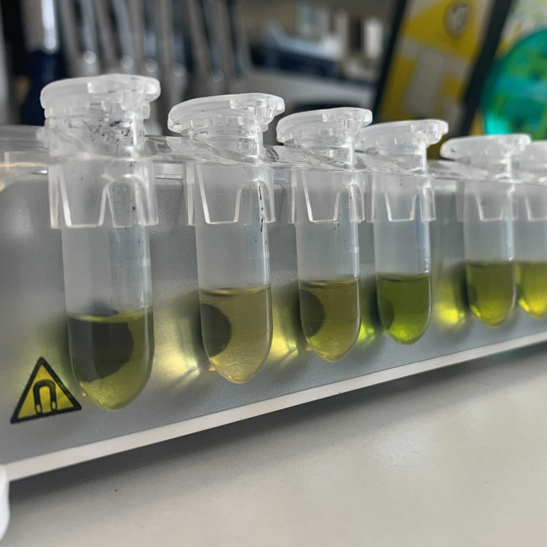
Place the tubes on the magnetic rack and allow 2–5 minutes for the beads to migrate (more viscous samples will take longer). Remove the supernatant and discard.
Remove the tubes from the magnetic rack and add 700 µL Buffer MW1 directly to the bead pellet, then invert the tube 10 to 20 times to ensure the beads are suspended in the lysate.
Spin down tubes in a mini centrifuge for 1–2 seconds, then place the tubes back on the magnetic rack and allow 2–5 minutes for the beads to migrate (more viscous samples will take longer). Remove the supernatant and discard.
Repeat the MW1 wash for a total of two washes (steps 17 & 18).
Remove the tubes from the magnetic rack and add 700 µL Buffer PE directly to the bead pellet and invert 10 to 20 times to resuspend the beads.
Spin down tubes in a mini centrifuge for 1–2 seconds, then place the tubes back on the magnetic rack and allow 2–5 minutes for the beads to migrate (more viscous samples will take longer). Remove the supernatant and discard.
Repeat the PE wash for a total of two washes (steps 20 & 21).
Briefly centrifuge the tubes in a mini centrifuge and place the sample back on the magnetic rack. Use a small micropipette to remove any residual wash buffer.
Pipette 700 µL nuclease-free water onto the side opposite of the beads in the microcentrifuge tubes whilst the tubes are on the magnetic rack. Do not pipette the nuclease-free water directly onto the bead pellet. Incubate for exactly 1 minute then slowly aspirate and discard water from the tubes.
Repeat step 24 for a total of two washes.
Remove the samples from the magnetic rack and add 400 µL of Buffer AE directly to the bead pellet. Mix, either by gently flick mixing or using a wide-bore pipette tip, in order to dislodge the pellet from the tube.
Incubate samples overnight at room temperature.
Following overnight incubation, pipette-mix samples slowly 5 times with a wide-bore pipette tip, before briefly centrifuging samples in a mini centrifuge and then placing them on a magnetic rack to allow bead capture.
Using a 200 μL wide-bore pipette tip, carefully transfer the supernatant containing purified gDNA to a fresh microcentrifuge tube.
Proceed with a 0.45X SPRI, either manually (as described below) or automated on the KingFisher Apex (following the 0.45X SPRI steps in protocol Sanger Tree of Life HMW DNA Extraction: Automated MagAttract v.3).
Manual 0.45X SPRI
Set the heat block to 37 °C and label two sets of 1.5 mL microcentrifuge tubes for each sample.
Vortex AMPure PB beads for 30 seconds.
Immediately add 180 μL of AMPure PB beads to the 400 μL of DNA.
Mix the beads/DNA thoroughly by pipette-mixing 15 times with a wide-bore pipette tip. Do not flick the tube.
Spin down tubes in a mini centrifuge for 1–2 seconds to collect the beads.
Incubate the mix on the bench top for 5 minutes at room temperature.
Spin down tubes in a mini centrifuge for 1-2 seconds to collect beads.
Place the tubes in a magnetic bead rack and wait for the beads to pellet on the side of the tube. This may take up to 5 minutes or more depending upon the amount of DNA and beads within the sample.
Slowly pipette off cleared supernatant and save in the first labelled 1.5 mL microcentrifuge tube. Avoid disturbing the beads.
Wash beads with freshly prepared 80% ethanol.
Do not remove the tube from the magnetic rack.
Use a sufficient volume of 80% ethanol to fill the tube: 700–1000 µL for 1.5 mL tube is usually sufficient.
Do not disturb the beads – slowly dispense the 80% ethanol against the side of the tube opposite the beads.
After 30 seconds, pipette and discard the 80% ethanol.
Repeat step 40 for a total of two ethanol washes.
Spin down tubes in a mini centrifuge for 1–2 seconds, then return them to the magnetic rack to allow the beads to pellet. Aspirate and dispose of any remaining ethanol.
Check for any remaining ethanol droplets in the tube. If droplets are present, repeat step 42.
Take tubes off the magnetic rack and add 135 µL of EB buffer to the beads. Gently mix by slowly pipetting 15 times with a wide bore pipette tip. Do not flick the tube.
Incubate tubes on the heat block at 37 °C for 30 minutes at 350 rpm.
Briefly spin down the tubes for 1–2 seconds in a mini centrifuge and then place them back on the magnetic rack. Allow the beads to pellet - this may take up to 5 minutes or more depending upon the quantity and quality of DNA within the sample.
Slowly pipette off cleared supernatant and save in the second labelled 1.5 mL microcentrifuge tube. Avoid disturbing the beads.
Proceed samples directly to QC. If extraction has been successful, the supernatants saved in the first set of labelled 1.5 mL microcentrifuge tubes can now be discarded.
Store the extracted gDNA sample at 4 ºC.
Protocol references
MagAttract HMW DNA Handbook: MagAttract HMW DNA Handbook - QIAGEN
Sanger Tree of Life HMW DNA Extraction: Manual Plant MagAttract v.4 B. Jackson & C. Howard (2023)