Sep 29, 2025
Version 2
Sanger Tree of Life HMW DNA Extraction: Manual Modified Omega Bio-Tek E.Z.N.A.® V.2
- 1Tree of Life, Wellcome Sanger Institute, Hinxton, Cambridgeshire, CB10 1SA
- Tree of Life at the Wellcome Sanger Institute
- Earth BioGenome Project


Protocol Citation: Amy Denton, Caroline Howard 2025. Sanger Tree of Life HMW DNA Extraction: Manual Modified Omega Bio-Tek E.Z.N.A.®. protocols.io https://dx.doi.org/10.17504/protocols.io.36wgq3wj3lk5/v2Version created by Amy Denton
Manuscript citation:
License: This is an open access protocol distributed under the terms of the Creative Commons Attribution License, which permits unrestricted use, distribution, and reproduction in any medium, provided the original author and source are credited
Protocol status: Working
We use this protocol and it's working
Created: January 28, 2025
Last Modified: September 29, 2025
Protocol Integer ID: 119229
Keywords: HMW DNA extraction, Omega Bio-Tek, E.Z.N.A, magnetic bead extraction, SpeedBeads, manual DNA extraction, jellyfish, ASG, solid phase reversible immobilisation, reference genome, long read sequencing, metazoa, annelids, amphipods, decapods, isopods, molluscs, aquatic symbiosis genomics, sanger tree of life, tree of life programme, tunicates, sea squirts, sanger tree of life hmw dna extraction, life hmw dna extraction, aquatic symbiosis genomic, automated plant organic hmw gdna extraction, fresh frozen jellyfish sample, jellyfish species, insect dna kit protocol, insect dna kit, hmw dna, jellyfish sample, spri of hmw dna, hmw dna fragmentation, incubation temperatures for jellyfish sample, manual modified omega bio, combination of the omega bio, including mastigias papua, genome size of the species, omega bio, mastigias papua, arthropod, such as arthropod, pseudorhiza haeckeli, mollusc, manual extraction, aurelia spp, effective for the manual extraction, successful extraction, pelagia noctiluca, dna, other metazoa s
Funders Acknowledgements:
Wellcome Trust
Grant ID: 206194
Wellcome Trust
Grant ID: 218328
Gordon and Betty Moore Foundation
Grant ID: GBMF8897
Abstract
This protocol describes the manual extraction and SPRI of HMW DNA from fresh frozen jellyfish samples intended for long-read sequencing, using a combination of the Omega Bio-Tek E.Z.N.A.® Mollusc & Insect DNA kit and Sera-Mag™ SpeedBeads. It is effective for approximately 50 to 60% of jellyfish species covered by the Tree of Life Programme for the Aquatic Symbiosis Genomics (ASG) project. This protocol has resulted in successful extractions for a number of species including Mastigias papua, Mastigias albipunctatus, Rhopilema esculentum, Catostylus mosaicus, Cassiopea ornata, Cassiopea xamachana, Mertensia ovum, Bolinopsis mikado, Pseudorhiza haeckeli, Aurelia spp. and Pelagia noctiluca.
This protocol is also effective for the manual extraction and SPRI of HMW DNA from other metazoa samples, such as arthropods, including isopods, decapods and amphipods, annelids, sea squirts and molluscs.
The output of this protocol is HMW DNA, which depending upon yield and genome size of the species, can be directed towards either HMW DNA Fragmentation: Diagenode Megaruptor®3 for LI PacBio or HMW DNA Fragmentation: Covaris g-Tube for ULI PacBio.
This protocol was developed through R&D by the Tree of Life Core Laboratory; it was primarily adapted from the Omega Bio-Tek E.Z.N.A.® Mollusc & Insect DNA kit protocol by combining the initial lysis and precipitation (steps 1 to 10) with reduced incubation temperatures for jellyfish samples, omission of vortexing steps and the replacement of the spin-columns with a bead-based extraction using Sera-Mag™ SpeedBeads, as utilised in the Sanger Tree of Life HMW DNA Extraction: Automated Plant Organic HMW gDNA Extraction (POE) protocol.
Acronyms
HMW: high molecular weight
LI: low input
ULI: ultra-low input
Guidelines
- Input amounts of 100–200 mg of fresh frozen jellyfish tissue are required for this protocol. Smaller input amounts can be used, however the yields of DNA obtained may be lower.
- Input amounts of 25–35 mg of either whole fresh frozen or cryogenically disrupted tissue for arthropods and molluscs are required for this protocol. Smaller input amounts can be used, however the yields of DNA obtained may be lower.
- Ideally fresh frozen samples should be used for this protocol. Ethanol preserved samples can also be processed using this protocol, however the yields of DNA obtained may be lower.
- Keep samples on dry ice until the lysis buffer is ready to be added to them to maintain temperature and prevent nucleic acid degradation.
- An experienced operator can expect to comfortably process 8 samples, with approximately 3 to 4 hours handling time over a start to finish period of 4 to 5 hours. This estimation excludes subsequent QC checks.
Additional notes:
- FluidX tubes are used throughout the Tree of Life programme in order to track samples, therefore rather than the microcentrifuge tubes which have been mentioned in this protocol for DNA storage, all routine DNA extracts are stored in FluidX tubes.
Materials
- 1.5 mL DNA Lo-Bind microcentrifuge tubes (Eppendorf Cat. no. 0030108418)
- 2 mL DNA Lo-Bind microcentrifuge tubes (Eppendorf Cat. no. 0030108078)
- 1.5 mL BioMasher II tubes and pestles (sterile) (Cat. no. 9791a)
- 15 mL or 50 mL centrifuge tubes
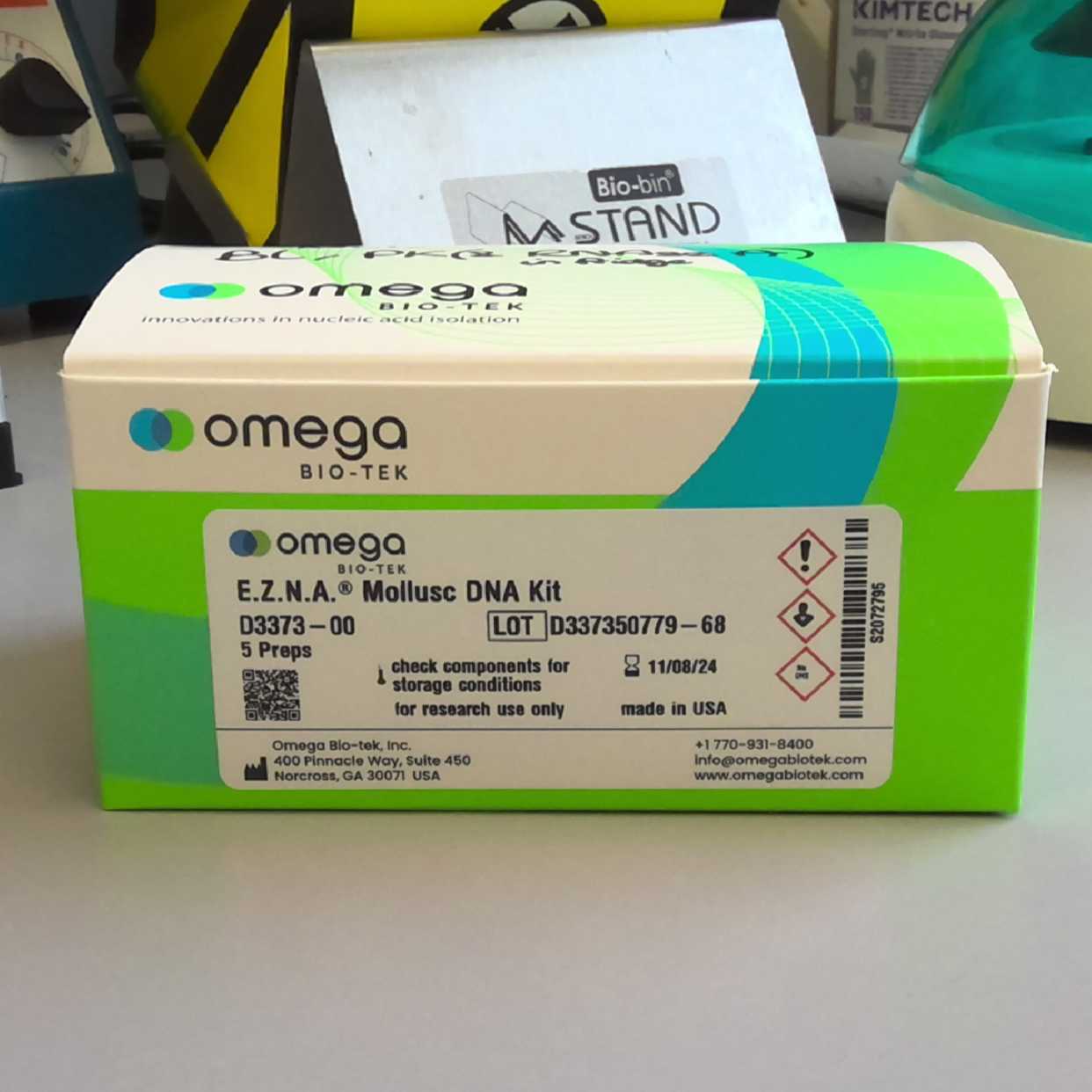
- Omega Bio-Tek EZNA® Mollusc & Insect DNA kit (Omega Bio-Tek Cat. no. D3373-00)
- Qiagen MagAttract HMW DNA extraction kit (Qiagen Cat. no. 67563)
- Sera-Mag™ magnetic carboxylate modified particles (Cat. no. GE24152105050250)
- AMPure PB beads (Pacific Biosciences Cat. no. 100-265-900)
- Buffer EB (Qiagen Cat. no. 19086)
- 100% absolute ethanol
- Chloroform:isoamyl alcohol (24:1, v/v) (Cat. no. 25666-100ML)
- Nuclease free water (Cat. no. AM9932)
- PEG 8000 (Cat. no. P5413-500-G)
- Tris-HCl (1 M stock concentration, pH 8.0)
- EDTA (0.1 M stock concentration, pH 8.0)
- NaCl (5 M stock concentration) (Cat. no. 59222C-500ML)
- Tween-20
- Dry ice
- Terumo™ 3-Part 50mL Luer Lock Syringes (Cat. no. 15349067)
- Merck Millex™-HP Sterile Polyethersulfone Syringe Filter Units, 0.45 μm (Cat. no. 16427565)
- Weighing boats (SLS Cat. no. bal1820sp)
Equipment:
- Pipettes for 0.5–1000 μL and filtered tips
- Wide-bore tips (200 μL, filtered if available)
- Diagnocine PowerMasher II tissue disruptor (Product no. 891300)
- Eppendorf ThermoMixer C (Cat. no. 5382000031)
- Eppendorf SmartBlock 2.0 mL (Cat. no. 5362000035)
- Eppendorf SmartBlock 50 mL (Cat. no. 5365000028)
- Mini centrifuge (Cat. no SS-6050)
- Eppendorf Centrifuge 5425/5425 R (Cat. no. 5405000263)
- HulaMixer Sample Mixer (Cat. no. 15920D)
- DynaMag™-2 magnetic rack (Cat. no. 12321D)
- Mettler Toledo Analytic Balance ME204 (Material No. 30029066)
- Timer
- Chemical Fume Hood
Reagent Recipes (taken directly from the Sanger Tree of Life HMW DNA Extraction: Automated Plant Organic HMW gDNA Extraction (POE) protocol):
50% PEG 8000
| Reagent | Target concentration | Molecular weight (g/mol) | Stock concentration | Input from stock (15 mL total) | |
| PEG 8000 | 50% (w/v) | 8000 | Powder | 7.5 g | |
| Nuclease-free water | - | - | - | 6 mL | |
| Incubate for 60 minutes, 75 °C at 600 rpm, routinely vortexing until fully dissolved. | |||||
| Nuclease-free water | - | - | - | Up to 15 mL | |
| Should be prepared fresh and allowed to cool before use in the SpeedBead Binding solution | |||||
10% Tween-20
| Reagent | Target concentration | Molecular weight (g/mol) | Stock concentration | Input from stock (50 mL total) | |
| Nuclease-free water | - | - | - | 44 mL | |
| Tris-HCl, pH 8.0 | 20 mM | 157.60 | 1 M | 1 mL | |
| Tween-20 | 10% (v/v) | 1,227.54 | 100% (v/v) | 5 mL | |
| Place on a tube rotator for 30 minutes, 20 rpm, ensuring Tween is dissolved. | |||||
| Store protected from the light at room temperature for up to 1 year (replace if solution becomes yellowed). | |||||
SpeedBead Wash Suspension:
| Reagent | Target concentration | Molecular weight (g/mol) | Stock concentration | Input from stock | |
| Sera-Mag™ SpeedBead stock solution, 4 °C | 0.2% (w/v) | - | 0.5% (w/v) | 800 μL | |
| Wash beads 4 times with nuclease free water before use to remove sodium azide | |||||
| Nuclease-free water | - | - | - | Up to 2.0 mL | |
| This should be prepared fresh before use in the Sera-Mag™ SpeedBead solution. | |||||
- Allow Sera-Mag™ SpeedBeads aliquot to reach room temperature (~30 minutes).
- Vortex thoroughly to resuspend the beads.
- Pipette 800 μL of Sera-Mag™ SpeedBead stock solution into a 2 mL Lo-Bind tube on a magnetic stand and wait for the beads to migrate to the magnet.
- When the supernatant is completely clear, remove and discard the supernatant from the tube without disturbing the beads.
- Add 1000 μL nuclease-free water to the tube.
- Vortex the tube to resuspend beads.
- Centrifuge briefly to remove droplets from tube lid.
- Place the tube on a magnetic stand until the supernatant is completely clear and beads are bound towards the magnet.
- Remove and discard the supernatant without disturbing beads.
- Repeat steps 5 to 9 three times.
- Add nuclease-free water up to 2 mL.
- Vortex tube to resuspend beads.
- Centrifuge briefly to remove droplets from tube lid.
- SpeedBead wash suspension can now be used for the Sera-Mag™ SpeedBead solution.
SpeedBead Binding Solution:
| Reagent | Target concentration | Molecular weight (g/mol) | Stock concentration | Input from stock (40 mL total) | |
| Tris-HCl, pH 8.0 | 10 mM | 157.60 | 1 M | 400 μL | |
| EDTA, pH 8.0 | 1 mM | 292.24 | 0.1 M | 400 μL | |
| NaCl | 1.6 M | 58.44 | 5 M | 12.8 mL | |
| Tween-20 | 0.05% (v/v) | 1,227.54 | 10% (v/v) | 200 μL | |
| PEG 8000 | 18% (w/v) | 8000 | 50 % (w/v) | 14.4 mL | |
| Nuclease-free water | - | - | - | up to 40 mL | |
| Filter sterilise through a 0.45 μM filter into a fresh 50 mL falcon. SpeedBead binding solution should be prepared fresh before use in the Sera-Mag™ SpeedBead solution. | |||||
Ensure that the exact volume of 50% PEG 8000 is added, as this is crucial for gDNA binding.
Sera-Mag™ SpeedBead Solution:
| A | B | C | D | E | |
| SpeedBead Binding Solution | - | - | - | 38 mL | |
| SpeedBead Wash Suspension | 0.01% (v/v) | - | 0.2% (v/v) | 2 mL | |
| Store at 4 °C in the dark for up to 3 months. | |||||
40 mL of Sera-Mag™ SpeedBead solution is enough for 50 - 65 samples.
Protocol PDF:  Sanger Tree of Life HMW DNA Extraction_ Manual Modified Omega Bio-Tek E.Z.N.A.® v2.pdf319KB
Sanger Tree of Life HMW DNA Extraction_ Manual Modified Omega Bio-Tek E.Z.N.A.® v2.pdf319KB Safety warnings
- The operator must wear a lab coat, powder-free nitrile gloves and safety specs to perform the laboratory procedures in this protocol. Cotton glove liners are strongly recommended when handling the samples on dry ice.
- Eye protection and silver shield/chemical resistant gloves should be worn when handling chloroform, with all handling performed in a chemical fume hood.
- Waste needs to be collected in a suitable container (e.g. plastic screw-top jar or Biobin) and disposed of in accordance with local regulations.
- Liquid waste needs to be collected in a suitable container (e.g. glass screw-top jar) and disposed of in accordance with local regulations.
Before start
- Set a heat block to 60 °C (non-jellyfish) or 25 °C (jellyfish) depending upon sample type.
- Ensure that an adequate amount of Sera-Mag™ SpeedBead solution has been prepared prior to initiating the protocol. A volume of 600 to 800 μL is needed per sample, and requires 50% PEG 8000, 10% Tween-20, SpeedBead wash suspension and SpeedBead binding solution to be prepared prior to initiating the protocol (details in the Materials section).
- Remove the prepared Sera-Mag™ SpeedBead solution from the fridge 30 minutes before starting the extraction step so they can be brought to room temperature.
- Remove the AMPure PB beads from the fridge 30 minutes before starting the 0.45X SPRI step to allow them to be brought to room temperature.
Sample Lysis & Precipitation
Set heat block to the required temperature for the samples being processed:
For non-jellyfish samples, set a heat block to 60 °C.
For jellyfish samples, set a heat block to 25 °C.
Prepare tubes for the samples being processed depending upon the method of disruption:
For samples that require powermashing, label 1.5 mL BioMasher tubes for each sample being processed. Save the BioMasher pestles for later use in their sterile packet, also labelling them for each sample.
For samples that have been cryogenically disrupted (e.g. via bead beating), label 2 mL microcentrifuge tubes for each sample being processed.
Transfer the samples into their corresponding 1.5 mL BioMasher tube or 2 mL microcentrifuge tube.
Add 350 μL of ML1 buffer to all samples.
The volume of Proteinase K solution to be added is dependent on the sample type being processed:
For non-jellyfish samples, add 25 μL Proteinase K solution
For jellyfish samples, add 10 μL Proteinase K solution.
Homogenise samples within the lysis buffer according to sample type and disruption method:
For non-jellyfish samples which have been cryogenically disrupted, pipette mix samples gently with a wide-bore pipette tip 10 times to homogenise.
For non-jellyfish samples which require powermashing, use the BioMasher pestle and Diagnocine PowerMasher II tissue disruptor to disrupt the sample within the buffer, following Sanger Tree of Life Sample Homogenisation: PowerMash protocol.
For jellyfish samples, use the BioMasher pestle to manually disrupt the sample within the buffer. Make sure to retain these pestles for further sample disruption at a later stage by returning them to their labelled sterile packets.
Following disruption of samples, incubate for the required time and temperature according to sample type:
Incubate non-jellyfish samples for at least 30 minutes at 60 °C. If samples are not completely solubilised after 30 minutes, allow for a longer incubation (45 minutes to 1 hour).
Incubate jellyfish samples for 15 minutes at 25 °C.
Halfway through this incubation, return to the samples and manually disrupt any tissue using the retained BioMasher pestles.
After incubation, transfer any samples in 1.5 mL BioMasher tubes to fresh 2 mL microcentrifuge tubes using a wide-bore pipette tip – label these 2 mL microcentrifuge tubes for each sample.
Samples that are already in 2 mL microcentrifuge tubes do not need to be transferred into new tubes.
Transfer samples to the fume hood for chloroform separation, then add 350 μL of chloroform:isoamyl (24:1) to each sample.
Transfer samples to the HulaMixer™, rotating at 8 rpm for 5 minutes.
Centrifuge samples at 10,000 g for 2 minutes at room temperature. The samples will then have three distinct phases as seen below in Figure 1: an upper aqueous phase containing the gDNA, a milky interphase containing inhibitors and contaminants, and a lower organic phase containing denatured proteins.
Centrifuged sample with three distinct phases.
Return samples to the fume hood in order to transfer the upper aqueous phase to a clean 2 mL microcentrifuge tube using a wide-bore pipette tip. Avoid transferring the milky interphase containing contaminants and inhibitors.
Add 1 X volume of BL buffer to each sample. For example, if 400 μL aqueous upper phase was recovered in Step 12, add 400 μL of BL buffer.
The volume of RNase A to be added is dependent on the sample type being processed:
For non-jellyfish samples, add 10 μL RNase A to each sample.
Pipette mix samples 15 times with a wide-bore pipette tip.
For jellyfish samples, add 5 μL RNase A to each sample.
Pipette mix samples 15 times with a wide-bore pipette tip.
Incubate samples for the required time and temperature according to sample type:
For non-jellyfish samples, incubate for 10 minutes at 70 °C.
For jellyfish samples, incubate for 10 minutes at 25 °C.
Extraction
Add 600 to 800 μL Sera-Mag™ SpeedBead solution to each sample, depending upon the volume remaining in the 2 mL microcentrifuge tube - the Sera-Mag™ SpeedBead solution should be vortexed before use to ensure that the beads are resuspended.
Invert the sample tubes 10 to 20 times to ensure that beads are suspended in the lysate, then allow at least 5 minutes for binding.
Briefly centrifuge the sample in a mini-centrifuge to collect at the bottom of the tubes.
Place the tubes on the magnetic rack and allow 2–5 minutes for the beads to migrate. Be aware that more viscous samples will take longer. Remove the supernatant and discard.
Add 1 mL 80% ethanol opposite the bead pellet, incubate for 30 seconds, then aspirate and discard.
Repeat for a total of two washes.
Remove the tubes from the magnetic rack and add 200 μL Buffer AE directly to the beads. Mix 15 times using a wide-bore pipette tip in order to dislodge the beads from the side of the tube.
Incubate samples for 15 minutes at room temperature, with a gentle mix halfway through and again at the end.
Briefly centrifuge the samples in a mini-centrifuge and place on a magnetic rack, allowing 2–5 minutes for bead migration.
Using a 200 μL wide-bore pipette tip, carefully transfer the supernatants containing purified gDNA to fresh 1.5 mL microcentrifuge tubes.
Remove samples from the magnetic rack and add 200 μL Buffer AE directly to the bead pellet. Incubate samples on the heat block for 3 minutes at 25 °C, shaking at 1,000 rpm.
Centrifuge samples briefly in a mini-centrifuge and place on a magnetic rack, allowing 2–5 minutes for bead migration.
Using a wide-bore pipette tip, carefully transfer the supernatants containing purified gDNA to the same 1.5 mL microcentrifuge tubes as in Step 25. Pipette-mix 5 to 10 times to homogenise the gDNA. The total eluate volume for each sample will now be 400 μL.
0.45X SPRI
Add 0.45X AMPure PB beads to the eluates (180 μL of beads for 400 μL eluate). Mix 15 times with a wide-bore pipette tip.
Incubate samples for 5 minutes at room temperature.
Centrifuge samples in a mini-centrifuge for 1–2 seconds, then place on a magnetic rack. Allow 2–5 minutes for the beads to migrate.
Remove and discard the supernatant.
Keep samples on the magnetic rack and add 1 mL 80% ethanol. Incubate for 30 seconds and then remove and discard.
Repeat for a total of two 80% ethanol washes.
Remove samples from the magnetic rack and add 130 μL EB Buffer. Pipette-mix 15 times with a wide-bore pipette.
Incubate samples on a heat block for 30 minutes at 37 °C, shaking at 350 rpm.
Return samples to the magnetic rack and transfer the supernatant containing the purified and SPRI'ed gDNA into a new 1.5 mL microcentrifuge tube.
Incubate the DNA at room temperature overnight and perform the required QC the following morning.
Store the DNA at 4 °C.
Protocol references
Jackson, B. and Howard, C. (2024) Sanger Tree of Life HMW DNA Extraction: Automated Plant Organic HMW gDNA Extraction (POE)