Collection Citation: Jocelyn Y. Kishi, Sylvain W. Lapan, Brian J Beliveau, Emma R. West, Allen Zhu, Hiroshi M. Sasaki, Sinem Saka, Yu Wang, Constance L Cepko, Peng Yin 2020. SABER-FISH – Signal amplification for multiplexed fluorescence in situ hybridization assays. protocols.io https://dx.doi.org/
Manuscript citation:
Kishi, J.Y., Lapan, S.W., Beliveau, B.J. et al. SABER amplifies FISH: enhanced multiplexed imaging of RNA and DNA in cells and tissues. Nat Methods 16, 533–544 (2019). https://doi.org/10.1038/s41592-019-0404-0
License: This is an open access collection distributed under the terms of the Creative Commons Attribution License, which permits unrestricted use, distribution, and reproduction in any medium, provided the original author and source are credited
Protocol status: Working
Created: July 06, 2020
Last Modified: August 21, 2020
Collection Integer ID: 38918
Keywords: multiplexed fluorescence, signal amplification for multiplexed fluorescence, multiplexing with saber, fluorescence imaging, iteritave rounds of fluorescence imaging, fish multiplexed signal amplification, signal amplification in fluorescence, short secondary fluorescent hybridization, saber technology the signal amplification, situ fluorescence, fluorophores on complementary imager, situ stainings for rna, hybridization of new imager strand, fluorescence, saber technology, expensive microscopy setup, new imager strand, separated fluorophore, saber, tissue sample, concatemerized probe, staining experiment, complementary to dna, cell basis for quantitative analysis, imager exchange, rapid tissue mapping, old imager strand, fluorescent, exchange imaging, multiplexed signal amplification, situ hybridization, vitro reaction, probe binding, fish signal, cell segmentation, transcriptomic loci, underlying probe binding, cell segmentation with puncta counting
Abstract
The SABER Technology
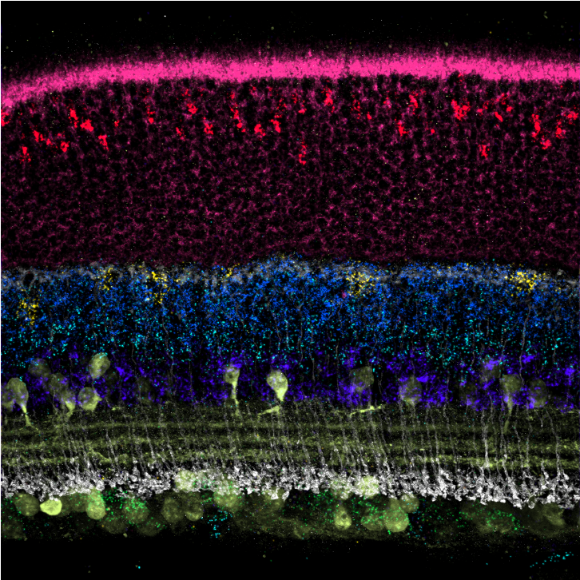
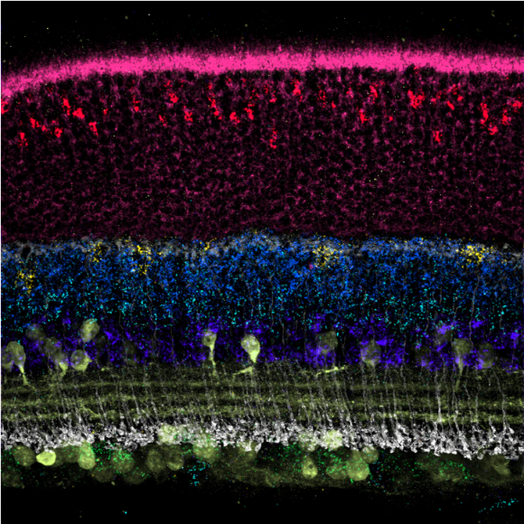
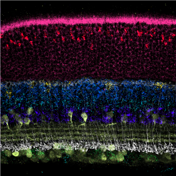
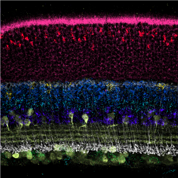
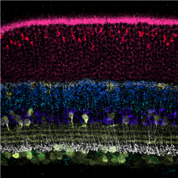
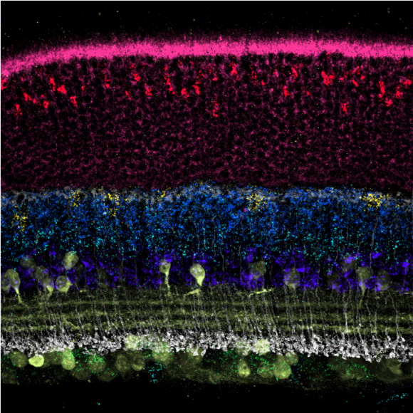
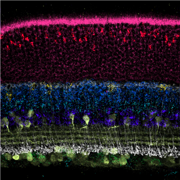
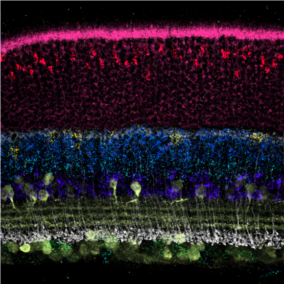
The Signal Amplification By Exchange Reaction (SABER) method is used for amplifying signal from multiplexed in situ fluorescence staining experiments. Developed by the Yin and Cepko labs at Harvard University and the Wyss Institute, the technique uses Primer Exchange Reactions (PERs) to generate three-letter (A, T, C) concatemeric sequences in bulk in vitro reactions. These concatemers can then be in situ hybridized to fixed cells and tissues and act as scaffolds that localize fluorescent 'imager' strands. The method can further be paired with DNA-Exchange Imaging (DEI) to increase multiplexing via rapid stripping of old imager strands and hybridization of new imager strands (imager exchange) and/or cell segmentation with puncta counting on a per cell basis for quantitative analyses.
See the following references and resources below for further information. SABER provides a scalable and cost-effective way to amplify multiplexed in situ stainings for RNA/DNA (SABER-FISH) and protein targets (Immuno-SABER, website, collection on protocols.io).
SABER-FISH
Multiplexed signal amplification enables rapid tissue mapping by increasing the number of targets that can be visualized per sample and reducing the exposure times required to see signals. Signal amplification in fluorescence in situ hybridization (FISH) assays can also increase sensitivity, potentially allowing smaller genomic and transcriptomic loci to be imaged, fewer probes to be applied, and/or less expensive microscopy setups to be utilized. SABER-FISH uses in vitro Primer Exchange Reactions (PERs) to synthesize long repetitive 'concatemer' sequences onto the 3' end of probes designed to be complementary to DNA and RNA targets of interest. After concatemer extension, probes are in situ hybridized to fixed cell and tissue samples, followed by a short secondary fluorescent hybridization that binds 20nt fluorophore-conjugated strands ('imagers') to the concatemers.
Multiplexing with SABER-FISH is achieved through the use of orthogonal concatemer sequences appended to probe sets, which can be read out to spectrally separated fluorophores on complementary imagers. Imager sequences can also be stripped from one set of concatemers without disrupting the underlying probe binding, which allows new sets of imagers targeting different loci to be imaged in iteritave rounds of fluorescence imaging. This process of stripping and hybridization is referred to as DNA-Exchange Imaging (DEI). SABER-FISH signal can further be enhanced with a branching strategy, where multiple rounds of concatemerized probe binding create branched structures in situ. Below are a number of resources intended to help adoption of the technology, including references, animations, and protocols.
Pardue, M. L. & Gall, J. G. Molecular hybridization of radioactive DNA to the DNA of cytological preparations. Proc. Natl Acad. Sci. USA 64, 600–604 (1969).
Riegel, M. Human molecular cytogenetics: from cells to nucleotides. Genet. Mol. Biol. 37, 194–209 (2014).
Bolzer, A. et al. Three-dimensional maps of all chromosomes in human male fibroblast nuclei and prometaphase rosettes. PLoS Biol. 3, 0826–0842 (2005).
Femino, A. M., Fay, F. S., Fogarty, K. & Singer, R. H. Visualization of single RNA transcripts in situ. Science 280, 585–590 (1998).
Raj, A., van den Bogaard, P., Rifkin, S. A., van Oudenaarden, A. & Tyagi, S. Imaging individual mRNA molecules using multiple singly labeled probes. Nat. Methods 5, 877–879 (2008).
Schröck, E. et al. Multicolor spectral karyotyping of human chromosomes. Science 273, 494–497 (1996).
Lubeck, E. & Cai, L. Single-cell systems biology by super-resolution imaging and combinatorial labeling. Nat. Methods 9, 743–748 (2012).
Jungmann, R. et al. Multiplexed 3D cellular super-resolution imaging with DNA-PAINT and Exchange-PAINT. Nat. Methods 11, 313–318 (2014).
Schueder, F. et al. Universal super-resolution multiplexing by DNA exchange. Angew. Chem. Int. Ed. Engl. 56, 4052–4055 (2017).
Wang, Y. et al. Rapid sequential in situ multiplexing with DNA exchange imaging in neuronal cells and tissues. Nano Lett. 17, 6131–6139 (2017).
Wang, S. et al. Spatial organization of chromatin domains and compartments in single chromosomes. Science 353, 598–602 (2016).
Bintu, B. et al. Super-resolution chromatin tracing reveals domains and cooperative interactions in single cells. Science 362, eaau1783 (2018).
Codeluppi, S. et al. Spatial organization of the somatosensory cortex revealed by osmFISH. Nat. Methods 15, 932–935 (2018).
Lubeck, E., Coskun, A. F., Zhiyentayev, T., Ahmad, M. & Cai, L. Single-cell in situ RNA profiling by sequential hybridization. Nat. Methods 11, 360–361 (2014).
Chen, K. H., Boettiger, A. N., Moffitt, J. R., Wang, S. & Zhuang, X. Spatially resolved, highly multiplexed RNA profiling in single cells. Science 348, aaa6090 (2015).
Levesque, M. J. & Raj, A. Single-chromosome transcriptional profiling reveals chromosomal gene expression regulation. Nat. Methods 10, 246–248 (2013).
Shah, S. et al. Dynamics and spatial genomics of the nascent transcriptome by intron seqFISH. Cell 174, 363–376 (2018).
Kerstens, H. M., Poddighe, P. J. & Hanselaar, A. G. A novel in situ hybridization signal amplification method based on the deposition of biotinylated tyramine. J. Histochem. Cytochem. 43, 347–352 (1995).
Player, A. N., Shen, S. P., Kenny, D., Antao, V. P. & Kolberg, J. A. Single-copy gene detection using branched DNA (bDNA) in situ hybridization. J. Histochem. Cytochem. 49, 603–611 (2001).
20. Wang, F. et al. RNAscope: a novel in situ RNA analysis platform for formalin-fixed, paraffin-embedded tissues. J. Mol. Diagn. 14, 22–29 (2012).
Beliveau, B. J. et al. Single-molecule super-resolution imaging of chromosomes and in situ haplotype visualization using Oligopaint FISH probes. Nat. Commun. 6, 7147 (2015).
Lizardi, P. et al. Mutation detection and single-molecule counting using isothermal rolling-circle amplification. Nat. Genet. 19, 225–232 (1998).
Dirks, R. M. & Pierce, N. A. Triggered amplification by hybridization chain reaction. Proc. Natl Acad. Sci. USA 101, 15275–15278 (2004).
Choi, H. M. T. et al. Programmable in situ amplification for multiplexed imaging of mRNA expression. Nat. Biotechnol. 28, 1208–1212 (2010).
Choi, H. M., Beck, V. A. & Pierce, N. A. Next-generation in situ hybridization chain reaction: higher gain, lower cost, greater durability. ACS Nano 8, 4284–4294 (2014).
Shah, S. et al. Single-molecule RNA detection at depth via hybridization chain reaction and tissue hydrogel embedding and clearing. Development 92, 2862–2867 (2016).
Rouhanifard, S. H. et al. ClampFISH detects individual nucleic acid molecules using click chemistry–based amplification. Nat. Biotechnol. 37, 84–89 (2018).
Nagendran, M., Riordan, D. P., Harbury, P. B. & Desai, T. J. Automated cell-type classification in intact tissues by single-cell molecular profiling. eLife 7, e30510 (2018).
Wang, X. et al. Three-dimensional intact-tissue sequencing of single-cell transcriptional states. Science 361, eaat5691 (2018).
Kishi, J. Y., Schaus, T. E., Gopalkrishnan, N., Xuan, F. & Yin, P. Programmable autonomous synthesis of single-stranded DNA. Nat. Chem. 10, 155–164 (2018).
Beliveau, B. J. et al. Versatile design and synthesis platform for visualizing genomes with Oligopaint FISH probes. Proc. Natl Acad. Sci. USA 109, 21301–21306 (2012).
Lee, C. S., Davis, R. W. & Davidson, N. A physical study by electron microscopy of the terminally repetitious, circularly permuted DNA from the coliphage particles of Escherichia coli 15. J. Mol. Biol. 48, 1–22 (1970).
Beliveau, J. et al. OligoMiner provides a rapid, flexible environment for the design of genome-scale oligonucleotide in situ hybridization probes. Proc. Natl Acad. Sci. USA 115, E2183–E2192 (2018).
Xu, Q., Schlabach, M. R., Hannon, G. J. & Elledge, S. J. Design of 240,000 orthogonal 25mer DNA barcode probes. Proc. Natl Acad. Sci. USA 106, 2289–2294 (2009).
Dirks, R. M. & Pierce, N. A. A partition function algorithm for nucleic acid secondary structure including pseudoknots. J. Comput. Chem. 24, 1664–1677 (2003).
Dirks, R. M. & Pierce, N. A. An algorithm for computing nucleic acid base-pairing probabilities including pseudoknots. J. Comput. Chem. 25, 1295–1304 (2004).
Dirks, R. M., Bois, J. S., Schaeffer, J. M., Winfree, E. & Pierce, N. A. Thermodynamic analysis of interacting nucleic acid strands. SIAM Rev. 49, 65–88 (2007).
Carpenter, A. E. et al. CellProfiler: image analysis software for identifying and quantifying cell phenotypes. Genome Biol. 7, R100 (2006).
Macosko, E. Z. et al. Highly parallel genome-wide expression profiling of individual cells using nanoliter droplets. Cell 161, 1202–1214 (2015).
Shekhar, K. et al. Comprehensive classification of retinal bipolar neurons bysingle-cell transcriptomics. Cell 166, 1308–1323 (2016).
Mosaliganti, K. R., Noche, R. R., Xiong, F., Swinburne, I. A. & Megason, S. G. ACME: automated cell morphology extractor for comprehensive reconstruction of cell membranes. PLoS Comput. Biol. 8, e1002780 (2012).
Solovei, I. et al. Nuclear architecture of rod photoreceptor cells adapts to vision in mammalian evolution. Cell 137, 356–368 (2009).
Shah, S., Lubeck, E., Zhou, W. & Cai, L. In situ transcription profiling of single cells reveals spatial organization of cells in the mouse hippocampus. Neuron 92, 342–357 (2016).
Emerson, M. M. & Cepko, C. L. Identification of a retina-specific Otx2 enhancer element active in immature developing photoreceptors. Dev. Biol. 360, 241–255 (2011).
ENCODE Project Consortium. An integrated encyclopedia of DNA elements in the human genome. Nature 489, 57–74 (2012).
Matsuda, T. & Cepko, C. L. Electroporation and RNA interference in the rodent retina in vivo and in vitro. Proc. Natl Acad. Sci. USA 101, 16–22 (2004).
Saka, S. K. et al. Highly multiplexed in situ protein imaging with signal amplification by Immuno-SABER. Nat. Biotechnol. (in the press).
Frieda, K. L. et al. Synthetic recording and in situ readout of lineage information in single cells. Nature 541, 107–111 (2017).
Yildirim, E., Sadreyev, R. I., Pinter, S. F. & Lee, J. T. X-chromosome hyperactivation in mammals via nonlinear relationships between chromatin states and transcription. Nat. Struct. Mol. Biol. 19, 56–61 (2011).
Kent, W. J. et al. The Human Genome Browser at UCSC. Genome Res. 12, 996–1006 (2002).
Quinlan, A. R. & Hall, I. M. BEDTools: a flexible suite of utilities for comparing genomic features. Bioinformatics 26, 841–842 (2010).
Langmead, B. & Salzberg, S. L. Fast gapped-read alignment with Bowtie 2. Nat. Methods 9, 357–359 (2012).
Marçais, G. & Kingsford, C. A fast, lock-free approach for efficient parallel counting of occurrences of k-mers. Bioinformatics 27, 764–770 (2011).
Casanova, M. et al. Heterochromatin reorganization during early mouse development requires a single-stranded noncoding transcript. Cell Rep. 4, 1156–1167 (2013).
Altschul, S. F., Gish, W., Miller, W., Myers, E. W. & Lipman, D. J. Basic local alignment search tool. J. Mol. Biol. 215, 403–410 (1990).
Beliveau, B. J., Apostolopoulos, N. & Wu, C. Visualizing genomes with Oligopaint FISH probes. Curr. Protoc. Mol. Biol. 2014, 14.23.1–14.23.20 (2014).
Ran, F. A. et al. Genome engineering using the CRISPR–Cas9 system. Nat. Protoc. 8, 2281–2308 (2013).
Schneider, C. A., Rasband, W. S. & Eliceiri, K. W. NIH Image to ImageJ: 25 years of image analysis. Nat. Methods 9, 671–675 (2012).
Schindelin, J. et al. Fiji: an open-source platform for biological-image analysis. Nat. Methods 9, 676–682 (2012).
McQuin, C. et al. CellProfiler 3.0: next-generation image processing for biology. PLoS Biol. 16, 1–17 (2018).
Linkert, M. et al. Metadata matters: access to image data in the real world. J. Cell Biol. 189, 777–782 (2010).
Yushkevich, P. A. et al. User-guided 3D active contour segmentation of anatomical structures: significantly improved efficiency and reliability. Neuroimage 31, 1116–1128 (2006).
Marr, D. & Hildreth, E. Theory of edge detection. Proc. R. Soc. Lond. B Biol. Sci. 207, 187–217 (1980).
Plaisier, S., Taschereau, R., Wong, J. & Graeber, T. Rank-rank hypergeometric overlap: identification of statistically significant overlap between geneexpression signatures. Nucleic Acids Res. 38, e169 (2010).
Hunter, J. D. Matplotlib: a 2D graphics environment. Comput. Sci. Eng. 9, 90–95 (2007).
Oliphant, T. E. A Guide to NumPy (Trelgol Publishing, 2006).
McKinney, W. Data structures for statistical computing in Python. in Proc. 9th Python in Science Conference (eds. van der Walt, S. & Millman, J.) 51–56 (SciPy, 2010).
Cock, P. J. A. et al. Biopython: freely available Python tools for computational molecular biology and bioinformatics. Bioinformatics 25, 1422–1423 (2009).