Oct 29, 2021
RNA Quality Control Non-Denaturing Agarose "Bleach" Gel
- Lynn Doran1
- 1Realizing Increased Photosynthetic Efficiency (RIPE)
- GEGC lab UIUC
- UIUC Long Lab


Protocol Citation: Lynn Doran 2021. RNA Quality Control Non-Denaturing Agarose "Bleach" Gel. protocols.io https://dx.doi.org/10.17504/protocols.io.bzjzp4p6
Manuscript citation:
Aranda, P. S., LaJoie, D. M., & Jorcyk, C. L. (2012). Bleach gel: a simple agarose gel for analyzing RNA quality. Electrophoresis, 33(2), 366–369. https://doi.org/10.1002/elps.201100335
License: This is an open access protocol distributed under the terms of the Creative Commons Attribution License, which permits unrestricted use, distribution, and reproduction in any medium, provided the original author and source are credited
Protocol status: Working
We use this protocol and it’s working
Created: October 29, 2021
Last Modified: July 11, 2023
Protocol Integer ID: 54617
Keywords: RNA, QC, RNA Quality Control, Agarose Gel Electrophoresis, Bleach Agarose Gel, quality of rna, rna quality, rna integrity, rna fragment, rna, denaturing gel electrophoresis system, gel electrophoresis system, denaturing agarose, true fragment size determination, gel, bleach
Funders Acknowledgements:
Realizing Increased Photosynthetic Efficiency (RIPE) that is funded by the Bill & Melinda Gates Foundation, Foundation for Food and Agriculture Research, and the U.K. Foreign, Commonwealth & Development Office
Grant ID: OPP1172157
Abstract
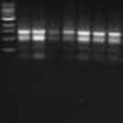
To evaluate the quality of RNA without the use of toxic chemicals, samples are run in a non-denaturing agarose "bleach" gel and product segregation is used to estimate RNA integrity.
Be aware that in a non-denaturing gel, the RNA will not segregate strictly on particle size due to secondary structures of the molecules. This method is for an estimation of quality only and the location of the banding in relation to a base pair ladder does not allow confident determination of the size of the RNA fragment. If true fragment size determinations are required, RNA quality should be evaluated on a denaturing gel electrophoresis system.
Materials
Reagents
- TAE, 0.5X (the concentration of TAE buffer will affect the voltage required to run the gel at the same speed)
- Sybrsafe, Invitrogen™ SYBR™ Safe DNA Gel Stain
- 6X Loading Dye, Thermo Scientific™ DNA Gel Loading Dye (6X)
- Bleach, 6% sodium hypochlorite
- 1 kb ladder, Thermo Scientific™ GeneRuler 1 kb DNA Ladder
- Dry ice
- Regular ice
Materials
- Gel electrophoresis tank, Bio-Rad Sub-Cell GT Cell
- Gel casting system, Bio-Rad Sub-Cell GT Gel Caster#1704412
- Gel combs, Bio-Rad 20-Well Comb#1704447
- Microwave
- Graduated cylinder, 100 mL
- Graduated cylinder, 1000 mL
- Erlenmeyer flask
- Analytical balance
- Weigh paper
- Kim wipe
- Hot pad
- Micropipette, 100-1000 ul
- Pipette tips, 100-1000 ul
- Micropipette 1-10 ul
- Pipette tips, 1-10 ul
- Ice buckets
- PCR tubes, Fisherbrand™ 0.2mL PCR Tubes
- UV Gel Imaging System, Bio-RAD Gel Doc XR and Quantity One software
Safety warnings
- Sybrsafe binds to nucleic acids. Proper PPE is recommended.
- Heating household bleach may release chlorine gas. Use caution and if preparing large volumes of agarose bleach gels, consider moving the melting and gel setting step to a chemical fume hood.
Before start
RNA degradation due to RNases is an ever present environmental risk to the samples. Best lab practices regarding RNA handling including using RNase free plasticware, autoclaving tips and materials, cleaning the lab workspace, and wearing appropriate and clean lab coats and gloves should be followed at all times during this protocol.
Empty and rinse the gel electrophoresis tank with distilled water at least three times.
Dry the tank with paper towels.
Treat the gel electrophoresis tank, the gel casting system, the gel tray, and the gel comb with RNase Away. Set up the gel tray and the gel casting system.
Prepare fresh 0.5X TAE.
Note
Measure the appropriate volume of 0.5X TAE for the gel cast size you have selected.
- 100 mL for a 10 x 15 cm tray.
Weigh molecular grade agarose to 1% of the volume of 0.5X TAE for the gel cast size you have selected.
- 1 g agarose for 100 mL of 0.5X TAE for a 10 x 15 cm tray.
Pour the 0.5X TAE and the weighed agarose into an erlenmeyer flask.
Measure 1% bleach for the volume of 0.5X TAE for the gel cast size you have selected
- 1 mL 6% sodium hypochlorite bleach for 100 mL of 0.5X TAE for a 10 x 15 cm tray.
Swirl the flask containing the 0.5X TAE, agarose, and bleach. Allow it to incubate atRoom temperature for 00:05:00
5m
Place a kim wipe in the top of the flask to reduce evaporation and microwave on high until the solution is boiling, the agarose has dissolved, and no floaties or striations are visible in the solution.
Remove the kim wipe and allow the solution to cool to a temperature where the very top of the flask can be handled without hot pads. Gently swirling the flask will help speed cooling but be careful to avoid bubble formation.
Note
If the solution is poured into the gel cast too hot, it can cause melting or warping of the plastic. If the solution is poured too cold into the gel cast, it may begin to set and will not form a smooth glassy surfaced gel.
Optional: Level the gel cast before pouring the gel if images will be used in publications.
Gently pour the agarose solution into the gel cast. Try to minimize bubble formation.
Pipette the appropriate volume of 1:20,000 Sybrsafe DNA stain for the gel cast size you have selected directly into the agarose solution.
- 5 µL Sybrsafe DNA Stain for 100 mL of 0.5X TAE for a 10 x 15 cm tray.
Note
Sybr Safe Manufacturer recommendation is 1:10,000 dilution. Clear visualization of nucleic acids, except at very low concentrations, is possible at 1:20,000 dilution.
Mix the Sybrsafe DNA Stain into the liquid agarose solution using the pipette tip. Try to evenly distribute the Sybrsafe throughout the gel.
Pop or push any bubbles that form to the sides of the gel cast using the pipette tip.
Insert the gel comb and gently rock it side to side to release any air bubbles trapped under the comb.
Allow the gel to set, approximately 20 minutes. The gel will turn from clear to milky and from liquid to semi-solid when it is set.
Remove the gel comb.
Release the gel tray from the gel casting system and place the gel and gel tray in the gel electrophoresis tank.
Fill the gel electrophoresis tank with fresh 0.5X TAE until the gel is covered by a few centimeters and no air bubbles remain in the wells of the gel where the comb was removed.
Prepare your RNA samples by adding 5 µL of RNA to 1 µL of 6X loading dye in a PCR tube. Keep your prepared samples On ice .
Note
Keep your RNA samples on dry ice whenever you are not actively working with them to inhibit RNases. RNases have been shown to retain activity even at temperatures as low as -20 °C
Load 6 µL of a prepared 1 kb nucleic acid ladder in the first well.
Load 6 µL of your prepared RNA with loading dye into subsequent wells.
Run the gel at 200 V until desired separation is achieved.
- For a 100 mL gel in 0.5X TAE, approximately 45-60 min.
Note
The addition of bleach affects the ionic buffering capacity of the running buffer. Additional voltage above what is typically used for a standard agarose gel may be needed when using a bleach agarose gel.
Time will vary depending on strength of TAE, size of gel, percent agarose, voltage selected, and amount of RNA fragment separation is required. If parameters outside this protocol are used, visually check separation of gel using the visible loading dye until desired separation is achieved.
If after visualization on the imaging system desired separation is not achieved, the gel can be returned to its tray, placed back in the gel electrophoresis system, and ran for a longer time period.
Remove the gel from the gel electrophoresis system, slide the gel out of its tray on to the imaging tray of the Bio-Rad Gel Doc XR Imager (or equivalent).
Visualize the gel under UV imaging using Quantity One software.