Mar 29, 2018
RNA Imaging with MERFISH - Sample Preparation and Staining
- Jeffrey R. Moffitt1,
- Xiaowei Zhuang1
- 1Harvard University, Cambridge, Massachusetts, 02138 USA
- Human Cell Atlas Method Development Community
- Neurodegeneration Method Development Community


External link: https://www.ncbi.nlm.nih.gov/pubmed/27241748
Protocol Citation: Jeffrey R. Moffitt, Xiaowei Zhuang 2018. RNA Imaging with MERFISH - Sample Preparation and Staining. protocols.io https://dx.doi.org/10.17504/protocols.io.merc3d6
Manuscript citation:
Jeffrey R. Moffitt, Xiaowei Zhuang. RNA Imaging with Multiplexed Error Robust Fluorescence in situ Hybridization. Methods Enzymol. 2016; 572: 1–49. doi: 10.1016/bs.mie.2016.03.020
License: This is an open access protocol distributed under the terms of the Creative Commons Attribution License, which permits unrestricted use, distribution, and reproduction in any medium, provided the original author and source are credited
Protocol status: Working
We use this protocol and it's working
Created: December 27, 2017
Last Modified: March 29, 2018
Protocol Integer ID: 9393
Keywords: In situ hybridization; RNA; Single cells; Single molecules; Single-molecule imaging; Transcriptomics, rna imaging with merfish, merfish staining, rnase contamination, rna imaging, rnase, merfish, rna, smfish, staining of sample, sample preparation, staining
Abstract
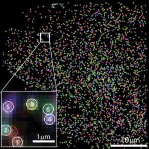
The preparation and staining of samples for MERFISH follows closely the typical protocols used for smFISH (Raj et al., 2008). However, there are a few places in which we have modified these protocols to optimize MERFISH staining. Again, RNase contamination can destroy samples, so care should be taken to work in an RNase-free environment.
Attachments

merFISH.pdf
1.4MB
Guidelines
The protocol workflow is as follows:
1. Fixation and Permeabilization of Cells (Steps 1-5)
The first step in most FISH protocols is to fix the cells and then to permeabilize the membrane using either an overnight incubation in 70% ethanol or a brief exposure to surfactant. We prefer the latter approach since this protocol requires less time; however, we have had reasonable performance with both approaches.
2. Hybridization of Encoding Probes (Steps 5-23)
This protocol will require the following reagents and buffers:
1. Fixation buffer (4% PFA in 1XPBS)
- 1 mL 32% paraformaldehyde (PFA; Electron MicroscopySciences; 15714)
- 0.8 mL 10X Phosphate Buffered Saline (PBS; Ambion; AM9625)
- 6.2 mL nuclease-free water
- Store at −20°C in single use aliquots
2. Permeabilization buffer (0.5% v/v Triton X-100 in 1XPBS)
- 5 mL 10X PBS
- 45 mL nuclease-free water
- 250 μL Triton X-100 (Sigma; T8787)
- Store at room temperature
3. 2X SSC buffer
- 5 mL 10X PBS
- 45 mL nuclease-free water
- 250 μL Triton X-100 (Sigma; T8787)
- Store at room temperature
4. 1X PBS buffer
- 5 mL 10X PBS
- 45 mL nuclease-free water
- Store at room temperature
5. 20% w/v Dextran sulfate
- In an RNase-free bottle (such as an empty nuclease-free water bottle), mix the following
- 100 g dextran sulfate (Fisher Scientific; bp1585100)
- 300 mL nuclease-free water
- Stir with a stir bar cleaned with D/RNaseFree while heating gently on a hot plate (37 °C) until dissolved
- Add nuclease-free water to produce a 500 mL total volume
- Store at room temperature
6. Encoding probe hybridization buffer
- 300 μL 100% deionized formamide (Ambion; AM9342)
- 500 μL 20% w/v dextran sulfate (above)
- 100 μL 20X SSC
- 90 μL nuclease-free water
- 1 mg yeast tRNA (Life Technologies; 15401-011)
- 10 μL 200 mM Vandyl Ribonucleoside Complex (VRC; New England Biolabs; S1402S)
- Store at −20°C
7. Encoding probe wash buffer
- 15 mL 100% deionized formamide
- 5 mL 20X SSC
- 29.5 mL nuclease-free water
- 0.5 mL 200 mM VRC
- Store at 4°C in the dark and use within a few days
8. 40-mm diameter, No. 1.5 coverslips (Bioptechs; 40-1313-0319)
9. 60-mm diameter, Cell culture petri dish (Corning; 353802)
10. 1” x 3” microscope slides
11. Parafilm (VWR; 52858-076)
12. Orange FluoSphere carboxylate-modified beads, 0.1-um-diameter (ThermoScientific; F-8800)
This protocol requires the following equipment:
1. Equipment for cell culture
2. 37°C incubator
3. 47°C incubator
4. Laboratory rocker
Materials
MATERIALS
Please see Before starting section for required materials
Troubleshooting
Fixation and Permeabilization of Cells
Culture cells on a suitable coverglass. The culture substrate for cells should be the same coverglass that will be used for imaging. We utilize 40-mm diameter No. 1.5 coverslips from Bioptechs since these coverslips fit within the flow chamber that we use for imaging. We typically culture cells in sterile, 60-mm-diameter petri dishes with one coverslip placed in the bottom of each dish.
Fix cells. Allow the fixation buffer described in the Before Starting section to warm to room temperature. Aspirate culture medium from cells then gently decant 2-3 mL of fixation buffer into the petri dish that holds the coverslip. Cover and rock gently for 15 minutes at room temperature.
00:15:00 Rocking gently at room temperature
Aspirate the fixation buffer and gently decant 2-3 mL of 1X PBS into the petri dish to wash the cells. Immediately aspirate this buffer. Repeat this 1X PBS wash for a total of three times.
3 µL 1X PBS
Permeabilize the cells. Aspirate any residual 1X PBS from the petri dish and decant 2-3 mL permeabilization buffer into the petri dish. Incubate the cells at room temperature with gentle rocking for 2 minutes.
3 µL Permeabilization buffer
00:02:00 Incubation at RT while gentle rocking
Aspirate out the permeabilization buffer and wash the cells three times with 1X PBS as described in Step 3.
Hybridization of Encoding Probes
Exchange buffer on cells. Aspirate any residual 1X PBS, and gently decant 2-3 mL of room temperature encoding probe wash buffer into each petri dish. Rock the sample at room temperature for 5 minutes.
3 µL Probe wash buffer
00:05:00 Rocking at room temperature
Note
This step insures that any residual buffer left on the coverslip will not significantly dilute the formamide in the hybridization buffer in the next step.
Add hybridization buffer with probes. Place a layer of fresh parafilm on a 1” x 3” microscope slide. Dilute the encoding probes made in the Probe Construction protocol into Encoding Probe Hybridization Buffer to the desired concentration, typically somewhere between 10 and 200 μM depending on the number of unique encoding probes in the probe set.
Add 30 μL of this probe solution to the parafilm surface.
30 µL Probe solution
Aspirate any residual encoding probe wash buffer from the coverslip and gently place the coverslip, cell-side-down, onto the droplet of encoding probes. Press gently to create a thin layer of encoding probe.
Note
The probe concentration needs to be optimized for each probe set: Higher probe concentrations produce brighter spots but a higher level of non-specifically bound probe background. We recommend titrating the concentration of probe across a ~40-fold concentration range (5 – 200 μM) to identify the concentration that produces the clearest signal relative to the background. We find that the optimal probe concentration only needs to be established once per probe set.
Hybridization of Encoding Probes
Incubation. Create a humidity chamber by pouring nuclease-free water into the base of an old pipette tip box.
Place the sample within this box and seal with parafilm.
Note
The sample will be sitting on the upper plastic level and should not come into contact with the water reservoir below.
Place this box into a 37 °C incubation chamber and incubate for at least 12 hours.
Note
In some cases, we find that longer incubations (~36 hours) increase the quality of staining. Again, we recommend varying the incubation time for each probe set to identify the optimal incubation time.
37 °C Incubation chamber
12:00:00 Incubation
Wash away residual encoding probes. Preheat 6 mL of encoding probe wash buffer per sample to 47 °C in a water bath.
6 µL Encoding probe wash buffer per sample
47 °C Water bath
Slowly peel the coverslip off of the microscope slide taking care not to crack the coverslip. If it appears to be stuck, immerse the assembly in a layer of encoding probe wash buffer for 5 minutes to loosen.
00:05:00 Immersion
Place the coverslip, cell-sideup, in a fresh 60-mm-diameter petri dish and add 3 mL of preheated encoding probe wash buffer.
3 µL Preheated encoding probe wash buffer
Place the petri dish in a 47-°C incubator for 30 minutes.
47 °C Incubator
00:30:00 Incubation
Aspirate the encoding probe wash buffer, add 3 mL of fresh encoding probe wash buffer, and repeat the 30-minute, 47-°C incubation.
3 µL Encoding probe wash buffer
47 °C Incubator
00:30:00 Incubation
Aspirate the wash buffer and wash the sample in 2X SSC by adding 3 mL of 2X SSC and then immediately aspirating this buffer. Repeat this SSC wash for a total of three times.
9 µL 2X SCC
Addition of fiducial beads. Prepare a 100X solution of fluorescent beads by adding 10 μL of 0.1-nm-diameter Orange Fluospheres beads to 10 mL 2X SSC. Vortex to mix. This stock solution can be stored at 4 °C in the dark for several months. Further, dilute this stock solution 1 to 100 in 2X SSC to create 3 mL of bead solution for each sample.
10 µL 0.1-nm-diameter Orange Fluospheres beads
10 µL 2X SSC
Aspirate the 2X SSC from the samples and add the bead solution to each petri dish. Gently rock at room temperature for 15 minutes.
Note
We choose orange-colored beads (561-nm excitation) because we prefer to use the red imaging channel (641-nm excitation) for our smFISH images; however, the color of these beads could be changed to suit different imaging demands. We aim to have ~20 beads per field of view; thus, if different beads are used, the concentration should be adjusted to produce a similar final density of beads on the sample.
00:15:00 Rocking at room temperature
Post-fixation. To fix the fiducial beads in place and to prevent subtle changes in the shape of the cell induced by the repeated washing that will occur in the MERFISH Imaging protocol, we perform an additional fixation of the samples. Aspirate the bead solution and add 3 mL of fixation buffer prewarmed to room temperature to each petri dish.
3 µL Fixation buffer
Gently rock the sample at room temperature for 10 minutes.
00:10:00 Gently rocking at RT
Aspirate the fixation buffer, and wash the sample three times with 2X SSC.
Note
If the sample was prepared in an RNase-free environment, it can now be stored in 2XSSC at 4°C for several days. We do not recommend prolonged storage.