Jun 12, 2026
RNA Extraction From Human Tissue Specimens for RNA-Seq
- Matthew Sapio1,
- Michael Dunkelberg1,
- Mark Weiss1,2,3,
- Geoffroy Laumet4,
- Michael Iadarola1,
- Andrew J Mannes1
- 1National Institutes of Health, Clinical Center, Department of Perioperative Medicine, Bethesda, MD 20892, USA;
- 2Department of Anatomy and Physiology, Kansas State University, Manhattan, KS 66506, USA;
- 3Midwest Institute of Comparative Stem Cell Biotechnology, Kansas State University, Manhattan, KS 66506, USA;
- 4Michigan State University, Departments of Physiology and Neuroscience, East Lansing, MI, 48823, USA
- Matthew Sapio: To whom correspondence should be addressed, [email protected];


External link: https://linktr.ee/matthew.sapio
Protocol Citation: Matthew Sapio, Michael Dunkelberg, Mark Weiss, Geoffroy Laumet, Michael Iadarola, Andrew J Mannes 2026. RNA Extraction From Human Tissue Specimens for RNA-Seq. protocols.io https://dx.doi.org/10.17504/protocols.io.4r3l21933g1y/v1
License: This is an open access protocol distributed under the terms of the Creative Commons Attribution License, which permits unrestricted use, distribution, and reproduction in any medium, provided the original author and source are credited
Protocol status: Working
We use this protocol and it's working
Created: August 30, 2025
Last Modified: June 12, 2026
Protocol Integer ID: 226067
Keywords: RNA extraction, RNA-Seq, Human tissue, Fresh frozen, rna extraction from human tissue specimen, rna extraction, extraction of rna, quality rna suitable for downstream rna, quality rna, rna, downstream rna, human tissue specimen, seq, variety of human tissue, extraction, perioperative medicine at the nih clinical center, perioperative medicine, seq this protocol, seq application, human tissue, tissue
Funders Acknowledgements:
NIH Intramural Research Program, Clinical Center
Grant ID: ZIACL090034-09, ZIACL090035-08, ZIACL0033-09
Office of Behavioral and Social Sciences Research
Grant ID: N/A
Bench to Bedside Funding, NIH Intramural Research Program
Grant ID: N/A
Disclaimer
This research was supported by the Intramural Research Program of the National Institutes of Health (NIH) Clinical Center. The contributions of the NIH author(s) are considered Works of the United States Government. The findings and conclusions presented in this paper are those of the author(s) and do not necessarily reflect the views of the NIH or the U.S. Department of Health and Human Services.
Abstract
This protocol is optimized for extraction of RNA from a variety of human tissues, and is based on previous experience summarized in the referenced publications from the Department of Perioperative Medicine at the NIH Clinical Center. These procedures will result in high-quality RNA suitable for downstream RNA-Seq applications.
Image Attribution
Thumbnail image depicts nucleic acid drawn by ChatGPT5.2
Materials
Supplies
RNeasy Mini Kit (Qiagen, Hilden, Germany; Cat No. 74104).
QIAzol lysis reagent (Qiagen; Cat No. 79306).
Chloroform.
RNse Away (Thermo Fisher Scientific, Cat. No. 7003PK) and/or RNase Zap (Thermo Fisher Scientific, Cat. No. AM9782).
RNase free filter pipette tips.
RNase free centrifuge tubes (1.5-2 mL).
Homogenization tubes for rotor-stator homogenization (2 mL tubes with gently sloping or flat conical bottom).
Homogenization tubes for MP-Bio bead beating homogenization (Lysing Matrix D, 1169130-CF).
Equipment
Refrigerated centrifuge (such as Eppendorf 5430R).
Bead beating homogenizer (FastPrep-24, MP Biomedicals).
Rotor-stator homogenizer (such as PROScientific PRO200 with Multi-Gen 7XL Generators).
Note: these tips are detachable from the driver barrel, and can be switched to increase sample processing throughput.
Safety warnings
This protocol involves human tissues and other potentially hazardous reagents. All procedures must be approved by the Institutional Biosafety Committee (IBC) and performed in compliance with institutional and regulatory safety guidelines.
Ethics statement
All experiments involving human tissues must be approved in advance by the appropriate Institutional Review Board (IRB) and conducted in accordance with applicable institutional policies and local, state, and federal regulations.
Preparation and setup: Before starting complete the following preparation steps to ensure smooth progress during the protocol.
Graphical Abstract. Drawn by Michael G. Dunkelberg with assistance from Octapost (octapoist.ai).
Set up an RNase free area using heavy duty aluminum foil, and RNase Zap or a similar reagent. Blot excess RNase removal reagent using a clean Kimwipe.
Note
A cleaner Kimwipe can be achieved by discarding or setting aside the first wipe and using the second wipe (no air/dust contact).
Spray hands, bench, instruments (forceps, scissors, etc.) and tube holders with RNase Zap to remove contamination, and blot dry to remove excess detergents and/or rinse in RNase free water.
Arrange and label homogenization tubes (2 mL , use tubes with a gently sloping conical bottom if using rotor-stator, or bead beating homogenization tubes). Additionally, prepare 2x 1.5 mL conical centrifuge tubes.
Prepare 70% ethanol by mixing 35 mL 100% ethanol with 15 mL ultrapure (RNase free) water.
Note
Note: Label bottle ‘70% v/v, RNA-grade’ and record prep date, using NIH official hazard sticker (flammable).
Set centrifuge to 6 °C .
Note
Note: this is set to 6°C to avoid accidental freezing of the samples if the refrigerated benchtop centrifuge overshoots. If excessive frost is observed, the temperature should be confirmed to be ≥4 °C . Freezing temperatures inside the centrifuge can critically impact this procedure.
Citation
LINK
Set up insulated buckets with dry ice and wet ice.
Ensure DNase stock is reconstituted. Remove DNase buffer from 4 °C ref rigerator.
Remove samples from -80 °C storage. Determine the homogenization tube that will be used subsequently. If the tube is not in the homogenization tube at this stage, chill the target tube on dry ice for ≥00:10:00 , chill the tips of forceps in dry ice, and transfer the samples taking care not to defrost them.
Note
1. Even momentary defrosting of samples is enough to degrade the samples substantially.
2. Samples may be brittle and "springy" at -80 °C . If they do not immediately come loose warm them to at least -40 °C to prevent samples from launching when becoming dislodged. Samples can have high velocity when broken or dislodged forcibly at -80 °C .
Determine the volume of QIAzol to use. The ratio of QIAzol to chloroform must be maintained at 5:1. This protocol is for 1 mL of QIAzol and 200 µL of chloroform.
Creation of "frozen slurry" to ensure stability of RNA during homogenization
Add 200 µL QIAzol to sample tubes and return to dry ice to ensure a flash freeze. Frozen QIAzol should fully envelop the frozen sample before proceeding.
Citation
LINK
Remove samples from dry ice and add 800 µL of room temperature QIAzol and move immediately to next step. At this stage the frozen tissue and frozen QIAzol should remain at the bottom of the tube, and room temperature QIAzol should form a liquid layer on top of the samples.
Homogenization and phenol-chloroform extraction (bead-beating method)
Citation
LINK
Homogenization and phenol-chloroform extraction (rotor-stator method)
8m 15s
Homogenize the tissue by rotor-stator, moving the rotor end up and down in the liquid
Qiazol to ensure proper mixing. Do not homogenize for an extended period of
time, as whipping in air bubbles can sheer and emulsify phenol, which makes the
interface less clean.
If you have multiple samples, wash the homogenizer with RNase ZAP and RNase free
water between samples. Alternatively, you can have multiple rotor-stator
attachments pre-cleaned and stored in the RNase-free benchtop field to swap out
between samples.
Rest the tube for 00:05:00 at room temperature.
5m
Transfer the samples to the fume hood and carefully add 200 µL of chloroform per 1 mL of Qiazol (in this case, 200 µL ).
Manually invert with shaking for 00:00:15 and let sit for 00:03:00 . Vortexing here can risk shearing.
3m 15s
Sample should start to separate into 2 layers immediately.
Centrifuge at 12000 x g, 6°C, 00:15:00 using desktop centrifuge.
Note
6 °C is set (instead of 4°C as directed in the Qiagen protocol) because the centrifuge may overshoot on temperature and if your sample freezes the extraction will fail.
Make sure setting is at “g” not RPM.
Make sure to balance centrifuge.
In the meantime, label Qiagen spin columns and 1.5 mL sample tubes.
Turn back to Room temperature when done and leave open.
Transport centrifuged sample tubes back to RNA bench. Take care not to disturb layers.
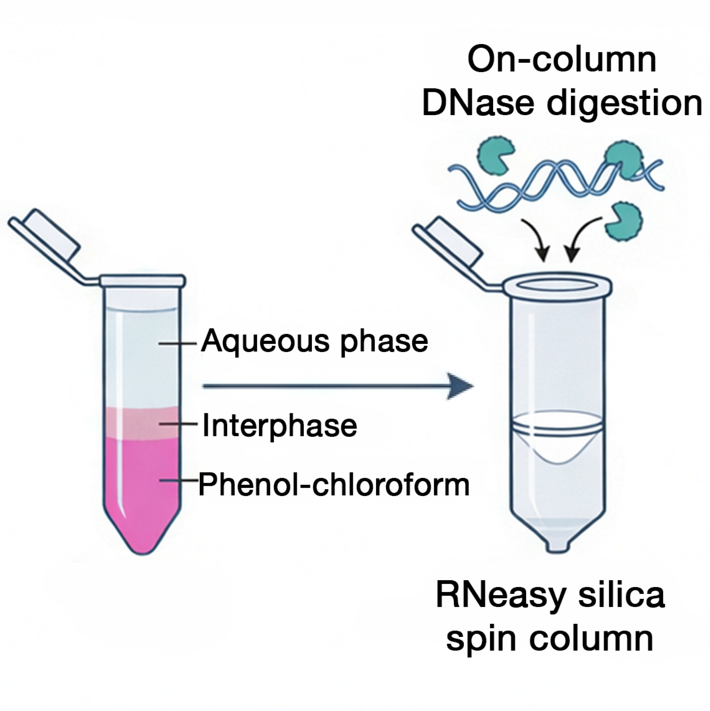
Carefully pipette up to 400 µL of clear supernatant (the top aqueous phase) into new 1.5 mL sample tubes.
Note
Take care not to touch the interface. If interface is interrupted by pipetting, re-spin the sample tube. Err on the side of removing less volume of supernatant than touching the interface as yield is generally not prioritized over purity for this procedure.
We suggest the following in order to extract the greatest possible volume while reducing the risk of disturbing the interface:
a. Drawing 4 volumes of 100 µL using a P100
b. Drawing 1 volume of 200 µL using a P1000 followed by 2 volumes of 100 µL using a P100
Add an approximately equal volume of 70% ethanol to sample tube and vortex for 5 sec on low intensity. Do not let ethanol sit for too long before binding, as guanidium/phenol can precipitate and stick to RNA.
RNeasy RNA purification chromatography
1m
Transfer up to 700 µL to an RNeasy spin column placed in a 2 mL collection tube (supplied).
Note
This maximum volume is the maximum capacity of the centrifuge tube. If you have more than this maximum volume, a second binding step can be performed to increase yield, but may also increase presence of contaminants.
Note that for most RNA-Seq applications increasing yield is secondary to obtaining high quality purification.
Place the spin column in centrifuge. Ensure balance, and centrifuge at 8000 x g, 22°C for 00:00:15 .
Discard flow-through.
Re-use collection tube in the next step.
Perform on-column DNAse digestion
Add 700 µL Buffer RW1 to the spin column and let stand for 00:01:00 before centrifuging for 8000 x g, 22°C, 00:00:15 , and discard flow through.
1m
In an extra sample tube, add 10 µL of DNase solution to 70 µL Buffer RDD per sample. Mix gently by inverting and spin down in a minifuge.
Note
1. Prepare 1.1x the amount calculated per total number of samples (i.e. for 10 samples prepare enough for 11 samples to ensure adequate amount).
Quality control assessment using Nanodrop
Using ultrapure RNase-free water and 70% ethanol, clean the stage of the Nanodrop gently. Lightly wet a Kimwipe with 70% ethanol and gently clean the top and bottom of the stage. Let evaporate briefly. Lightly wet a Kimwipe with ultrapure water, and repeat. Gently dry with a Kimwipe.
Blank the Nanodrop using elution buffer (water in this protocol). Take care that the same aliquot/bottle of water is used.
Note
Plasticizers and other substances inside most commercial tubes can be extracted in water and may be detectable on the Nanodrop, although generally minimally impact readings. Use of the exact same bottle increases accuracy by ensuring a "clean" blank.
Measure 2 µL of the sample.
The target for this reading is to obtain ≥2 for both 260/230 (salt) and 260/280 (protein). The most
common issue is generally salt contamination (high 230 absorption.) Samples with readings below 1.8 should be considered for additional purification steps such as sodium acetate precipitation (next section).
Note
Notes for increasing QC results:
1. If salt carryover is suspected, you may increase the volume of Buffer RPE (ex. 600 mL) or add one more wash with a 00:00:30 on-column incubation before centrifugation to remove excess salt.
2. If yield is low, you can recycle the eluate through a column a second time to modestly increase yield. Note that yield should not generally be a concern for most RNA-Seq applications, and purity should generally be prioritized over yield.
Cleanup using sodium acetate precipitation (optional)
20m
Raise the sample volume to at least 100 µL using RNase free water. To your RNA solution, add 1/10 volume of 3M sodium acetate (pH 5.2); 10 µL sodium acetate solution if using 100 µL .
Add 2.5 volumes of 100% ethanol (ice-cold, pre-chilled on ice). 250 µL if using 100 µL above. Mix by gentle inversion or pipetting.
Incubate at -20 °C overnight in a frost free freezer.
Centrifuge at 17500 x g, 4°C, 00:30:00 to pellet RNA.
Carefully discard the supernatant. Remove most of the volume at an angle using a P-1000 pipette. Point the hinge upwards to avoid disturbing the pelleted RNA. Note that RNA may be difficult or impossible to visualize at this step. Remove any remaining liquid carefully with a smaller pipette. Work slowly to avoid dislodging.
Wash the pellet by adding 900 µL of 70% ethanol (ice cold) to the pellet. Vortex briefly by hand or flick the tube to wash the pellet in ethanol solution.
Centrifuge at 17500 x g, 4°C, 00:30:00 to pellet RNA and remove supernatant, as before.
Air-dry pellet 00:10:00 . Do not overdry, as this may make pellet more difficult to resuspend. If visible, pellet should appear slightly translucent.
10m
Dissolve pellet in RNase-free water (in this case 50 µL ). Incubate at 60 °C for 00:10:00 if pellet is hard to dissolve.
10m
Quality control assessment using Bioanalyzer 2100
Perform RNA quality assessment using the Agilent Bioanalyzer 2100 according to the manufacturer’s instructions.
RNA integrity numbers (RIN) and electropherogram traces must be recorded for downstream sequencing. Samples with low RNA integrity (RIN ≤ 4) are generally unsuitable for PolyA+ selection and should instead be processed using ribosomal RNA depletion–based library preparation or confirmed using both methods.
Protocol references
These procedures are adapted from the following studies:
1. Kim, J. J. et al. Transcriptional Activation, Deactivation and Rebound
Patterns in Cortex, Hippocampus and Amygdala in Response to Ketamine Infusion
in Rats. Front Mol Neurosci 15, 892345 (2022). https://doi.org/10.3389/fnmol.2022.892345
2. Sapio, M. R. et al. Comparative Analysis of Dorsal Root, Nodose and Sympathetic Ganglia for the Development of New Analgesics. Front Neurosci 14, 615362 (2020). https://doi.org/10.3389/fnins.2020.615362
3. Rosenstein, R. K. et al. Host-Pathogen Interactions in Human Polyomavirus 7‒Associated Pruritic
Skin Eruption. J Invest Dermatol 141, 1344-1348 e1348 (2021). https://doi.org/10.1016/j.jid.2020.09.014
4. LaPaglia, D. M. et al. RNA-Seq investigations of human post-mortem trigeminal ganglia. Cephalalgia38, 912-932 (2018). https://doi.org/10.1177/0333102417720216
5. Sapio, M. R. et al. Pain control through selective chemo-axotomy of centrally projecting TRPV1+ sensory neurons. J Clin Invest 128, 1657-1670 (2018). https://doi.org/10.1172/JCI94331
6. Sapio, M. R. et al. The Persistent Pain Transcriptome: Identification of Cells and Molecules Activated by Hyperalgesia. J Pain 22, 1146-1179 (2021). https://doi.org/10.1016/j.jpain.2021.03.155
7. Sapio, M. R. et al. Longitudinal human transcriptomic and spatial gene profiling at the incisional edge during long surgical procedures. Comms Bio (2025). https://doi.org/10.1038/s42003-025-09366-0
Citations
Step 10
LaPaglia DM, Sapio MR, Burbelo PD, Thierry-Mieg J, Thierry-Mieg D, Raithel SJ, Ramsden CE, Iadarola MJ, Mannes AJ. RNA-Seq investigations of human post-mortem trigeminal ganglia.
https://doi.org/10.1177/0333102417720216Step 3
Kim JJ, Sapio MR, Vazquez FA, Maric D, Loydpierson AJ, Ma W, Zarate CA Jr, Iadarola MJ, Mannes AJ. Transcriptional Activation, Deactivation and Rebound Patterns in Cortex, Hippocampus and Amygdala in Response to Ketamine Infusion in Rats.
https://doi.org/10.3389/fnmol.2022.892345Step 8
Sapio MR, Vazquez FA, Loydpierson AJ, Maric D, Kim JJ, LaPaglia DM, Puhl HL, Lu VB, Ikeda SR, Mannes AJ, Iadarola MJ. Comparative Analysis of Dorsal Root, Nodose and Sympathetic Ganglia for the Development of New Analgesics.
https://doi.org/10.3389/fnins.2020.615362