Oct 14, 2021
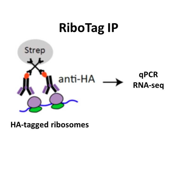
RiboTag Immunoprecipitation
- Benjamin Hobson1,
- Peter Sims1
- 1Columbia University


External link: https://doi.org/10.1016/j.celrep.2021.110208
Protocol Citation: Benjamin Hobson, Peter Sims 2021. RiboTag Immunoprecipitation. protocols.io https://dx.doi.org/10.17504/protocols.io.by37pyrn
Manuscript citation:
Hobson, B.D., Kong, L., Angelo, M.F., Lieberman, O.J., Mosharov, E.V., Herzog, E., Sulzer, D., and Sims, P.A. (2021). Subcellular and regional localization of mRNA translation in midbrain dopamine neurons. BioRxiv 2021.07.30.454065.
License: This is an open access protocol distributed under the terms of the Creative Commons Attribution License, which permits unrestricted use, distribution, and reproduction in any medium, provided the original author and source are credited
Protocol status: Working
We use this protocol and it’s working
Created: October 14, 2021
Last Modified: October 14, 2021
Protocol Integer ID: 54111
Keywords: RiboTag, method for ribotag immunoprecipitation, ribotag immunoprecipitation this protocol, ribotag immunoprecipitation, immunoprecipitation, mouse brain tissue
Funders Acknowledgements:
ASAP
Grant ID: ASAP-000375
Abstract
This protocol describes an optimized method for RiboTag Immunoprecipitation from mouse brain tissue.
Guidelines
Maintain a clean workspace decontaminated of RNAse. Use filter tips and proper technique to avoid contamination of samples. All buffers should be prepared with nuclease free water.
Materials
Materials
- Brain matrix for adult mouse brain (Zivic Instruments)
- Glass dounce homogenizer (e.g., MilliporeSigma, catalog #D9063)
- RNEasy MinElute kit (Qiagen, catalog #74204)
- Magnetic rack
- Temperature-controlled benchtop centrifuge
- Streptavidin T1 Dynabeads (ThermoFisher, catalog #65601)
- Biotinylated Rabbit anti-HA (Abcam, ab26228)
Buffers
- Dissection Buffer: 0.32 M sucrose buffer with 5 mM HEPES pH 7.4, 10 mM MgCl2, and 100 µg/mL cycloheximide (CHX)
- Lysis Buffer: 20 mM HEPES pH 7.4, 150 mM KCl, 10 mM MgCl2, 0.5 mM DTT, 100 µg/mL cycloheximide (CHX), 1x EDTA-free protease inhibitors (Roche), and 100 U/ml SUPERaseIN
- High Salt Buffer: 20 mM HEPES pH 7.4, 350 mM KCl, 10 mM MgCl2, 1% Igepal CA-630, 0.5 mM DTT, 100 µg/mL cycloheximide (CHX), 1x EDTA-free protease inhibitors (Roche), and 100 U/mL SUPERaseIN
- Ribosome Release Buffer: 20 mM HEPES pH 7.4, 50 mM EDTA, 100 U/mL SUPERaseIN
Troubleshooting
Safety warnings
Cycloheximide is toxic and should be considered hazardous waste. Consult with your local Environmental Health and Safety office for proper handling and disposal.
Before start
For tissue dissection, chill the brain matrix and dissection buffer for at least 30 minutes on ice.
For RiboTag IP, pre-cool the benchtop centrifuge to 4C. Chill the dounce homogenizer and lysis buffer for at least 30 minutes on ice.
Tissue Dissection
From the moment of euthanasia onward, changes in translation can occur rapidly. The dissection procedure should be conducted on ice and as quickly as possible. The following steps are suitable for mouse brain tissue; modification may be required for other tissues.
Sacrifice the mouse by cervical dislocation, rapidly extract the brain, and submerged it in ice-cold Dissection Buffer. After ~10 seconds, placed the brain on an ice-cold brain matrix and separate into 0.5-1.0 mm sections using ice cold razor blades.
Dissect the relevant areas of brain tissue. Dissected brain tissue must either be immediately homogenized (see below) or immediately frozen**.
**Note: In order to facilitate the simultaneous processing of many experimental replicates (mice), we typically prefer to freeze tissues from large cohorts of mice.
If freezing tissue, place the dissected tissue in a 1.5 mL tube, seal tightly, and flash freeze on liquid nitrogen. Store tissue at -80°C until further use.
If proceeding to homogenization, place dissected tissue in the glass-glass dounce homogenizer on ice and immediately homogenize as described below.
RiboTag Immunoprecipitation (IP)
Thaw frozen tissues on ice in a glass-glass dounce homogenizer with 1-1.5 mL of ice-cold Lysis Buffer. Homogenize tissues on ice using 30 strokes each with A and B pestles. Use slow strokes and take care to avoid foaming.
Transfer lysates to pre-chilled Eppendorf tubes and centrifuge at 1,000 xg 4°C for 10 minutes. Transfer the supernatant to a new tube.
If desired, the crude nuclear pellet (P1) from this spin can be stored at -80°C for downstream genotyping.
Add 1/9th the volume of 10% Igepal CA-630 to the lysates (final concentration 1%) and rotate at 4°C for 15 minutes.
Clarify the lysates by centrifuging at 20,000 xg 4°C for 10 minutes and transferred to a new tube.
Reserve 5% of the clarified lysate as 'Input' and store at -80°C. Add biotinylated rabbit anti-HA antibody** to the lysates and rotate overnight at 4°C.
**Note: The amount of anti-HA antibody must be optimized for the target cells of interest. A shortage of antibody will decrease the yield, while excess antibody will increase background. We typically use 6 µg of antibody for approximately 20,000-30,000 target cells.
The following day, prepare the appropriate amount of Streptavidin T1 Dynabeads** by washing several times in ice-cold Lysis Buffer.
**Note: The amount of T1 Dynabeads should correspond to the amount of anti-HA antibody added to the lysates. We use 5 µL of T1 beads per µg of biotinylated antibody.
Add Streptavidin T1 Dynabeads to the lysates and rotate for 30 minutes** at 4°C.
**Note: Because of the extremely high affinity of the streptavidin-biotin interaction, a short incubation is desirable. We found that >95% yield was achieved in 30 minutes, with longer incubations resulting in increased background. This may depend on the expected yield and should be optimized for each experiment.
Capture beads on a magnetic rack and discard the supernatant ('flow through', alternatively, reserve flow-through to ensure complete capture of target material). Resuspend the beads in 500 µL of ice-cold High Salt Buffer and transfer to a new tube.
Rotated for 30 minutes at 4°C, then capture on a magnetic rack; Discard the supernatant. Complete 3 more washes with 500 µL of ice-cold High Salt Buffer (four washes total over two hours).
RNA Purification
After the last wash, resuspend beads in 100 µL of Ribosome Release Buffer and incubate for 10 minutes at room temperature. Capture beads on a magnetic rack for ~2 minutes.
Transfer the supernatant (eluate), which contains the released mRNAs, to a new tube. If desired, freeze the tube with the dry beads** on liquid nitrogen and store at -80°C.
**Note: The EDTA in the ribosome release buffer causes dissociation of the bound ribosomes and therefore releases the mRNA into the supernatant. However, the eL22-HA protein itself remains bound to the beads, and can be separately analyzed by western blot.
Proceed with RNA purification. For ultra-low yield samples, we typically use the RNEasy MinElute kit (Qiagen, catalog #74204) according to the manufacturer’s instructions. Trizol extraction or other column-based purification kits are also suitable and can be tailored based on the expected RNA yield.
When using the RNEasy MinElute kit, we elute RNA from the column in 14 µL of nuclease free water supplemented with 20 U/mL SUPERaseIN. RNA should be immediately stored at -80°C.
Downstream Analysis
RNA yield can be determined using any of the following techniques, listed in order from the least to most sensitive: Qubit RNA HS, Agilent 2100 RNA Pico BioAnalyzer, or qRT-PCR.
Depending on the yield, RNA sequencing libraries can be prepared using a variety of commercial protocols (e.g., SMARTer Stranded Total RNA-Seq Kit v3, Pico Input Mammalian, Takara Bio, catalog no. 634485). If the yield is extremely low (<500 pg total RNA), RNA sequencing protocols designed for single cells, with unique molecular identifiers (UMIs) are recommended.