Sep 08, 2025
REVA #8: Phosphotungstic Acid (PTA) Staining of Cadaveric Human Vagus Nerves
- Aniruddha Upadhye1,
- Jichu Zhang1,
- Noa B. Nuzov1,
- Anandakumar Shunmugavel1,
- Constantinos Tsipsis1,
- Justin Chin1,
- Chaitanya Kolluru1,
- Megan C. Thompson1,
- Eleana Cintron1,
- Jennifer J. Coleman1,
- Michael W. Jenkins1,2,3,
- David L. Wilson1,4,
- Nicole A. Pelot5,
- Andrew J. Shoffstall1,6
- 1Department of Biomedical Engineering, Case Western Reserve University, Cleveland, OH, USA, 44106;
- 2FES Center, Louis Stokes Cleveland VA Medical Center, Cleveland, OH, USA;
- 3Department of Pediatrics, Case Western Reserve University, Cleveland, OH, 44106;
- 4Department of Radiology, Case Western Reserve University, Cleveland, OH 44106;
- 5Department of Biomedical Engineering, Duke University, Durham, NC, USA, 27708;
- 6APT Center, Louis Stokes Cleveland Department of Veterans Affairs Medical Center, Cleveland, OH
- Aniruddha Upadhye: ORCID: 0000-0002-9303-5872;
- Jichu Zhang: ORCID: 0009-0005-9791-8786;
- Noa B. Nuzov: ORCID: 0000-0001-8187-2115;
- Anandakumar Shunmugavel: ORCID: 0000-0001-8230-7361;
- Justin Chin: ORCID: 0009-0008-1482-0735;
- Chaitanya Kolluru: ORCID: 0000-0002-3211-7794
- Megan C. Thompson: ORCID: 0009-0008-6134-2625
- Eleana Cintron: ORCID: 0009-0001-4412-158X
- Jennifer J. Coleman: ORCID: 0000-0001-6702-729X
- Michael W. Jenkins: ORCID: 0000-0002-8908-5383
- David L. Wilson: ORCID: 0000-0001-9763-1463
- Nicole A. Pelot: ORCID: 0000-0003-2844-0190
- Andrew J. Shoffstall: ORCID: 0000-0002-0881-2180


Protocol Citation: Aniruddha Upadhye, Jichu Zhang, Noa B. Nuzov, Anandakumar Shunmugavel, Constantinos Tsipsis, Justin Chin, Chaitanya Kolluru, Megan C. Thompson, Eleana Cintron, Jennifer J. Coleman, Michael W. Jenkins, David L. Wilson, Nicole A. Pelot, Andrew J. Shoffstall 2025. REVA #8: Phosphotungstic Acid (PTA) Staining of Cadaveric Human Vagus Nerves. protocols.io https://dx.doi.org/10.17504/protocols.io.5qpvod95xg4o/v1
Manuscript citation:
Upadhye, A.R., Cintron, E., Zhang, J., Coleman, J., Kolluru, C., Jenkins, M.W., Wilson, D., Pelot, N.A., Shoffstall, A.J., 2025. Phosphotungstic Acid Staining to Visualize the Vagus Nerve Perineurium Using Micro-CT. J Neuroimaging 35, e70040. https://doi.org/10.1111/jon.70040
Zhang, J., Lapierre-Landry, M., Kalpatthi, H.K., Jenkins, M.W., Wilson, D.L., Pelot, N.A., Shoffstall, A.J., 2026. Automated 3D segmentation of human vagus nerve fascicles and epineurium from micro-computed tomography images using anatomy-aware neural networks. J Neural Eng. https://doi.org/10.1088/1741-2552/ae33f6
License: This is an open access protocol distributed under the terms of the Creative Commons Attribution License, which permits unrestricted use, distribution, and reproduction in any medium, provided the original author and source are credited
Protocol status: Working
We use this protocol and it's working
Created: July 15, 2025
Last Modified: September 08, 2025
Protocol Integer ID: 225840
Keywords: Human anatomy, Gross anatomy, Vagus nerve, Cranial nerves, Peripheral nervous system, Autonomic nervous system, Neuroanatomy, Computed tomography, CT, Microcomputed tomography, MicroCT, Tissue staining, staining of human cadaveric vagus nerve, cadaveric human vagus nerve, cadaveric human vagus nerves this protocol detail, human cadaveric vagus nerve, cadaveric human vagus nerves this protocol, phosphotungstic acid, pta, epineurium in computed tomography, tissue contrast between the fascicle, tissue contrast, surrounding epineurium, nerve, computed tomography
Funders Acknowledgements:
NIH SPARC
Grant ID: 75N98022C00018
NIH SPARC
Grant ID: OT2 025340
NIH
Grant ID: R01 EB033403
Disclaimer
DISCLAIMER – FOR INFORMATIONAL PURPOSES ONLY; USE AT YOUR OWN RISK
The protocol content here is for informational purposes only and does not constitute legal, medical, clinical, or safety advice, or otherwise; content added to protocols.io is not peer reviewed and may not have undergone a formal approval of any kind. Information presented in this protocol should not substitute for independent professional judgment, advice, diagnosis, or treatment. Any action you take or refrain from taking using or relying upon the information presented here is strictly at your own risk. You agree that neither the Company nor any of the authors, contributors, administrators, or anyone else associated with protocols.io, can be held responsible for your use of the information contained in or linked to this protocol or any of our Sites/Apps and Services.
Abstract
This protocol details the preparation and staining of human cadaveric vagus nerves with phosphotungstic acid (PTA) to provide tissue contrast between the fascicles and surrounding epineurium in computed tomography (CT) imaging.
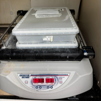
Image Attribution
Photo acquired by Aniruddha Upadhye at Case School of Engineering on May 20th, 2025. The photo shows the boxes in which the nerves are stained placed on a shaker manufactured by Orbital Genie.
Guidelines
N/A
Materials
1. Fume hood
- Company: VELCRO, sold by Staples. Size: 20 feet long, 2 inches wide. Item number: 464675.
3. Five plastic containers with airtight lids (two for formalin, two for PTA, one for storing samples in fridge at the end of the protocol) (Link)
- 6 L capacity container (VWR Catalog number: 75812-744): 15.5” long x 11” wide x 2 5/8” tall
- Lid for the 6 L container (VWR Catalog number: 75812-774)
4. Razor blades
- Company: Fisherbrand, sold by Fisher Scientific. Name: Razor Blades.
5. 10% neutral buffered formalin
6. Dissection probe tool
- Company: Fisherbrand, sold by Fisher Scientific. Name: Seeker with Bent-End
7. Gorilla® Micro Precise Super Glue (Prod. #102812)
8. Deionized (DI) water (VWR BDH1168-5GL)
9. Phosphotungstic acid (PTA) (10%) (Sigma-Aldrich-HT152-250ml), diluted in DI water (v/v) to 3% PTA
10. Phosphate buffered saline (PBS) 10X (Fisher-BP399-4), diluted in DI water to 1X PBS
11. Shaker for staining
12. Duct tape
13. Gauze or cheesecloth
Safety warnings
This protocol might include items and/or substances that may pose hazards (e.g., chemical, physical, biological, or otherwise) to your health upon use or exposure. Before engaging in the processes described in this protocol, familiarize yourself with and follow the safety data sheets, manufacturer safety recommendations, and local regulations.
Ethics statement
Be sure to seek approval for or an exemption from human subjects research from your local regulatory body(ies) as required by local and/or institutional regulations before initiating studies.
This study was determined to be exempt from IRB oversight by the Case Western Reserve University Institutional Review Board (IRB) because it involved de-identified cadaveric tissue and no protected health information was collected from the donors.
Before start
See the protocols for dissecting (dx.doi.org/10.17504/protocols.io.yxmvmb976g3p/v1) and removing (dx.doi.org/10.17504/protocols.io.n2bvje9r5gk5/v1) the human vagus nerve from embalmed cadavers, resulting in the vagal complex (from brainstem to abdomen) glued to a series of acrylic boards, each 3 cm wide and up to 9 cm long. The nerve on each acrylic board is termed a “sample”. The acrylic boards are secured with Velcro to a large acrylic sheet in a large container.
Section 1: PTA Staining
1d
Line the inside bottom of each container with the Velcro, sufficient to attach the samples.
Replace the containers used for PTA staining after three uses because the PTA degrades the plastic.
Cut between the distal-most thoracic sample and proximal-most esophageal plexus on the left and right. Use a fresh razor blade and a fast downward motion (not sawing) for each cut.
Immediately after cutting, apply a small amount of glue to both ends of the sample to ensure it is stable with minimal movement. Use a dissection probe to gently push the nerve down on top of the glue while it dries.
Snap the acrylic boards (where they are pre-thinned) between the thoracic and esophageal plexus samples, as described in dx.doi.org/10.17504/protocols.io.3byl46w72go5/v1.
Place the samples in a single container.
In a fume hood:
Place the cervical and thoracic samples of the nerve in container 1 of 5 and the esophageal-subesophageal samples of the nerve in container 2 of 5. Fill each container with 1 L of 10% formalin or until the nerve is fully submerged and place in the fridge for 12 hours to 4 days.
12h
To prepare for PTA staining, move the cervical and thoracic samples of the nerve into container 3 of 5, and the esophageal-sub esophageal samples of the nerve into container 4 of 5. The samples for one or two cadavers can be placed in a single container.
Fill the container with 1.5 L of 3% PTA (or sufficient volume to fully submerge all nerve sections).
Place the container on a shaker set at 35 rpm, 24:00:00 (cervical and thoracic samples) or 48 hours (esophageal and sub-esophageal samples).
Carefully decant the PTA for waste disposal.
Rinse the nerve with DI water 3 times.
Place the acrylic boards in container 5 of 5.
Cover the nerve in PBS-soaked gauze or cheesecloth.
Place the nerve in its container in the fridge until it’s time for imaging (12 hours (Overnight) to 4 days).
12h
Acknowledgements
The authors thank the donors and staff of the Case Western Reserve University Anatomical Gift Program. Without the selfless donations of the donors and the tireless efforts of the staff of this program, the methods described in this protocol and the insights and advancements that result from studies conducted according to this protocol would not be possible.