Sep 08, 2025
REVA #2: Computed Tomography (CT) of Embalmed Cadaver
- Noa B Nuzov1,
- Nicole A Pelot2,
- Andrew J. Shoffstall1,3
- 1Department of Biomedical Engineering, Case Western Reserve University, Cleveland, OH, USA, 44106;
- 2Department of Biomedical Engineering, Duke University, Durham, NC, USA, 27708;
- 3APT Center, Louis Stokes Cleveland Department of Veterans Affairs Medical Center, Cleveland, OH
- Noa B Nuzov: ORCID: 0000-0001-8187-2115;
- Nicole A Pelot: ORCID: 0000-0003-2844-0190;
- Andrew J. Shoffstall: ORCID: 0000-0002-0881-2180


Protocol Citation: Noa B Nuzov, Nicole A Pelot, Andrew J. Shoffstall 2025. REVA #2: Computed Tomography (CT) of Embalmed Cadaver. protocols.io https://dx.doi.org/10.17504/protocols.io.81wgbwr1ogpk/v1
Manuscript citation:
License: This is an open access protocol distributed under the terms of the Creative Commons Attribution License, which permits unrestricted use, distribution, and reproduction in any medium, provided the original author and source are credited
Protocol status: Working
We use this protocol and it's working
Created: July 14, 2025
Last Modified: September 08, 2025
Protocol Integer ID: 225833
Keywords: Computed tomography, CT, Human anatomy, Gross anatomy, Embalmed cadaver, embalmed cadaver this protocol, embalmed cadaver, embalmed human cadaver, human cadaver, computed tomography, cadaver, parameters for computed tomography, reva, ct, procedure
Funders Acknowledgements:
NIH SPARC REVA
Grant ID: 75N98022C00018
Disclaimer
DISCLAIMER – FOR INFORMATIONAL PURPOSES ONLY; USE AT YOUR OWN RISK
The protocol content here is for informational purposes only and does not constitute legal, medical, clinical, or safety advice, or otherwise; content added to protocols.io is not peer reviewed and may not have undergone a formal approval of any kind. Information presented in this protocol should not substitute for independent professional judgment, advice, diagnosis, or treatment. Any action you take or refrain from taking using or relying upon the information presented here is strictly at your own risk. You agree that neither the Company nor any of the authors, contributors, administrators, or anyone else associated with protocols.io, can be held responsible for your use of the information contained in or linked to this protocol or any of our Sites/Apps and Services.
Abstract
This protocol describes the procedures and parameters for computed tomography (CT) of embalmed human cadavers.
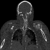
Image Attribution
Coronal plane of bone-enhanced CT scan of cadaver (SR060). Data collected by Noa Nuzov on 06/06/2025.
Guidelines
N/A
Materials
1. Siemens SOMATOM Definition Flash Dual-Source CT Scanner
2. Disinfectant wipes
Safety warnings
This protocol might include items and/or substances that may pose hazards (e.g., chemical, physical, biological, or otherwise) to your health upon use or exposure. Before engaging in the processes described in this protocol, familiarize yourself with and follow the safety data sheets, manufacturer safety recommendations, and local regulations.
Ethics statement
Be sure to seek approval for or an exemption from human subjects research from your local regulatory body(ies) as required by local and/or institutional regulations before initiating studies.
This study was determined to be exempt from IRB oversight by the Case Western Reserve University Institutional Review Board (IRB) because it involved de-identified cadaveric tissue and no protected health information was collected from the donors.
Before start
See the protocol for embalming and preparing a human cadaver for imaging (dx.doi.org/10.17504/protocols.io.kxygx4wm4l8j/v1), including placing it in a vacuum-sealed bag on a dissection cart for transport. On the day of the scan, before transporting the cadaver, ensure that the vacuum seal bag is still tight and no air has entered the bag; if air has entered the vacuum seal bag, use an electric vacuum to remove it.
Section 1: CT Scan
Remove the secondary opaque bags.
Place the cadaver (in a vacuum-sealed mattress bag) on the CT scanner bed in the supine position with the head closest to the scanner’s bore. Use the sheet that is under the cadaver for transfer.
Perform a “scout” scan to confirm the position of the cadaver.
The cadaver should be positioned as straight as possible.
The legs should be straight. If they are bowed or the knees are bent, a velcro strap, rope, or any fabric without metal can be used to tie the legs together.
If the cadaver is not oriented properly on the scout scan, return to the scanner and adjust its position.
Select the desired field of view using the scout scan.
Scan from the top of the head to the bottom of the toes, and as wide as possible to capture the entire rib cage.
Scan the sample with a slice thickness of 0.3 mm and the number of pixels in the X and Y directions as 512 by 512. In an axial cross section, the pixels are isotropic, but their physical size will vary depending on the width and height of the field of view.
Transfer the cadaver to a dissection cart to be transported from the CT room.
Use disinfectant wipes to clean the CT scanner bed, control panels, and any other surfaces that were touched by the cadaver or other personnel.
Acknowledgements
The authors thank the donors and staff of the Case Western Reserve University Anatomical Gift Program. Without the selfless donations of the donors and the tireless efforts of the staff of this program, the methods described in this protocol and the insights and advancements that result from studies conducted according to this protocol would not be possible.