Jul 02, 2026
Recombinant Human Interleukin-6 (IL6) (CSB-EP011664HU) Cell Proliferation Assay Protocol
- Rosie Liu1
- 1CUSABIO
- CUSABIO TECHNOLOGY LLC


External link: https://www.cusabio.com/
Protocol Citation: Rosie Liu 2026. Recombinant Human Interleukin-6 (IL6) (CSB-EP011664HU) Cell Proliferation Assay Protocol. protocols.io https://dx.doi.org/10.17504/protocols.io.5qpvojye9g4o/v1
License: This is an open access protocol distributed under the terms of the Creative Commons Attribution License, which permits unrestricted use, distribution, and reproduction in any medium, provided the original author and source are credited
Protocol status: Working
We use this protocol and it's working
Created: July 02, 2026
Last Modified: July 02, 2026
Protocol Integer ID: 320216
Keywords: Recombinant human Interleukin-6 (IL6); Human TF-1 cell line; Cell Proliferation Assay; ED₅₀ (half-maximal effective dose); cell viability
Abstract
This protocol aims to determine the biological activity of Recombinant Human IL6 (CSB-EP011664HU) by quantifying its dose-dependent stimulatory effect on the proliferation of human TF-1 cells, with the half-maximal effective dose (ED50) as the activity indicator.
Image Attribution
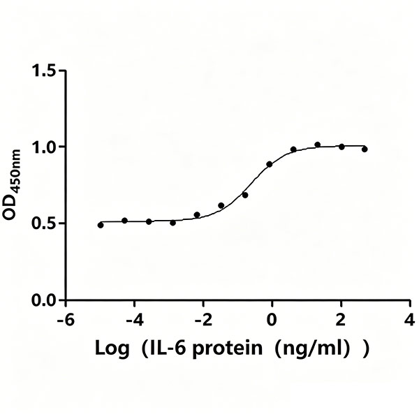
The ED50, as determined by the dose-dependent stimulation of the proliferation of human TF-1 cells, is 0.1518 - 0.3987 ng/mL.
Guidelines
Principle:
TF-1 is a human erythroleukemia cell line whose proliferation depends on hematopoietic cytokines such as GM-CSF. In the absence of GM-CSF, exogenous IL-6 binds to its specific receptor on the surface of TF-1 cells, activates downstream JAK-STAT and other signaling pathways, and promotes cell proliferation in a dose-dependent manner. The CCK-8 assay quantifies viable cell number by detecting the orange formazan product generated from WST-8 reduction via intracellular dehydrogenases; the absorbance at 450 nm is directly proportional to the number of viable cells. By fitting the dose-response curve of IL-6 concentration and OD value, the ED50 can be calculated to represent the biological activity of the recombinant IL-6 protein.
Operational Notes:
1. Maintain strict aseptic technique throughout the entire experiment to prevent bacterial or fungal contamination of cells and reagents.
2. Only use TF-1 cells in the logarithmic growth phase with high viability; over-confluent or low-viability cells will significantly reduce assay stability and reliability.
3. Perform 5-fold serial dilutions of IL-6 protein with precise pipetting to ensure the accuracy of each concentration gradient.
4. Minimize bubble formation in wells during reagent addition (bubbles interfere with OD measurement).
5. Resuspend cells fully and evenly before seeding to minimize well-to-well variation in cell density. Ensure consistent liquid volume across all control and experimental groups.
6. Keep the CCK-8 incubation time consistent across all assay batches; over-incubation may cause OD values to exceed the linear detection range.
7. Read the absorbance promptly after CCK-8 incubation to avoid signal drift caused by prolonged reaction at room temperature.
Materials
Cell Line:
- Human TF-1 cell line (BNCC)
Proteins and Reagents:
- Recombinant Human Interleukin-6 (IL6) (CUSABIO; code: CSB-EP011664HU; E.coli-expressed; Expression Region:
30-212aa; N-terminal 6xHis-tagged; purity>95%; Endotoxin<1.0 EU/ug)
- RPMI-1640 basal medium
- Fetal Bovine Serum (FBS)
- Recombinant human GM-CSF (rhGM-CSF)
- Cell Counting Kit-8 (CCK-8) (Beyotime, code: C0038)
- Sterile deionized water
- Glycerol (for long-term storage of reconstituted protein)
Consumables:
- 15 mL sterile centrifuge tubes
- 96-well cell culture plate (Corning, code: 3599)
- Sterile pipette tips of various specifications
Instruments:
- Automated cell counter
- Microplate reader (compatible with 450 nm detection)
- CO₂ cell culture incubator (37°C, 5% CO₂)
- Benchtop centrifuge
- Class II biosafety cabinet
Troubleshooting
Problem
Low overall OD values and weak proliferative response (Possible Causes: Poor cell viability before seeding; loss of IL-6 protein activity)
Solution
Verify cell viability (>90%) and logarithmic growth phase before the experiment; use freshly reconstituted protein and avoid repeated freeze-thaw cycles.
Problem
No clear dose-dependent trend in results (Possible Causes: Inaccurate gradient dilution; protein degradation after long-term storage)
Solution
Perform serial dilutions with calibrated pipettes; prepare protein dilutions freshly before each assay; confirm proper protein storage conditions.
Problem
Large well-to-well variation (Possible Causes: Uneven cell seeding; partial well contamination)
Solution
Fully resuspend cells before seeding and add cell suspension quickly; maintain strict aseptic operation; inspect wells for microbial contamination.
Problem
OD values exceed the upper linear detection limit (Possible Causes: Excessively long CCK-8 incubation; too high cell seeding density)
Solution
Reduce CCK-8 incubation time; optimize cell seeding density according to cell growth rate.
Problem
High background absorbance (Possible Causes: Contaminated CCK-8 reagent; impurities in culture medium)
Solution
Use sterile, unused detection reagent; set blank control wells (medium only + CCK-8) to subtract background signal.
Problem
Abnormally low OD in vehicle control (Possible Causes: Cell damage during washing steps; excessive centrifugation force)
Solution
Control centrifugation speed and time; handle cell pellets gently during resuspension.
Safety warnings
1. All cell handling operations must be performed in a biosafety cabinet to ensure personnel biosafety and prevent sample contamination.
2. Avoid repeated freeze-thaw cycles of reconstituted IL-6 protein, as this will cause protein denaturation and loss of biological activity. Aliquot the protein stock into single-use portions immediately after reconstitution.
3. CCK-8 reagent is light-sensitive; store, aliquot, and use it protected from direct light.
4. Dispose of all biological waste (spent culture medium, used pipette tips, discarded cell plates) in accordance with local laboratory biosafety regulations.
5. Do not use any reagents or consumables past their expiration date or with damaged sterile packaging.
Ethics statement
This protocol involves the use of animal blood samples. Users must obtain prior approval from their Institutional Animal Care and Use Committee (IACUC) or equivalent ethics committee before performing this protocol. All procedures must comply with applicable institutional and governmental regulations regarding the ethical use of animals.
Before start
Prior to the assay, equilibrate all frozen reagents to room temperature under sterile conditions. Confirm TF-1 cells are in the logarithmic growth phase with viability above 90%. Reconstitute the lyophilized IL-6 protein per the manufacturer’s instructions, and prepare all media and dilutions aseptically in a biosafety cabinet to avoid microbial contamination. Reserve sufficient basal medium for blank and vehicle control groups to ensure consistent reaction volume across all wells.
Reagent Preparation
Complete Culture Medium: Supplement RPMI-1640 basal medium with 10% (v/v) fetal bovine serum (FBS) and 2.5 ng/mL recombinant human GM-CSF (rhGM-CSF). Store at 2–8°C for short-term use.
Basal Culture Medium: Prepare RPMI-1640 medium supplemented with 10% (v/v) FBS. Store at 2–8°C after preparation. This medium is also used for protein dilution and control group preparation.
IL-6 Protein Gradient Dilutions:
Reconstitute the lyophilized Human IL6 with sterile deionized water to a stock concentration of 0.1–1.0 mg/mL (e.g., 0.5 mg/mL).
Perform a 5-fold serial gradient dilution using basal culture medium to generate 12 working concentrations, fully covering the range of 0.00002 ng/mL to 1000 ng/mL. The 12 working concentration gradients are listed as follows: 1000 ng/mL, 200 ng/mL, 40 ng/mL, 8 ng/mL, 1.6 ng/mL, 0.32 ng/mL, 0.064 ng/mL, 0.0128 ng/mL, 0.00256 ng/mL, 0.000512 ng/mL, 0.0001024 ng/mL, and 0.00002048 ng/mL (≈ 0.00002 ng/mL). Prepare all dilutions freshly immediately before the assay.
CCK-8 Detection Reagent: Thaw the CCK-8 solution at room temperature, and protect it from light before use.
Assay Procedures
TF-1 Cell Maintenance:
- Culture TF-1 cells (BNCC) in complete culture medium at 37°C in a humidified 5% CO₂ incubator.
- Subculture the cells at a split ratio of 1:4 to 1:5, 2–3 times per week to maintain continuous logarithmic growth.
Cell Preparation:
- Collect TF-1 cells in the logarithmic growth phase into a sterile 15 mL centrifuge tube.
- Centrifuge at 1000 rpm for 5 minutes at room temperature, then discard the supernatant completely.
- Resuspend the cell pellet with 5 mL of basal culture medium and centrifuge again. Repeat this wash step 3 times to remove residual rhGM-CSF from the culture system.
- Resuspend the washed cell pellet in basal culture medium and adjust the cell concentration to 4×10^5 cells/mL using a cell counter.
Cell Seeding:
- Use a 96-well cell culture plate and mark wells for blank control, vehicle control, and IL-6 concentration groups in advance. Set at least 3 technical replicates for each group.
- Blank control wells: Add 50 µL of basal culture medium (no cells) to serve as the background control for medium and CCK-8 reagent.
- Vehicle control wells and experimental wells: Seed 50 µL of the adjusted TF-1 cell suspension into each well.
Adding Protein Dilutions:
- For blank control wells: add 50 µL of basal culture medium (no IL-6 protein) to maintain a total volume of 100 µL per well.
- For vehicle control wells: add 50 µL of basal culture medium (no IL-6 protein) to reflect the baseline proliferation of TF-1 cells without cytokine stimulation.
- For experimental wells: add 50 µL of the 5-fold serially diluted IL-6 protein solutions (12 gradients in total) to the corresponding wells. After equal-volume mixing, the final concentration gradient of IL-6 in the culture system ranges from 0.00001 ng/mL to 500 ng/mL.
- Gently tap the plate edge to mix the solutions evenly. Place the 96-well plate in a 37°C, 5% CO₂ humidified incubator and incubate for 72 hours.
CCK-8 Viability Detection:
- After 72 hours of incubation, add 10 µL of CCK-8 solution to each well under sterile conditions. Avoid generating bubbles during pipetting to prevent interference with absorbance reading.
- Return the plate to the incubator and culture for an additional 2–4 hours at 37°C with 5% CO₂.
Absorbance Reading:
- Immediately transfer the plate to a microplate reader after incubation, and measure the optical density (OD) value at a wavelength of 450 nm.
Result Analysis
Background correction: Calculate the mean OD value of blank control wells, and subtract this blank value from the OD values of all vehicle control and experimental wells to eliminate the background absorbance from the culture medium and CCK-8 reagent.
Baseline reference: The corrected OD value of the vehicle control group represents the baseline proliferation level of TF-1 cells in the absence of IL-6 stimulation.
Dose-response curve plotting:
Plot a sigmoidal dose-response curve with the logarithm of final IL-6 concentration (ng/mL) on the x-axis and the corresponding corrected OD₄₅₀ₙₘ value on the y-axis.
ED50 calculation: Calculate the ED50 via nonlinear regression analysis of the dose-response curve.
Reference Assay Result: The ED50 of recombinant human IL-6 determined by this TF-1 cell proliferation assay is 0.1518 – 0.3987 ng/mL, demonstrating its high-grade proliferative bioactivity in the TF-1 cell system. This activity level meets the quality standard of high-purity active recombinant IL-6 products, confirming that the protein maintains intact receptor binding ability and downstream signaling activation function and is suitable for downstream functional studies such as cell proliferation induction, immune regulation research, and signaling pathway exploration.
Protocol references
- Recombinant Human Interleukin-6 (IL6) (Active) (Cat. No. CSB-EP011664HU)
- TF-1 Cell (4×10^5 cells/mL; 50μl/well)
- Cell Counting Kit-8 (CCK-8) (Cat. No. C0038)
- 96-well cell culture plate (Cat. No. 3599)