Apr 12, 2026
Recombinant Human EGFR His and Myc Tagged, low endotoxin (CSB-MP007479HU) ELISA Protocol
- Rosie Liu1
- 1CUSABIO
- CUSABIO TECHNOLOGY LLC


Protocol Citation: Rosie Liu 2026. Recombinant Human EGFR His and Myc Tagged, low endotoxin (CSB-MP007479HU) ELISA Protocol. protocols.io https://dx.doi.org/10.17504/protocols.io.81wgbj8q1vpk/v1
License: This is an open access protocol distributed under the terms of the Creative Commons Attribution License, which permits unrestricted use, distribution, and reproduction in any medium, provided the original author and source are credited
Protocol status: Working
We use this protocol and it's working
Created: April 09, 2026
Last Modified: April 12, 2026
Protocol Integer ID: 314809
Keywords: EGFR protein, activity validation, ELISA, egfr recombinant antibody, elisa plate with the recombinant egfr protein, affinity of the cusabio recombinant human egfr protein, cusabio recombinant human egfr protein, recombinant human egfr, recombinant egfr protein, tested batch of immobilized egfr, immobilized egfr, functional elisa, recombinant antibody, elisa protocol, elisa protocol this protocol, secondary antibody, antibody, egfr, dilution series of the antibody, low endotoxin, elisa, elisa plate, sigmoidal dose
Abstract
This protocol quantitatively assesses the functional activity and binding affinity of the CUSABIO Recombinant Human EGFR protein (CSB-MP007479HU, His and Myc tagged) through a functional ELISA. The protocol provides a standardized method to coat the ELISA plate with the recombinant EGFR protein, incubate with a dilution series of the antibody, and detect the binding using an HRP-conjugated Goat anti-Mouse IgG secondary antibody. The key readout is the half-maximal effective concentration (EC50) derived from the sigmoidal dose-response curve, showing that the tested batch of immobilized EGFR (at 1 μg/ml) has an EC50 of 2.867 - 3.571 ng/ml for binding the EGFR recombinant antibody.
Image Attribution
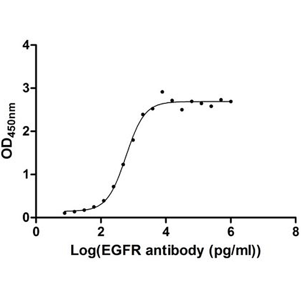
Measured by its binding ability in a functional ELISA. Immobilized EGFR at 1 μg/ml can bind Anti-EGFR recombinant antibody, the EC50 of human EGFR protein is 2.867-3.571 ng/ml.
Guidelines
All operations shall be performed under aseptic conditions to avoid contamination, with consistent timing and volume handling across all wells to ensure data repeatability. All incubation steps shall be performed in a humidified chamber to prevent reagent evaporation.
- Principle**: This indirect ELISA protocol is designed to validate the specific binding activity of low-endotoxin, His/Myc-tagged recombinant human EGFR protein (CSB-MP007479HU) to anti-EGFR recombinant antibody. It uses a standardized, optimized workflow of coating, blocking, gradient antibody incubation, and HRP colorimetric detection to accurately quantify binding affinity via EC50 calculation across a validated wide antibody concentration range.
- General**: This protocol is designed for the specific Human EGFR protein (CSB-MP007479HU) and its matched antibody. Performance may vary by lot or material.
- Coating**: Ensure the plate is coated evenly. The overnight incubation at 4°C is critical for optimal protein adsorption.
- Blocking**: Thorough blocking with 4% non-fat milk is essential to minimize non-specific binding in subsequent steps.
- Washing**: Aspirate or decant solutions completely between each step. The protocol emphasizes washing thoroughly (with PBST followed by PBS) to reduce background. After washing, invert the plate and tap it on clean paper towels to remove residual liquid.
- Incubation**: Maintain precise incubation times and temperatures (37°C for antibody steps) for reproducible results.
- Detection**: The TMB incubation time (10-20 min at 37°C) is a critical variable. Monitor the development of blue color in the wells containing the highest antibody concentration. Terminate the reaction with H2SO4 when a strong blue color develops in the positive-control/highest-concentration wells, but before saturation occurs in any well. Read the plate immediately after stopping.
- Bioactivity Preservation**: The recombinant EGFR protein and Anti-EGFR antibody are bioactive macromolecules. Avoid repeated freeze-thaw cycles, prolonged exposure to room temperature, or vigorous vortexing, which may cause protein denaturation and irreversible loss of binding activity.
- Sample Preparation and Storage**: All diluted protein and antibody solutions must be prepared fresh on the day of the experiment, and stored on ice or at 2-8°C before use.
- Experiment Operation**:
1. Use only calibrated pipettes for all liquid handling steps, and verify pipette accuracy before the experiment. Inconsistent pipetting volume is the primary cause of high data variability.
2. Use a new sterile pipette tip for each reagent and antibody dilution to avoid cross-contamination. Never reuse tips between different concentration gradients.
3. Do not touch or scratch the bottom of the ELISA plate wells with pipette tips, as this will interfere with the microplate reader's absorbance measurement and produce invalid data.
4. Use only ELISA-grade non-fat milk powder for blocking buffer preparation. Impure milk powder may contain non-specific binding proteins or phosphatase activity, which will cause a high background signal.
5. Ensure complete removal of liquid from the wells during all washing steps. Residual liquid will lead to a high background signal, non-specific binding, and inaccurate EC50 calculation.
6. The TMB substrate reaction is highly light-sensitive and temperature-dependent. Strictly control the incubation time and temperature, and protect the plate from direct light during substrate incubation to avoid over-development.
7. Read the OD value immediately after adding the stop solution, as the yellow color of the substrate reaction is unstable and will fade over time, leading to underestimated absorbance values and invalid results.
Materials
Target Protein & Antibodies:
• Recombinant Human Epidermal Growth Factor Receptor (EGFR), partial (Active) (Cat. No. CSB-MP007479HU, CUSABIO)
• Anti-EGFR Recombinant Antibody
• Goat anti-Mouse IgG Antibody, HRP Conjugated
Chemical Reagents:
• Sodium Bicarbonate (NaHCO₃), ACS grade
• Sodium Carbonate (Na₂CO₃), ACS grade
• ELISA-grade non-fat milk powder
• Sterile 1×PBS (pH 7.4)
• Tween-20, molecular biology grade
• Ready-to-use ELISA TMB substrate solution
• Concentrated sulfuric acid (H₂SO₄), ACS grade
Laboratory Consumables:
• 96-well high-binding polystyrene ELISA plate
• ELISA plate sealing film
• Calibrated single-channel and multi-channel pipettes (10 μl, 100 μl, 1000 μl)
• Sterile DNase/RNase-free pipette tips
• Disposable nitrile gloves
• Lint-free clean paper towels
• 0.22 μm and 0.45 μm sterile filter membranes
• Sterile polypropylene centrifuge tubes, beakers, and graduated cylinders
Laboratory Equipments:
• Microplate reader (calibrated, capable of absorbance reading at 450 nm)
• 4℃ refrigerator / cold room
• 37℃ constant temperature humidified incubator
• High-speed refrigerated centrifuge
• Calibrated pH meter (accuracy ±0.02 pH)
• Vortex mixer
• Chemical fume hood
• Ice maker/ice bucket
Troubleshooting
Problem
High background signal / Non-specific binding (Cause: Incomplete blocking; inadequate washing; excessive primary/secondary antibody concentration; TMB over-development; ELISA plate contamination)
Solution
Use fresh blocking buffer, extend blocking time to 2 h at 37℃, or overnight at 4℃. Strictly follow 3×PBST + 3×PBS washing protocol, ensure complete liquid aspiration after each wash. Verify antibody dilution ratios and reduce antibody concentration if necessary. Shorten TMB incubation time, strictly control incubation at 37℃, and monitor color development in real time. Use a new high-binding ELISA plate, and avoid touching the well bottoms with pipette tips.
Problem
Low Signal or No Signal (Cause: Inefficient EGFR coating; inactive detection antibody (HRP conjugate); expired or improperly prepared TMB substrate; incorrect incubation times/temperatures; incorrect microplate reader wavelength setting.)
Solution
Check protein concentration and coating buffer pH. Test a different lot of the detection antibody or use a positive control antibody. Ensure TMB is fresh and at room temperature before use. Verify the incubator temperature is accurately 37°C. Confirm the microplate reader is set to read absorbance at 450 nm.
Problem
High Variation Between Replicate Wells (Cause: Inconsistent pipetting; bubbles in wells during reagent addition; uneven incubation; inconsistent washing; edge effect.)
Solution
Calibrate pipettes before the experiment, fully mix all dilutions, and avoid bubbles during pipetting. Use a well-calibrated humidified 37℃ incubator, and add reagents quickly and in a consistent order. Standardize washing operations for all wells, and ensure complete residual liquid removal. Seal the plate tightly during incubation, and add PBS to unused edge wells to maintain humidity.
Problem
Poor Standard Curve/EC₅₀ Outside Expected Range (Cause: Errors in preparing the antibody dilution series; degradation of the primary antibody; incorrect fitting of the dose-response curve.)
Solution
Carefully re-prepare the dilution series. Aliquot and store the antibody properly to avoid freeze-thaw cycles. Ensure the curve fitting software uses the correct model (4-PL) and that the high and low concentration points form clear upper and lower plateaus.
Problem
Saturated OD signal (plateau at lowest antibody dilution) (Cause: Excessive EGFR coating amount; too high primary/secondary antibody concentration; TMB over-development.)
Solution
Verify EGFR coating concentration is 1 μg/ml (0.1 μg/well) and do not exceed the recommended coating amount. Reduce the starting concentration of the primary antibody gradient, confirm secondary antibody dilution is 1:10000. Shorten TMB incubation time, terminate the reaction once positive wells show a distinct blue color.
Safety warnings
Prepare all antibody dilutions fresh on the day of the experiment, and store diluted samples at 2-8°C protected from light before use to preserve antibody binding activity. Always add acid to water, never water to acid.
- Sulfuric Acid (2M H2SO4) Stop Solution**: Sulfuric acid is a strong corrosive and oxidizing agent. Always prepare the solution in a certified fume hood, and wear appropriate personal protective equipment (PPE), including nitrile gloves, a lab coat, and safety goggles. Never add water to concentrated sulfuric acid, as this will cause violent splashing and severe thermal burns. In case of skin or eye contact, flush the affected area immediately with copious amounts of water for at least 15 min and seek emergency medical attention.
- TMB Substrate Solution**: TMB is a potential irritant and suspected carcinogen. Avoid direct contact with skin, eyes, and mucous membranes. Wear PPE during handling, and dispose of waste TMB solution in accordance with local laboratory hazardous waste regulations.
- General Laboratory Safety**: Follow standard laboratory safety protocols. Treat all biological reagents as potentially hazardous.
- Waste Disposal**: Dispose of all chemical and biological waste according to your institution's safety regulations. Liquid waste from this assay (containing acid, TMB, and proteins) likely requires specific disposal procedures.
Before start
All protein and antibody samples shall be stored at -20°C (short-term) or -80°C (long-term) in single-use aliquots to avoid repeated freeze-thaw cycles. Before the experiment, equilibrate all samples to room temperature (18-25°C) on ice, and centrifuge at 10,000×g for 5 min at 4°C to remove precipitates or aggregates before dilution.
Pre-experiment Sample Processing
EGFR Coating Protein Processing:
• Dilute the Recombinant Human EGFR His and Myc Tag protein to a final working concentration of 1 μg/ml using pre-cooled pH 9.5 carbonate coating buffer, to achieve a final coating amount of 0.1 μg per well (100 μl/well).
• Prepare the protein dilution immediately before the coating step, and maintain the diluted solution on ice at all times to prevent protein denaturation and degradation.
Anti-EGFR Recombinant Antibody Sample Processing:
• Prepare serial gradient dilutions of the Anti-EGFR Recombinant Antibody using 4% non-fat milk PBS buffer as the diluent, with a final concentration range of 7.629 pg/ml to 1,000,000 pg/ml.
• Perform 2-fold to 18-fold serial gradient dilutions according to the experimental design, ensure complete mixing of each dilution by gentle pipetting, and avoid generating bubbles during the dilution process.
• Prepare all antibody dilutions fresh on the day of the experiment, and store diluted samples at 2-8℃ protected from light before use to preserve antibody binding activity.
• Set up blank control samples (4% non-fat milk PBS buffer without primary antibody) for background signal correction.
Detection Antibody Processing:
The Goat anti-Mouse IgG-HRP antibody must be diluted 1:10,000 in a solution of 4% non-fat milk powder in PBS. This dilution should be prepared fresh before the detection step.
Substrate and Stop Solution:
Equilibrate the TMB substrate solution to room temperature. Prepare a 2M H₂SO₄ solution for reaction termination, ensuring proper safety precautions.
Reagent Preparation
• Coating Buffer (Carbonate-Bicarbonate Buffer, pH 9.5): 0.035 mol/L Sodium Bicarbonate (NaHCO₃), 0.015 mol/L Sodium Carbonate (Na₂CO₃) dissolved in ultrapure water. Adjust pH to 9.5 at room temperature, then filter through a 0.22 μm sterile membrane.
• Wash Buffer (1×): Phosphate-Buffered Saline (PBS), pH 7.4.
• Wash Buffer (2×): 0.1% (v/v) Tween-20 added to 1×PBS (pH 7.4). Mix thoroughly to ensure uniform dispersion of Tween-20.
• Blocking Buffer: 4% (w/v) ELISA-grade non-fat milk powder dissolved in 1×PBS (pH 7.4). Mix thoroughly until fully dissolved, then filter through a 0.45 μm membrane to remove insoluble particles. Must be prepared fresh on the day of the experiment. Do not store for later use.
• Antigen Dilution: Human EGFR protein, diluted to 1 µg/mL in Coating Buffer.
• Primary Antibody Dilution Series: Anti-EGFR Recombinant Antibody, serially diluted to concentrations from 7.629 to 1,000,000 pg/mL.
• Detection Antibody Dilution: Goat anti-Mouse IgG, HRP conjugated, diluted 1:10,000 in Blocking Buffer (4% non-fat milk PBS). Must be prepared fresh immediately before use. Protect from light at 2-8℃ before plate addition.
• Substrate Solution: TMB (ready-to-use solution).
• Stop Solution: 2M Sulfuric Acid (H₂SO₄). Always add acid to water, never water to acid.
Assay Procedure
Coating
Coat the plate with 0.1 μg/well (1 μg/ml, 100 μl/well) EGFR protein at 4℃ overnight. The protein is diluted in pH 9.5 coating buffer, namely Carbonic acid buffer (0.035 mol/L NaHCO3, 0.015 mol/L Na2CO3)
Washing
Set the enzyme labelling board at room temperature for 10 min. Remove the protein solution by decanting or aspirating. Make sure that the solution in the wells is completely removed. Then wash the wells with 400 μl/well PBS (pH 7.4) once, aspirate the solution, invert the plate, and place it on multilayer clean paper towels for a while to remove the solution completely.
Blocking
Block the wells with 400 μl/well blocking buffer (4% Non-fat milk powder in PBS) at 37℃ for 2h. After blocking, aspirate the solution in the wells, invert the plate, and set it on multilayer clean paper towels for a while to remove the solution completely.
Adding the recombinant antibody
Add 100μl/well 7.629-1000000pg/ml (gradient dilution, 2x to 18x) Anti-EGFR Recombinant Antibody to each well, and seal the plate with sealing film. Then, put the plate at 37℃ and incubate for 1h.
Washing
First, wash the wells with 400 μl/well PBST (0.1% Tween-20 in PBST, pH 7.4) three times, and then wash the wells with 400 μl/well PBS three times. Then aspirate the solution, invert the plate, and set it on multilayer clean paper towels for a while to remove the solution completely.
Adding the detection antibody
Dilute the Goat anti-Mouse IgG antibody, HRP conjugated by 4% non-fat milk powder in PBS at 1:10000. Add 100 μl/well on the plate and incubate at 37℃ for 1h.
Washing
Repeat step 5
Adding substrate
Add 100μl TMB solution to each well and incubate at 37℃ for 10-20min.
Termination
Add 50 μl/well 2M H2SO4 solution to the plate to terminate the reaction.
Read OD
Immediately put in the enzyme label and read at 450 nm.
Result Analysis
Raw Data Preprocessing:
Blank Correction: Subtract the average OD450 value of the blank control wells from the OD450 value of each sample well to eliminate background absorbance from the plate and reagents.
Repeatability Validation: For duplicate/triplicate wells, calculate the coefficient of variation (CV) of the corrected OD values. Only data with CV ≤ 10% is considered statistically valid; discard data with excessive CV and repeat the experiment if necessary.
Outlier Elimination: Use Grubbs' test or Dixon's Q-test to identify and remove statistically significant outliers from the dataset to ensure the reliability of subsequent curve fitting.
Dose-Response Curve Fitting:
Plot a standard dose-response curve with the corrected OD450 values as the vertical (Y) axis, and the logarithmic value of the Anti-EGFR Recombinant Antibody concentration (Log[Ab], pg/ml) as the horizontal (X) axis.
Use a four-parameter logistic (4-PL) regression model (the gold standard for ELISA dose-response data analysis) for curve fitting, following the equation:
Y = Bottom + (Top - Bottom) / (1 + 10^( (EC50 - X) * Hill Slope ))
○ Y = Corrected OD450 value
○ X = Log10 of the Anti-EGFR antibody concentration (pg/ml)
○ Top = Maximum asymptotic OD value (saturation binding signal)
○ Bottom = Minimum asymptotic OD value (background signal)
○ Hill Slope = Slope factor of the sigmoidal curve
○ EC₅₀ = Half-maximal effective concentration (the antibody concentration at which the binding signal reaches 50% of the maximum, the core quantitative indicator of binding affinity)
A curve fit with an R² value ≥ 0.99 is required for reliable EC₅₀ calculation and bioactivity evaluation.
Experimentally Determined Result:
The EC₅₀ value of the specific binding reaction between Recombinant Human EGFR (CSB-MP007479HU) and Anti-EGFR Recombinant Antibody, obtained from this standardized ELISA assay, is in the range of 2.867-3.571 ng/ml. This is the final experimental result of this binding activity verification test, reflecting the actual binding performance between the target protein and the antibody under the protocol-specified experimental conditions.
Measured by its binding ability in a functional ELISA. Immobilized EGFR at 1 μg/ml can bind Anti-EGFR recombinant antibody, the EC50 of human EGFR protein is 2.867-3.571 ng/ml.
Result Interpretation:
The EC₅₀ value is the core quantitative indicator of binding affinity: a lower EC₅₀ value indicates higher binding affinity between the Anti-EGFR Recombinant Antibody and the target EGFR protein, and stronger specific binding activity of the recombinant EGFR protein.
The experimentally measured EC₅₀ range of 2.867-3.571 ng/ml confirms that the Recombinant Human EGFR (CSB-MP007479HU) exhibits clear, specific, and dose-dependent binding activity to the Anti-EGFR Recombinant Antibody.
In repeated verification assays, if the measured EC₅₀ value deviates significantly from the 2.867-3.571 ng/ml range obtained from this experiment, it indicates potential changes in the bioactivity of the EGFR protein/antibody or deviations in experimental operations that require troubleshooting and repeated testing.
Protocol references
○ Product Name: Recombinant Human Epidermal Growth Factor Receptor (EGFR), partial (Active)
○ Catalog Number: CSB-MP007479HU
○ Target Species: Homo sapiens (Human)
Target Analyte: EGFR