Jul 04, 2025
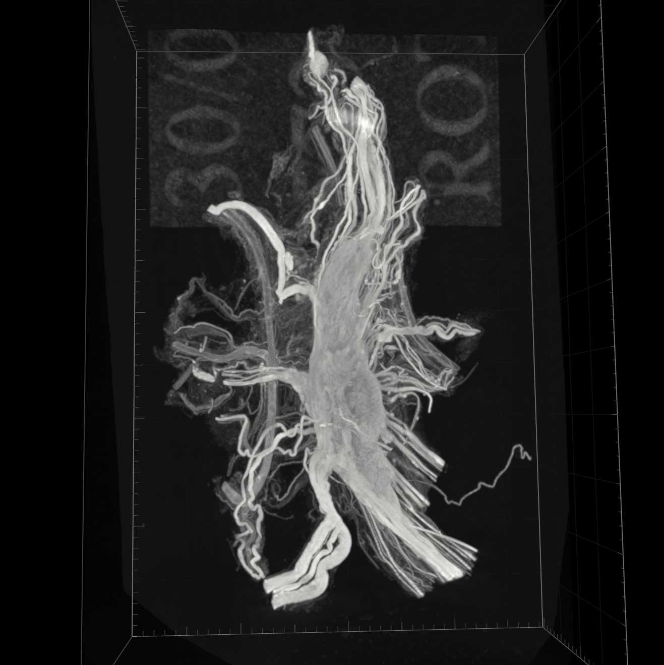
Rat pelvic nerve preparation for serial block-face scanning electron microscopy
- 1University of Melbourne
- SPARCTech. support email: [email protected]


Protocol Citation: John-Paul Fuller-Jackson, Peregrine B Osborne, Janet R Keast 2025. Rat pelvic nerve preparation for serial block-face scanning electron microscopy. protocols.io https://dx.doi.org/10.17504/protocols.io.8epv5r6jdg1b/v1
License: This is an open access protocol distributed under the terms of the Creative Commons Attribution License, which permits unrestricted use, distribution, and reproduction in any medium, provided the original author and source are credited
Protocol status: Working
Created: March 28, 2024
Last Modified: July 04, 2025
Protocol Integer ID: 97477
Keywords: electron microscopy, scanning block face electron microscopy, ultrastructure, peripheral nerve, rat pelvic nerve preparation for serial block, rat pelvic nerve for serial block, pelvic nerve preparation, pelvic nerve, electron microscopy this protocol, scanning electron microscopy, electron microscopy, peripheral nerve, suitable for any peripheral nerve
Funders Acknowledgements:
NIH SPARC
Grant ID: OT2OD023872
Abstract
This protocol describes the steps required to prepare rat pelvic nerve for serial block-face scanning electron microscopy. It is suitable for any peripheral nerve.
Materials
BenzyldimethylamineProSciTechCatalog #C054 Araldite 502ProSciTechCatalog #C041 Procure 812 mixProSciTech Dodecenyl succinic anhydrideProSciTechCatalog #C044 Lead nitrateBio-Rad LaboratoriesCatalog #4192 Aspartic acidMerckCatalog #100126 ThiocarbohydrazideProSciTechCatalog #EMS21900 Osmium TetroxideProSciTech Potassium ferricyanideBanksia Scientific Company (AJAX FineChem)Catalog #393-500G
Preparation for perfusion
Make up the following solutions:
Perfusion prewash solution: To 300 ml 0.9% sodium chloride (w/v) add 3.75 ml 1% sodium nitrite (w/v) and 0.11 ml heparin (5000 IU/ml). This is made up immediately prior to use.
Perfusion fixative: 1.25% glutaraldehyde and 2% paraformaldehyde in 0.1M phosphate buffer, pH 7.4. This is made up no longer than 48h prior to use, stored at 4 ºC and brought to room temperature on the day of perfusion.
Intracardiac perfusion
Induce anesthesia by an intraperitoneal injection of ketamine (100 mg/kg) and xylazine (10 mg/kg).
After opening the chest cavity, inject the left ventricle with mixture of 0.25 ml heparin (5000 IU/ml) and 0.5 ml 1% sodium nitrite.
A needle connected to tubing (T-connector to prewash and perfusion solutions), and a peristaltic pump (setting: 50 ml/min) is then inserted into the left ventricle and clamped into place using hemostats. Make a small incision in the right atrium to drain blood and perfusate during the procedure.
Perfuse with pre-wash solution until the fluid flowing from the right atrium is clear, the liver and extremities are pale (typically 2-3 min).
Perfuse with fixative for a minimum of 10 minutes, by which time the organs have stiffened and the neural tissues will be well preserved.
Dissect major pelvic ganglion with pelvic nerve attached and place in fixative for storage at 4 ºC (min 24h).
Solution preparation
Make up cacodylate buffer for additional tissue fixation to contain the following in double-distilled water:
- 0.1 M sodium cacodylate (0.214 g per ml H2O = 1 M)
- 1.5 % glutaraldehyde
- 1 % paraformaldehyde
Make 0.15 M cacodylate, 1.5% potassium ferrocyanide, 2% osmium tetroxide in double-distilled water.
Make 1% thiocarbohydrazide in double-distilled water, incubate in 60 °C for 1 h. Pre-heat water to 60 °C to speed up dissolution of solid.
Make Walton's lead aspartate by dissolving 76.3 mg of lead nitrate in 11.5 ml of 0.03 M aspartic acid. Adjust solution pH to 5-5.5 with droplets of 1 M potassium hydroxide. Keep warm at 60 °C for at least 30 min prior to use.
Major pelvic ganglion preparation
Place major pelvic ganglion in a petri dish containing 0.1 M phosphate-buffered saline under a dissection microscope.
Identify the pelvic nerve attached to the major pelvic ganglion.
Remove any excess tissue such as adipose tissue or blood vessels that may have been included in the dissection.
Immerse the ganglion in 1 ml of cacodylate buffer in 1.5 ml Eppendorf tube for at least 2 hours prior to further processing for electron microscopy.
Electron microscopy tissue processing
Rinse sample in 0.175 M cacodylate buffer 5 times for 3 min at room temperature.
Incubate sample in 0.15 M cacodylate, 1.5% potassium ferrocyanide, 2% osmium tetroxide for 1 h on ice.
Rinse sample in double-distilled water 5 times for 3 min at room temperature.
Incubate sample in 1% thiocarbohydrazide solution for 20 min at room temperature.
Rinse sample in double-distilled water 5 times for 3 min at room temperature.
Incubate sample in 2% osmium tetroxide solution for 30 min at room temperature.
Rinse sample in double-distilled water 5 times for 3 min at room temperature.
Incubate sample in 1% aqueous uranyl acetate overnight at 4 °C.
Rinse sample in double-distilled water 5 times for 3 min at room temperature.
Incubate sample in Walton's lead aspartate for 30 min at 60 °C.
Rinse sample in double-distilled water 5 times for 3 min at room temperature.
Resin embedding
Make resin by mixing 12.5 ml of Procure, 7.5 ml of Araldite and 27.5 ml of dodecenyl succinic anhydride. Keep the mixture at 60 °C for at least 2 h. Store mixture in oven for short term, freezer for long term.
Dehydrate sample in ethanol:
- 20% ethanol in double-distilled water for 5 min.
- 50% ethanol in double-distilled water for 5 min.
- 70% ethanol in double-distilled water for 5 min.
- 90% ethanol in double-distilled water for 5 min.
- 100% ethanol three times for 5 min.
- 50% ethanol and 50% acetone for 10 min.
- 100% acetone two times for 10 min.
Substitute sample by incubation in 50% acetone and 50% resin for 2 h.
Infiltrate sample by incubation in 100% resin twice for 12 h.
Add 27.4 µl benzyldimethylamine for every 1 ml of resin, mix for at least 2 h.
Embed sample in fresh resin and polymerise in appropriately sized Eppendorf tube at 60
Manually trim the resin block using a razor blade to a pyramidal shape containing the sample, keeping edges as parallel as possible.
Acknowledgements
We thank Prof Eric Hanssen and Dr Adam Blanch (Ian Holmes Imaging Centre, University of Melbourne) for expert advice about this method.