1Medical Research Council Brain Network Dynamics Unit, Nuffield Department of Clinical Neurosciences, University of Oxford, Mansfield Road, Oxford, OX1 3TH, United Kingdom;
2Aligning Science Across Parkinson's (ASAP) Collaborative Research Network, Chevy Chase, MD
License: This is an open access collection distributed under the terms of the Creative Commons Attribution License, which permits unrestricted use, distribution, and reproduction in any medium, provided the original author and source are credited
Protocol status: Working
We use this collection and it's working
Created: September 07, 2023
Last Modified: May 14, 2024
Collection Integer ID: 87520
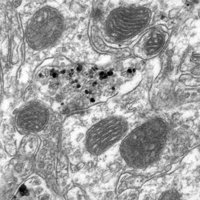
Keywords: Dopamine, Axon terminal, Striatum Tyrosine hydroxylase (TH), Transmission Electron Microscopy (TEM), Electron microscopy (EM), Substantia nigra pars compacta (SNc), Ventral Tegmental Area (VTA), Vesicle Synapse, Ultrastructure, Immuno-EM, Immunogold, Quantification, ImageJ, user process animal brain tissue for electron microscopy, marker of dopaminergic axon, dopaminergic axon terminals in the brain, tissue sections ready for electron microscope, dopaminergic axon, dopamine neuron axon, dopaminergic axon terminal, electron microscopy, electron microscope, user process animal brain tissue, transmission electron microscope, image ultrathin sections of tissue, microscope, brain tissue, axon, release of dopamine, quantitative analyses of the ultrastructural feature, tissue section, dopamine, ultrastructural feature, inappropriate dopamine release, tissue
Abstract
The release of dopamine from axons is critical for normative brain function and behaviour. Impaired or otherwise inappropriate dopamine release often correlates with changes in the ultrastructure of dopamine neuron axons that can assessed with electron microscopy. Here, we provide two protocols that can be used serially to, first, help the user process animal brain tissue for electron microscopy and, secondly, help the user undertake quantitative analyses of the ultrastructural features of dopaminergic axon terminals in the brain.
Protocol #1 describes how to prepare brain tissue, carry out pre-embedding immunohistochemistry for tyrosine hydroxylase as a marker of dopaminergic axons, and then make tissue sections ready for electron microscope.
Protocol #2 details how to examine and image ultrathin sections of tissue using a transmission electron microscope and then how to analyse the digital images.
Quantitative analyses of the ultrastructural features of dopaminergic axon terminals. Protocol #2: Acquisition and analysis of electron microscopy images