Sep 12, 2022
Quantitative analyses of the ultrastructural features of dopaminergic axon terminals. Protocol #2: Acquisition and analysis of electron microscopy images
- 11. MRC Brain Network Dynamics Unit, Nuffield Department of Clinical Neurosciences, University of Oxford, Mansfield Road, Oxford, OX1 3TH, United Kingdom;
- 2Linköping University;
- 3Aligning Science Across Parkinson's (ASAP) Collaborative Research Network, Chevy Chase, MD


External link: https://www.mrcbndu.ox.ac.uk/
Protocol Citation: Natalie Doig, Max Larsson, Peter Magill 2022. Quantitative analyses of the ultrastructural features of dopaminergic axon terminals. Protocol #2: Acquisition and analysis of electron microscopy images. protocols.io https://dx.doi.org/10.17504/protocols.io.x54v9d8ppg3e/v1
License: This is an open access protocol distributed under the terms of the Creative Commons Attribution License, which permits unrestricted use, distribution, and reproduction in any medium, provided the original author and source are credited
Protocol status: Working
We use this protocol and it's working
Created: September 12, 2022
Last Modified: December 26, 2022
Protocol Integer ID: 69846
Keywords: Dopamine, Axon terminal, Striatum Tyrosine hydroxylase (TH), Transmission Electron Microscopy (TEM), Electron microscopy (EM), Substantia nigra pars compacta (SNc), Ventral Tegmental Area (VTA), Vesicle Synapse, Ultrastructure, Immuno-EM, Immunogold, Quantification, ImageJ, ASAPCRN, user process animal brain tissue for electron microscopy, marker of dopaminergic axon, dopaminergic axon terminals in the brain, electron microscopy image, dopamine neuron axon, tissue sections ready for electron microscopy, digital electron microscope image, electron microscopy, analysis of electron microscopy image, dopaminergic axon, dopaminergic axon terminal, electron microscope, user process animal brain tissue, transmission electron microscope, brain tissue, axon, analyse electron micrograph, user to analyse electron micrograph, density of neurotransmitter vesicle, microscopic image analysis, including axon terminal size, axon terminal size, neurotransmitter vesicle, release of dopamine, quantitative analyses of the ultrastructural feat
Funders Acknowledgements:
Medical Research Council of the United Kingdom
Grant ID: MC_UU_12024/2
Medical Research Council of the United Kingdom
Grant ID: MC_UU_00003/5
Wellcome Trust Investigator Award
Grant ID: 101821
Abstract
The release of dopamine from axons is critical for normative brain function and behaviour. Impaired or otherwise inappropriate dopamine release often correlates with changes in the ultrastructure of dopamine neuron axons that can assessed with electron microscopy. Here, we provide two protocols that can be used serially to, first, help the user process animal brain tissue for electron microscopy and, secondly, help the user undertake quantitative analyses of the ultrastructural features of dopaminergic axon terminals in the brain.
This Protocol #2 details how to examine and image pre-prepared ultrathin sections of brain tissue using a transmission electron microscope, and then how to analyse the digital images.
In this protocol, digital electron microscope images are analysed. We describe a method involving the use of freely available open-source software; ImageJ (Rueden et al., 2017; Schindelin et al., 2012) and custom designed plugins for electron microscopic image analysis, PointDensity and PointDensitySyn (Anwar et al., 2011; Connor-Robson et al., 2019; Janezic et al., 2013;Kosillo et al., 2019; Larsson et al., 2015). This software allows the user to analyse electron micrographs and obtain data on multiple variables including axon terminal size, incidence of synapses, number and density of neurotransmitter vesicles, distance of vesicles to the cell membrane, distance of vesicles to the active zone (if present) and the inter-vesicle distance as an indicator of clustering (See Figure 1).
Figure 1. Schematic showing vesicle distribution parameters that can be analysed with this protocol. Left, Vesicle distribution parameters in non-synaptic axon terminals; inter-vesicle distance (a), shown for one vesicle and its distance to two other vesicles for clarity, and the vesicle distance to perimeter (b) measured as the shortest distance to the plasma membrane. Right, Vesicle distribution parameters in synapse-forming axon terminals; the vesicle distance to synaptic specialization (c) measured as the shortest distance to the delineated active zone (grey thickening of membrane on pre-synaptic side of synaptic cleft), and the length of the synaptic specialization (d), shown on the post-synaptic side of the synaptic cleft for clarity.
The partner Protocol #1 describes how to prepare brain tissue, carry out pre-embedding immunohistochemistry for tyrosine hydroxylase as a marker of dopaminergic axons, and then make tissue sections ready for electron microscopy.
Protocol #1 and #2 are part of the collection "Quantitative analyses of the ultrastructural features of dopaminergic axon terminals".
Materials
Equipment:
- TEM fitted with digital camera, Hitachi. We use a Hitachi HT7800 TEM with a XAROSA CMOS camera (EMSIS GmbH)
Software:
Before start
The analysis method presented in this protocol requires the user to manually delineate and register ultrastructural features in electron microscope images. It is likely that you will become more proficient at delineating axon terminals and marking vesicles with practice over time. It is important to account for this effect in your analysis. For example, if you were to begin your analysis with an animal in your control group, you may falsely get the impression that there are fewer vesicles in this dataset, as compared subsequent datasets obtained from other animals, simply because you got better at marking vesicles over time. One way to mitigate is to ‘train’ yourself with a random set of images before starting your intended analyses, and then to randomize the experimental animal you begin with, as well as the order in which you subsequently analyse data from additional animals. As the analysed images are saved as copies of the original micrographs with the recorded coordinates visible, it is also possible to go back and re-evaluate the accuracy of the analysis at a later time. All analyses should be performed blinded wherever possible.
Examination of Sections in the Electron Microscope
Sampling: One of the challenges for EM is to design a sampling strategy to ensure your data sample is representative, reflecting the true distribution/occurrence of a particular structure, and robust enough to detect effects of e.g., experimental manipulations.
The striatum is so densely packed with dopaminergic terminals that one could obtain hundreds of images from a very small volume of tissue, but this would not be representative of the striatum as a whole and could easily bias your data. Here, we describe a sampling strategy based on previous work (Kosillo et al., 2019). An example sampling strategy is laid out in Figure 2.
If you are examining dopaminergic axon terminals in the dorsolateral striatum, we would recommend re-embedding tissue from at least two sections at different rostrocaudal planes per animal. From each of these sections, you can re-embed tissue from at least two regions of interest, making two blocks. From each block, you would then sample from at least two grids, and then from each grid take multiple images from at least two ultrathin sections (Fig 2).
This is a guideline; the optimum strategy (where possible) would be to carry out a pilot study to acquire some initial data so that you can perform a power analysis and estimate the sample size required.
Figure 2. A guideline sampling strategy for analysis of the ultrastructure of dopaminergic (TH-immunopositive) axons in the striatum. For each mouse, two sections in the along rostrocaudal axis are examined with light microscopy for regions of interest. Two regions are then re-embedded from each section (to make blocks). Tissue on each block is then re-sectioned using an ultramicrotome. Ribbons of ultrathin sections are collected onto multiple grids. Grids are then analysed in the electron microscope and digital images are taken from two sections from each grid for further analysis.
Image Acquisition: This protocol describes a random systematic sampling strategy to acquire digital images for analysis using a transmission electron microscope (TEM) fitted with a digital camera.
Place grids into the specimen holder and insert into electron microscope.
Image the whole grid and choose 2 sections to sample from. Pick sections that are as distant as possible from each other (opposite ends of the ribbon) or examine different regions on the 2 different sections; this will decrease the likelihood of imaging the same terminals in both sections.
Determine a magnification at which you can locate immunolabelled axon profiles, but you cannot see individual synapses, this is your ‘searching magnification’. Define a criterion for these ‘immunopositive’ structures depending on the level of background labelling, e.g., a minimum of, for example, 5 silver-enhanced gold particles per profile.
Immunogold labelling has limited penetration into the tissue but is also non-specific at the tissue surface. In order to avoid imaging non-specific labelling, locate the surface of the tissue (where tissue meets the covering resin) and move a set distance (e.g., 2-5 µm) from there before starting with image acquisition.
Once at a safe distance from the surface, pick a random starting point and move in a straight line until you locate an immunopositive axon terminal, increase the magnification so that it is possible to visualize synapses and vesicles and take an image the profile. When imaging, keep your immunopositive terminal in the centre of the frame and make sure you do not crop off any of the plasma membrane.
Decrease to your searching magnification and move in a straight line until you locate the next immunopositive terminal, increase your magnification and image.
Repeat this until you have at least the required number of images from that section.
Move to the second ultrathin section on the grid that you have chosen to image from and repeat the procedure.
Once you have enough images from the sections on that grid, move to the next grid and repeat the steps above.
Analysis of Electron Micrographs
Software: All the software for electron micrograph analysis described here is open-source and freely available.
Each software tool is composed of two components:
A program written in Python.
As such, the software can run on any platform where ImageJ and Python are available, including Windows, macOS and Linux.
Installation of the ImageJ plugin entails downloading a .jar file, placing it in the plugins folder of ImageJ and restarting ImageJ. A Windows executable is available for the Python component, whereas for other platforms the program is run from the source code via the Python interpreter.
See https://liu.se/medfak/forskning/larsson-max/software or https://github.com/maxdl for download links and more detailed installation instructions, and (Larsson et al., 2015) for general usage.
Image Analysis: The first stage of the image analysis entails delineating the dopaminergic axon profiles, vesicles, and any synapses in ImageJ with the custom-built plugins. The second part of the analysis is to take the co-ordinates generated from the image files and to compute the various measurements of interest, such as axon terminal cross-sectional area, inter-vesicle distances, density of vesicles, distances of vesicles to profile borders etc. (see Fig. 1). All dopaminergic axon profiles are analysed with the PointDensity tools; and any profiles that are forming synapses can be analysed with the PointDensitySyntools. The order in which you analyse the images should be considered before beginning analyses (see “Before Start”).
Analysis of non-synaptic axon profiles:
Expected result
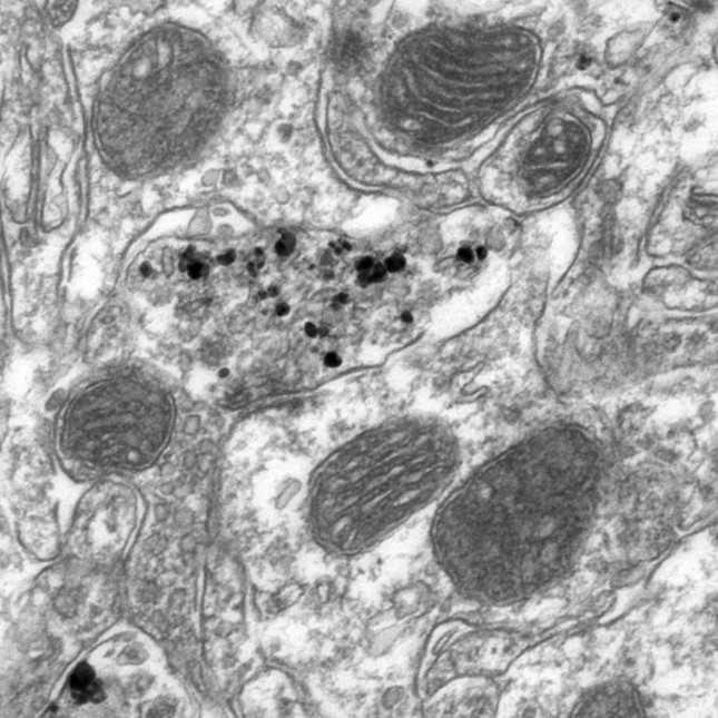
Figure 3. Analysis of a non-synaptic dopaminergic axon terminal using ImageJ and PointDensity plugin. A dopaminergic axon terminal is delineated (blue line) and the outline is defined as ‘Profile border’ using the PointDensity interface (left). Vesicles are then marked using the multi-point selection tool and defined as ‘Points’; a few vesicles are marked here for illustration (green).
Open the first EM image for analysis in ImageJ and load the PointDensity plugin.
Trace the plasma membrane of the immunopositive axon profile using the ImageJ polygon tool. Close the border. Designate the selection as ‘Profile border’ in the PointDensity panel (See Figure 3).
Place a point marker in the centre of each vesicle using the ImageJ multi-point tool. Vesicles should be counted if at least 50% of the vesicle membrane is visible. Define the selection as ‘Particles’ on the PointDensity panel (Fig 3).
Carefully name and save all data.
Analysis of synapse-forming axon profiles: All axon terminals that are forming synapses can be analysed with the PointDensitySyn tool. Dopaminergic terminals in the striatum normally form symmetric synapses (Gray’s type II) which are characterized by the presence of presynaptic and postsynaptic membrane specializations of similar thickness, a widened synaptic cleft and cleft material.
Expected result
Figure 4. Analysis of a synapse-forming dopaminergic axon terminal using ImageJ and PointDensitySyn plugin. An axon terminal is delineated (blue line) and defined as ‘Plasma membrane’ using the PointDensitySyn interface (left). Vesicles are then marked using the multi-point selection tool and defined as ‘Points’; a few vesicles are marked here for illustration (green). The pre-synaptic specialization is then delineated (yellow) and defined as ‘Postsynaptic density’ using the interface.
Open EM image in ImageJ and load the PointDensitySyn plugin.
As before, draw around and define the profile border and then mark and define all vesicles (See Figure 4).
Trace the length of the active zone(s) with the ImageJ segmented line tool. The active zone is defined as the length of plasma membrane directly opposing the postsynaptic density, on the presynaptic side of the synaptic cleft (Fig 4).
Name and save all data.
Processing coordinate files: To compute the values of interest from the co-ordinates saved for each analyzed EM image, the text files need to be processed with the second (Python) component (Larsson et al., 2015).
Once all images have been analysed in ImageJ, with either the PointDensity or PointDensitySyn plugins you can run the related Python component of the tool, i.e., PointDensity or PointDensitySyn.
Add all profile coordinate files to the input file list, check your options, and run (Larsson et al., 2015).
Values can be exported to Excel or tabulated text files depending on your preference.