May 12, 2026
Quantification of Nuclear Mitochondrial DNA Insertions (NUMTs) Using Fiber-FI
- Jeevan Labh1
- 1Manipal University
- Jeevan Labh: Currently graduated from Manipal University


Protocol Citation: Jeevan Labh 2026. Quantification of Nuclear Mitochondrial DNA Insertions (NUMTs) Using Fiber-FI. protocols.io https://dx.doi.org/10.17504/protocols.io.4r3l2ddmjg1y/v1
License: This is an open access protocol distributed under the terms of the Creative Commons Attribution License, which permits unrestricted use, distribution, and reproduction in any medium, provided the original author and source are credited
Protocol status: In development
We are still developing and optimizing this protocol
Created: May 11, 2026
Last Modified: May 12, 2026
Protocol Integer ID: 316819
Keywords: Numtogenesis, NUMTs, Hybridization, Western Blot, fi nuclear mitochondrial dna insertion, nuclear mitochondrial dna insertion, mitochondrial dna, detection of mtdna insertion, fragments of mitochondrial dna, labeled mtdna probe, quantification of mtdna integration event, mtdna probe, mtdna insertion, mitochondrial sequence, detailed guidance for mitochondrial isolation, stretched nuclear dna fiber, mtdna integration event, nuclear dna fiber, mitochondrial isolation, mtdna, nuclear genome, fish imaging, fluorescence in situ hybridization, reproducible application in mammalian cell system
Disclaimer
The images were computationally generated using multiple LLMs.
Abstract
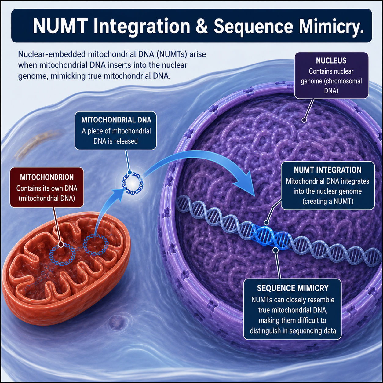
Nuclear mitochondrial DNA insertions (NUMTs) are fragments of mitochondrial DNA (mtDNA) integrated into the nuclear genome through an ongoing evolutionary and pathological process termed NUMTogenesis. Accurate visualization and quantification of NUMTs at the single-molecule level remain technically challenging due to the repetitive nature of mitochondrial sequences and limitations of short-read sequencing approaches. Here, we describe a Fiber-Fluorescence In Situ Hybridization (Fiber-FISH)-based protocol for the direct visualization and quantification of mtDNA integration events into stretched nuclear DNA fibers. This protocol combines differential mitochondrial and nuclear isolation, fluorescently labeled mtDNA probes, and high-resolution Fiber-FISH imaging to identify and quantify NUMT events. The method enables the detection of mtDNA insertions at the single-fiber level and can be adapted to evaluate NUMTogenesis under physiological or pathological conditions.
This protocol is designed for reproducible application in mammalian cell systems and provides detailed guidance for mitochondrial isolation, nuclear purification, probe preparation, slide preparation, hybridization, detection, and image analysis.
Image Attribution
Fragments of mitochondrial DNA embed themselves in the nuclear DNA, turning into NUMTs that mimic complete mitochondrial DNA.
Guidelines
Follow standard BSL 2 safety protocols to prevent contamination.
Materials
| Reagents | |
| Biotin-16-dUTP | |
| Bovine Serum Albumin (BSA) | |
| Centri-Sep Spin Column | |
| Chicken anti-rabbit Alexa Fluor 647 antibody | |
| Dextran sulfate | |
| Digoxigenin-11-dUTP | |
| DNase I/DNA Polymerase I Nick Translation Kit | |
| DNP-11-dUTP | |
| EDTA | |
| Ethanol | |
| Formamide | |
| Glacial acetic acid | |
| Goat anti-biotin antibody conjugated with avidin | |
| HEPES | |
| IGEPAL CA-630 | |
| Mannitol | |
| Mouse anti-digoxigenin antibody | |
| Phosphate-Buffered Saline (PBS) | |
| PMSF | |
| Poly-L-Lysine-coated microscope slides | |
| Rabbit anti-DNP antibody | |
| Rabbit anti-mouse Alexa Fluor 568 antibody | |
| Salmon sperm DNA | |
| Sarkosyl | |
| SDS | |
| SSC Buffer | |
| Streptavidin-Alexa Fluor 488 | |
| Sucrose | |
| Triton X-100 |
Table 1: List of Reagents used in this experiment.
| Equipment | |
| Refrigerated centrifuge | |
| Glass homogenizer | |
| Incubator | |
| Water bath | |
| Zeiss Axioplan 2 fluorescence microscope | |
| CCD camera | |
| Thermal cycler | |
| Microcentrifuge | |
| Humidified hybridization chamber |
Table 2: List of Equipment required for this experiment.
Troubleshooting
Problem
Faint band in nuclear lane of SDS-PAGE
Solution
This shows contamination in nuclear DNA. Repeat washing steps.
Isolation of Mitochondria
Harvest cultured cells by scraping into 5 ml ice-cold PBS.
Centrifuge at 800 × g for 5 min at 4 °C.
Discard the supernatant and resuspend the pellet in 5 ml Solution A.
Solution A: 20 mM HEPES-KOH, pH 7.6; 220 mM mannitol; 70 mM sucrose; 1 mM EDTA; 0.5 mM PMSF; 2 mg/ml BSA
Homogenize cells using a glass homogenizer with 20–30 gentle strokes on ice.
Centrifuge homogenate at 800 × g for 5 min at 4 °C.
Transfer the supernatant to a new tube.
Centrifuge at 10,000 × g for 10 min at 4 °C.
Discard the supernatant carefully.
Resuspend the mitochondrial pellet in Solution B.
Solution B: 20 mM Hepes-KOH pH 7.6; 220 mM mannitol; 70 mM sucrose; 1 mM EDTA; 0.5 mM PMSF
Perform a clarifying centrifugation at 800 × g for 5 min.
Transfer the supernatant and centrifuge again at 10,000 × g for 10–20 min.
Resuspend the final mitochondrial pellet in sucrose buffer.
Store on ice until slide preparation.
Isolation and Purification of Nuclei with minimal Mitochondrial contamination
Resuspend harvested cells in nuclear isolation lysis buffer containing 10% IGEPAL.
Nuclear Lysis Buffer: 10 mM HEPES, pH 7.9; 10 mM KCl; 0.1 mM EDTA
Incubate for 10 min at room temperature.
Centrifuge at 15,000 × g for 3 min.
Carefully discard the supernatant.
Repeat the lysis procedure 2–3 additional times to minimize mitochondrial contamination.
Resuspend purified nuclei in PBS.
Negative Quality Control (Western Blot for mitochondrial encoded COXII Protein)
Lyse the purified nuclei in RIPA Buffer.
RIPA Buffer: Tris-HCl, 50 mM; Sodium Chloride, 150 mM; Triton X-100, 1%; Sodium deoxycholate, 0.5%; Sodium lauryl sulphate, 0.1%
High powered sonication (3 sets of 10 seconds sonication at 20% amplitude) is required to break the nuclear envelope and shear the DNA.
BCA Assay is used to quantify the total Protein concentration.
Western Blotting Parameters:
4-20% Gradient SDS-PAGE on 0.2 μm PVDF membrane.
Use 5% non-fat milk in TBST for 1 hour for blocking.
Primary Antibody: Rabbit Polyclonal or Mouse Monoclonal Anti-COXII, 1:1000 in 5% BSA, overnight at 4℃.
Secondary Antibody: HRP-conjugated secondary; 1:5000 in 5% non-fat milk.
Strong band in mitochondrial lane, and no band in nuclear lane signifies purity. Use twice the protein load in nuclear lane for more accurate results.
Probe Preparation & Labeling
Probes were prepared using PCR primers as developed by Singh et al., 2018.
Verify PCR products by agarose gel electrophoresis.
Purify amplified products using Centri-Sep spin columns according to the manufacturer’s instructions.
Prepare the nick translation reaction mixture: 5 μl 0.2 mM dATP/dCTP/dGTP mixture; 1 μl labeled nucleotide (biotin-16-dUTP, digoxigenin-11-dUTP, or DNP-11-dUTP); 1 μg probe DNA; 5 μl DNase I/DNA polymerase mixture; Distilled water to 50 μl total volume
Mix gently and centrifuge briefly.
Incubate at 15 °C for 120 min.
Heat inactivate at 65 °C for 10 min.
Allow tubes to cool at room temperature for 15 min.
Add 6.25 μl 4 M ammonium acetate and 5 μl 100% ethanol.
Centrifuge at 12,000 rpm for 5 min.
Remove supernatant carefully.
Resuspend labeled probe pellet in 20 μl 100% formamide.
Store at −20 °C until use.
mtDNA Fiber Preparation
Apply 5 μl resuspended mitochondrial DNA to the center of a Poly-L-Lysine-coated slide.
Add 15 μl Slide Preparation Lysis Buffer 1. (2% Sarkosyl; 0.25% SDS; 50 mM Tris-HCl, pH 7.4; 50 mM EDTA, pH 8.0; 0.5% Triton X-100)
Incubate slides at 37 °C for 10 min.
Gently place a 22 × 22 mm coverslip over the suspension.
Fix fibers at 60 °C for 30 min.
Remove coverslips using a 3:1 ethanol:glacial acetic acid solution.
Dry slides at 60 °C for 15 min.
Nuclear DNA Fiber Preparation
Deposit 2 μl nuclei suspension on one end of a Poly-L-Lysine slide.
Allow the suspension to dry briefly.
Add 10 μl Slide Preparation Lysis Buffer 2. (0.5% SDS; 5 mM EDTA; 100 mM Tris-HCl, pH 7.0)
Incubate at room temperature for 4 min.
Extend DNA fibers using a coverslip.
Air-dry slides completely.
Fix slides in 3:1 ethanol:glacial acetic acid for 2 min.
Heat slides at 60 °C for 30 min.
Hybridization
Prepare hybridization mixture containing labeled probes. (50 ng DNA probe; 50% formamide; 10% dextran sulfate; 10 mM EDTA; 3 M NaCl; 5 μg salmon sperm DNA)
Apply probe mixture to prepared DNA fibers.
Cover with a coverslip and seal edges.
Incubate slides overnight at 37 °C in a humidified chamber.
Post-Hybridization Wash
Wash slides twice in 2× SSC for 5 min each at room temperature.
Wash in 50% formamide/2× SSC at 42 °C for 10 min.
Wash in 2× SSC at 42 °C for 10 min.
Air-dry slides protected from light.
Detection of Labeled Probes
For Digoxigenin labeled probes
- Incubate slides with mouse anti-digoxigenin antibody (1:200 dilution).
- Wash with PBS.
- Incubate with rabbit anti-mouse Alexa Fluor 568 antibody (1:200 dilution).
Biotin-labeled probes:
- Incubate slides with goat anti-biotin antibody conjugated with avidin (1:200 dilution).
- Wash with PBS.
- Incubate with streptavidin-Alexa Fluor 488 (1:200 dilution).
DNP-Labeled Probes:
- Incubate slides with rabbit anti-DNP antibody.
- Wash with PBS.
- Incubate with chicken anti-rabbit Alexa Fluor 647 antibody.
- Wash slides thoroughly and mount using antifade mounting medium.
Image Acquisition & Analysis
Acquire Fiber-FISH images using a Zeiss Axioplan 2 fluorescence microscope equipped with a CoolSNAP HQ2 CCD camera.
Capture images using AxioVision 4.8 software.
Process image contrast uniformly using Adobe Photoshop CS5.
Perform signal measurements and fiber length analysis using ImageJ software.
Quantify NUMT events by identifying mtDNA probe signals localized within nuclear DNA fibers.
Quantification Strategy
Count total nuclear DNA fibers analyzed.
Count fibers containing mtDNA integration signals.
Calculate NUMT frequency as: NUMT Frequency = (Number of fibers containing mtDNA signals / Total nuclear fibers analyzed) × 100
Measure insertion signal length where applicable.
Protocol references
Koo DH, Singh B, Jiang J, Friebe B, Gill BS, Chastain PD, Manne U, Tiwari HK, Singh KK. Single molecule mtDNA fiber FISH for analyzing numtogenesis. Anal Biochem. 2018 Jul 1;552:45-49. doi: 10.1016/j.ab.2017.03.015. Epub 2017 Mar 18. PMID: 28322800; PMCID: PMC5814351.
Singh, K. K., Choudhury, A. R., & Tiwari, H. K. (2017). Numtogenesis as a mechanism for development of cancer. Seminars in cancer biology, 47, 101–109. https://doi.org/10.1016/j.semcancer.2017.05.003
Schall, P. Z., Meadows, J. R. S., Ramos-Almodovar, F., & Kidd, J. M. (2024). Characterization of nuclear mitochondrial insertions in canine genome assemblies. openRxiv. https://doi.org/10.1101/2024.09.13.612826
Harutyunyan T. (2024). The known unknowns of mitochondrial carcinogenesis: de novo NUMTs and intercellular mitochondrial transfer. Mutagenesis, 39(1), 1–12. https://doi.org/10.1093/mutage/gead031
Acknowledgements
I would like to thank Dr. Sanjiban Chakrabarty for his guidance and for encouraging me to study NUMTs.