Feb 19, 2026
Version 2
Purification of SerMNucA (Benzonase/Turbonuclease) as 10xHis, MBP or myc-tag fusions V.2
- Lucas Rieubon1,
- Michael Lim2,
- Kelvin Lau1,
- Florence Pojer1
- 1EPFL SV PTPSP;
- 2King's College London
- EPFL SV PTPSP



Protocol Citation: Lucas Rieubon, Michael Lim, Kelvin Lau, Florence Pojer 2026. Purification of SerMNucA (Benzonase/Turbonuclease) as 10xHis, MBP or myc-tag fusions. protocols.io https://dx.doi.org/10.17504/protocols.io.261ged7xov47/v2Version created by Kelvin Lau
License: This is an open access protocol distributed under the terms of the Creative Commons Attribution License, which permits unrestricted use, distribution, and reproduction in any medium, provided the original author and source are credited
Protocol status: Working
We use this protocol and it's working
Created: February 17, 2026
Last Modified: February 19, 2026
Protocol Integer ID: 243545
Keywords: nuclease, endonuclease, dna, rna, benzonase, turbonuclease, dnase, rnase, purification, protein purification, purification of the sermnuca nuclease, protein purification alongside other commercial enzyme, sermnuca nuclease, purification of sermnuca, enzymatic digestion by nuclease, removal of nucleic acid, purification from either periplasm, common protein purification protocol, large release of nucleic acid, latter enzyme, other commercial enzyme, enzyme, nucleic acid, purified protein, use of the enzyme, nucleic acids research, enzymatic digestion, such as benzonase, stranded dna, rna with high activity, sermnuca endonuclease, untagged versions of the sermnuca endonuclease, protein from either fraction, tag fusions during the lysi, presence of dna, recombinant versions of the serratia, cleavable with tev protease, protein, lysis of cell
Funders Acknowledgements:
EPFL
Grant ID: SV Core Funding
Abstract
From the Author (KL) : all the plasmids are available here > > https://www.addgene.org/browse/article/28244500/
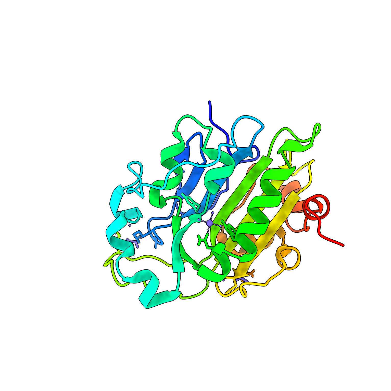
During the lysis of cells, in particular of eukaryotic origin, there is a large release of nucleic acids. The presence of DNA/RNA increases the viscosity of a lysate and may pose problems during downstream processes. Removal of nucleic acids can be performed in many ways, of which one is by enzymatic digestion by nucleases. DNase/RNase are commonly added during protein purification alongside other commercial enzymes such as Benzonase (Novagen) and Turbonuclease. The latter enzymes are industrially produced or recombinant versions of the Serratia marcescens nuclease. This enzyme, hereafter referred to as SerMNucA, is able to hydrolyze both single- and double-stranded DNA and RNA with high activity.
Reported methods have shown that the nuclease can be made as inclusion bodies followed by refolding or secreted in the extracellular media. Here we report the purification of the SerMNucA nuclease in a soluble form, followed by purification from either periplasm or cell lysate. We find that purified protein from either fraction are active in a salmon sperm degradation assay with the periplasmic fraction slightly more. One can choose to purify from either fraction or from the total lysate.
This protocol allows for a lab to independently produce tagged and untagged versions of the SerMNucA endonuclease in large quantities that may be required for certain experiments that may be cost prohibitive otherwise.
3 versions are reported here:
10x-His tagged
MBP tagged (From SGC Toronto/Oxford)
myc tagged
The use of the enzyme is in such small quantities that repurification with the tag should not be an issue. The MBP tagged version is cleavable with TEV protease (see also: 10x-His-SuperTEV). Finally the myc-tagged version should not interfere with any common protein purification protocols.
References:
Benedik and Strych. (1998) FEMS Microbiology Letters
Biedermann et al. (1989) Carlsberg Research Communications
Friedholl et al. (1996) Nucleic Acids Research
Guidelines
This protocol is written with the expectation of standard bacterial culture and basic purification knowledge
Materials
Plasmid #1
Plasmid #2
Plasmid #3
BL21 (DE3) cells (Lucigen)
TB + trace elements (Formedium)
Glycerol (Applichem)
Akta system (Cytiva)
HisPur NiNTA resin (Pierce/Thermo)
Amylose resin (NEB)
Hitrap Q HP 5 mL column (Cytiva)
12-14 kDa dialysis tubing (SpectraPor)
Dialysis clips (SpectraPor)
Sonicator
5 M NaCl
1 M HEPES, pH 7.5
1 M TRIS, pH 8.0
0.5 M EDTA, pH 8.0
2.5 M imidazole, pH 7.5 (We recommend, Sigma #56749-1KG for low background absorbance)
1 M MgCl2
1 M IPTG
Sucrose
4X LDS loading dye (Genscript)
NuPage 4-20% SDS-PAGE Gels (Thermofisher)
8M Urea
Salmon testes DNA (Rockland)
Plastic disposable or glass reusable columns (Biorad)
Before start
Recommended to have ready before starting alongside Materials.
-Autoclaved flasks and bottles
-Autoclaved LB media
-Autoclaved TB media
-Buffers
-Liquid nitrogen
Growing bacterial cultures (2L) and periplasmic extraction
4d
Transform the bacterial plasmid expressing the construct of interest (His, MBP or myc fusions) into BL21 (DE3) cells. Plate on to LB-Agar plates + Kanamycin. Grow Overnight ]at Temperature 37 °C or over the weekend at Temperature 25 °C .
1d
In an afternoon, pick a streak of cells and innoculate into 200 mL of LB + Kanamycin media. Note: for every 1 L of expression culture, you will require 10-20 mL of pre-culture. Grow Overnight at 37 °C
1d
The next morning, add MgCl2 to 2 millimolar (mM) , innoculate 20-40 mL of preculture into 1 flasks containing 2 L of TB media (2 L in a 5 L flask is appropriate). Shake in an incubator at 37 °C for 3-4 hours until OD600 ~0.8-1. Take a 1 mL sample of the culture, this will be used as a pre-induction control sample. Immediately change the temperature to 18 °C , add IPTG to 0.5 millimolar (mM) , continue shaking overnight (approximately 20:00:00 )
20h
Take a 1 mL sample of the culture and measure the OD600, this will be used as a whole cell post-induction sample. Harvest the culture by centrifuging at 5000 x g, 10°C, 00:30:00 .
30m
SerMNucA has been designed to be secreted into the oxidizing environment of the periplasm of E.coli to enable proper folding. Proteins from this particular compartment can be extracted by osmotic shock.
Prepare the following:
| A | B | C | |
| Buffer | Component | Concentration (mM) | |
| TES 1X | TRIS, pH 8 | 200 | |
| EDTA, pH 8 | 0.5 | ||
| Sucrose | 500 |
10m
For every 2 L of culture, add 20 mL of ice-cold 1X TES buffer and gently rotate or mix for at least 02:00:00 at 4 °C . This will resuspend the pellet without disrupting the cell membrane or cell wall.
Then mix 30 mL of ice-cold 0.25 X TES buffer (ie. 1X TES diluted with water to 0.25X) to the resuspend pellet above to generate the osmotic shock. This mixture is then rotated and mixed for another02:00:00 at 4 °C . The periplasmic shocked mixture was recovered by centrifuging at 20000 x g, 10°C, 00:30:00 . The supernatant contains the periplasmic extract and desired protein. Take a 1 mL sample. The pellet can be saved if one choose to purify from this compartment as well. Both fractions can be stored at -20 °C .
5h
Confirm expression by running an SDS-PAGE gel.
Recommended recipe to prepare SDS-PAGE samples. Mix 1:4, 4X LDS loading dye and 8 M Urea, to make a Urea loading dye.
For samples before and after induction with IPTG, calculate the amount of sample needed to prepare. We use the following formula:
1/OD600 x 100 uL = volume in uL to centrifuge down 14000 rpm, 00:01:00 .
Discard the supernatant, Keep the pellet.
Resuspend pellet in 40 uL of the Urea loading dye.
For the osmotic shock supernatant, mix 30 uL of sample and 10 uL of 4X loading dye
Load 10 uL on to a NuPage 4-12% bis-Tris SDS-PAGE gel.
Expected result
Typical expression pattern and periplasmic extracts are shown here
1h
Prepare purification buffers
From stock solutions prepare the following as needed :
Filter 0.22 or 0.45 um
| A | B | C | D | E | |
| Buffer | Component | Concentration (mM) | |||
| Wash buffer (1 L) | NaCl | 700 | |||
| HEPES, pH 7.5 | 20 | ||||
| Dialysis buffer | NaCl | 100 | |||
| TRIS, pH 8 | 100 | ||||
| MgCl2 | 10 | ||||
| If performing Ni-NTA | |||||
| Ni-NTA Elution buffer (100 mL) | NaCl | 700 | |||
| HEPES, pH 7.5 | 20 | ||||
| Imidazole, pH 7.5 | 500 | ||||
| If performing Amylose | |||||
| Amylose Elution buffer (100 mL) | NaCl | 700 | |||
| HEPES, pH 7.5 | 20 | ||||
| Maltose | 10 | ||||
| If performing anion-exchange | |||||
| Q-Low buffer (1 L) | HEPES, pH 7.5 | 20 | |||
| Q-High (1 L) | NaCl | 1000 | |||
| HEPES, pH 7.5 | 20 | ||||
Buffer lists for purification
Option 1 : Purification by Ni-NTA (10xHis construct)
2h 40m
Defrost the pellet, and resuspend in40 mL of wash buffer and glycerol to 10% v/v. Cells are lysed by sonication and clarified by 20000 x g, 4°C, 00:30:00 . Filter the supernatant with a 0.45 um membrane and supplement with imidazole to 25 millimolar (mM) .
Defrost the periplasmic extract and supplement with imidazole to 25 millimolar (mM) .
40m
Add 5 mL of settled NiNTA resin already equilibrated in wash buffer to the supernatant (SN) and the periplasmic extract. Mix the resin gently for 02:00:00 at 4 °C
2h
Transfer the resin to a column and collect the flowthrough (FT). The resin is then washed and the target protein eluted as follows:
| A | B | C | |
| Step | Concentration imidazole (mM) / %Elution buffer | Volume (mL) | |
| Wash 1 | 20 mM / 4% | 4 x 25 mL | |
| Wash 2 | 50 mM / 10% | 4 x 12.5 mL | |
| Wash 3 | 100 mM / 20% | 2 x 12.5 mL | |
| Elute 1 | 300 mM / 60% | 5 x 10 mL | |
| Elute 2 | 500 mM / 100% | 1 x 15 mL | |
Wash and Elute buffers are made by mixing proportionally from the stock Wash and Elution buffers
1h
Fractions from each step are run on a gel and the purest fractions are pooled. A typical purification is as follows.
Fractions containing the target protein at a desired purity can be pooled and dialyzed into dialysis buffer overnight at 4 °C .
Expected result
Typical SDS-PAGE gel of a purification from both periplasmic extract and cell pellet. Additional bands purified from the cell pellet could be potential contaminants requiring additional purification steps or higher order oligomers.
40m
After dialysis, the sample was concentrated to around 1 mg/mL diluted with glycerol to 50 % (v/v) and run on a gel to verify integrity and purity. The purified SerMNucA-10xHis can be stored at -20 °C .
Expected result
Sample after dialysis and dilution with glycerol
Note
1) Increased purity can be achieved by adding additional steps. (See anion exchange section or by size-exclusion chromatography)
2) Both the periplasmic and cell pellet gives active protein (See activity assay section). However the purified protein from the cell pellet is observed to be forming higher-order oligomeric states. This is most likely mediated by improper disulfides formed within the cytosol. The periplasmic extract does not contain these species.
30m
Option 2 : Purification by Amylose (MBP construct)
4h 10m
Defrost the pellet, and resuspend in40 mL of wash buffer and glycerol to 10% v/v. Cells are lysed by sonication and clarified by 20000 x g, 4°C, 00:30:00 . Filter the supernatant with a 0.45 um membrane.
Defrost the periplasmic extract.
30m
Add 5 mL of settled amylose resin already equilibrated in wash buffer to the supernatant (SN) and the periplasmic extract. Mix the resin gently for 02:00:00 at 4 °C
2h
Transfer the resin to a column and collect the flowthrough (FT). The resin is then washed and the target protein eluted as follows:
| A | B | C | |
| Step | %Elution buffer | Volume (mL) | |
| Wash | 0 | 4 x 25 mL | |
| Elute | 100 | 5 x 10 mL | |
Wash and Elute buffers are made by mixing proportionally from the stock Wash and Elution buffers
1h
Fractions from each step are run on a gel and the purest fractions are pooled. A typical purification is as follows.
Fractions containing the target protein at a desired purity can be pooled and dialyzed into dialysis buffer overnight at 4 °C .
Expected result
Typical SDS-PAGE gel of a purification from both periplasmic extract and cell pellet.
40m
After dialysis, the sample was concentrated to around 1 mg/mL diluted with glycerol to 50 % (v/v) and run on a gel to verify integrity and purity. The purified MBP-SerMNucA can be stored at -20 °C .
Expected result
Sample after dialysis and dilution with glycerol
Note
1) We observed degradation of the fusion construct. Increased purity can be achieved by adding additional steps. (See anion exchange section or by size-exclusion chromatography).
2) TEV cleavage by TEV protease can also be performed. It should be noted that one should remove the protease if downstream use are with protein samples that contain TEV or 3C protease cleavage sites.
3) Both the periplasmic and cell pellet gives active protein (See activity assay section).
Option 3 : Purification by Anion-Exchange Chromatography (myc tagged)
30m
Defrost the pellet, and resuspend in40 mL of Q-Low buffer and glycerol to 10% v/v. Cells are lysed by sonication and clarified by 20000 x g, 4°C, 00:30:00 . Filter the supernatant with a 0.45 um membrane.
or
Defrost the periplasmic extract.
30m
Using an AKTA system, load the lysate sample or periplasmic extract on to a 5 mL HiTrap Q column (5 mL = 1 CV) equilibrated with Q-Low buffer.
Once loaded, run a gradient from 0 to 100% Q-High buffer over 16 CV.
The protein should elute between 20-30 mS/cm of conductivity
Fractions from each step are run on a gel and the purest fractions are pooled. A typical purification is as follows.
Fractions containing the target protein at a desired purity can be pooled and dialyzed into dialysis buffer overnight at 4 °C .
Expected result
Typical chromatograph and gel of fractions
After dialysis, the sample was concentrated to around 1 mg/mL diluted with glycerol to 50 % (v/v) and run on a gel to verify integrity and purity. The purified SerMNucA-2xmyc can be stored at -20 °C .
Expected result
Sample after dialysis and dilution with glycerol
Note
1) The purification from the pellet is not shown. What is shown in the final sample was an anion exchange step as for the periplasmic fraction, followed by a size exclusion on fractions that were enriched with the band of correct size.
2) Both the periplasmic and cell pellet gives active protein (See activity assay section).
Activity Assay
30m
1) Prepare Assay buffer (10 mL) : 50 millimolar (mM) TRIS, pH 8, 1 millimolar (mM) MgCl2
2) Dilute Salmon testes DNA to 2 mg/mL in Assay Buffer
3) Dilute enzyme to 0.005 mg/mL in Assay Buffer
4) Each reaction contains 25.6 µL of Salmon Testes DNA + in increasing concentrations of enzyme, total volume was 50 µL of reaction completed with buffer. In this example, His-tagged nuclease purified from either the periplasm or the pellet fraction.
5) Incubate at 37 °C for 00:30:00
6) Visualize the DNA bands on a 1% Agarose gel
Expected result
His tagged version
Expected result on an agarose gel showing the degradation to smaller DNA pieces in the presence of increasing concentrations of enzyme (His-tagged)
Expected result
myc tagged version
Expected result on an agarose gel showing the degradation to smaller DNA pieces in the presence of increasing concentrations of enzyme (myc-tagged)
Expected result
MBP tagged version
Expected result on an agarose gel showing the degradation to smaller DNA pieces in the presence of increasing concentrations of enzyme (MBP-tagged)
30m
Additional Validation versus "Vendor" Nuclease
Data from Michael Lim (Kings College). His tagged protein was purified via gravity flow (no batch binding) and assayed with indicated conditions compared to a commercial vendor's enzyme using their indicated activity units
Expected result
Acknowledgements
We thank Jesse Coker, Michael Fairchild, Eleanor Williams and SGC Toronto/Oxford for providing the MBP (Benzonase) plasmid. All plasmids have been deposited under https://www.addgene.org/browse/article/28244500/