Jun 15, 2026
Protocol for Hematoxylin and Eosin Staining in Vaginal Cytology
- Maria Flávia Santos Donadeli1,
- Patrícia Passaglia2,
- Luiz Guilherme de Siqueira Branco3
- 1Faculdade de Medicina de Ribeirão Preto-Universidade de São Paulo (FMRP-USP);
- 2Faculdade de Odontologia de Ribeirão Preto- Universidade de São Paulo (FORP-USP);
- 3Faculdade de Odontologia de Ribeirão Preto-Universidade de São Paulo (FORP-USP)
- Maria Flávia Donadeli


Protocol Citation: Maria Flávia Santos Donadeli, Patrícia Passaglia, Luiz Guilherme de Siqueira Branco 2026. Protocol for Hematoxylin and Eosin Staining in Vaginal Cytology. protocols.io https://dx.doi.org/10.17504/protocols.io.j8nlk75z1g5r/v1
License: This is an open access protocol distributed under the terms of the Creative Commons Attribution License, which permits unrestricted use, distribution, and reproduction in any medium, provided the original author and source are credited
Protocol status: Working
We use this protocol and it's working
Created: June 12, 2026
Last Modified: June 15, 2026
Protocol Integer ID: 319073
Keywords: Hematoxylin and eosin, vaginal cytology and estrous cycle, murine reproductive cycle, estrous cycle in rodent, gonadotropic hormone, luteinizing hormone, ovarian steroid, menopause induction, endocrine change, progesterone, main endocrine change, estrous cycle, reproductive cycle, vaginal cytology, hormonal interaction, vaginal cytology vaginal cytology, reproductive alteration, morphology of the vaginal epithelium, ovariectomy effectiveness, crucial tool in murine model, assessment of ovariectomy effectiveness, estradiol, hormonal interactions under various pathological condition, vaginal epithelium, murine model, stimulating hormone, stage of the cycle, alterations in the cellular composition, cycle, follicle, fluctuations in serum level, predominant cell types in each phase, estrus, murine
Funders Acknowledgements:
CAPEs
Grant ID: 88887.110827/2025-00
Abstract
Vaginal cytology is a rapid and effective method for assessing the estrous cycle in rodents. It is widely used not only to investigate reproductive alterations but also to evaluate hormonal interactions under various pathological conditions. Additionally, it is a crucial tool in murine models of menopause induction, enabling the assessment of ovariectomy effectiveness. The murine reproductive cycle has four phases: proestrus, estrus, metestrus, and diestrus. Fluctuations in serum levels of ovarian steroids such as 17β-estradiol and progesterone, and gonadotropic hormones, including luteinizing hormone (LH) and follicle-stimulating hormone (FSH), regulate transitions between these phases. These endocrine changes manifest as alterations in the cellular composition and morphology of the vaginal epithelium. Here, we describe a simple and effective protocol for determining the estrous cycle in rodents using hematoxylin and eosin (H�26E) staining. We detail the predominant cell types in each phase, their morphological features, and the main endocrine changes associated with each stage of the cycle.
Image Attribution
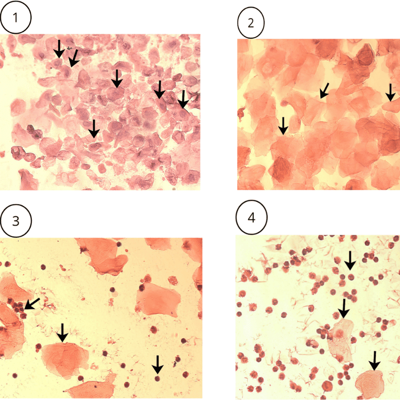
Fig. 3. The images above show the phases of the estrous cycle in female mice, the laminae
were stained with HE (40X magnification). (Image 1) Proestrus: cluster of nucleated epithelial cells
highlighted by arrows. (Image 2) Estrus: predominant presence of cornified squamous epithelial cells and
anucleated (black arrows). (Image 3) Metaestrus: simultaneous presence of nucleated cells, epithelial cells
anucleated cornified cells and leukocytes shown by arrows. (Image 4) Diestrus: predominance of leukocytes and rare
nucleated and cornified epithelial cells as shown by arrows.
Guidelines
Animal handling as well as tissue collection for this protocol requires prior approval from the user's Institutional Animal Care and Use Committee (IACUC) or equivalent ethics committee.
Materials
- Microscope slides
- Coverslips
- Sterile 0.9% saline solution
- Calibrated pipette for 30 µL
- Disposable pipette tips
- Hematoxylin solution (Product Reference: H9627-25G-Sigma)
- Eosin solution (Product Reference: E6003-25G-Sigma)
- Milli-Q water
- Falcon tubes (50 mL)
- Xylene
- Mounting medium (Entellan®)
- Pasteur pipette
- Optical microscope
**Preparation of Staining Solutions**
**Harris Hematoxylin Solution**
- Hematoxylin Crystals: 5g
- Absolute Ethanol: 50mL
- Potassium or Ammonium Alum: 100g
- Mercuric Oxide (Red): 2.5g
- Distilled Water: 1000mL
**Acidified Alcoholic Eosin Y Solution**
- Eosin Y (yellowish): 5g
- Distilled Water: 10mL
- 95% Ethanol: 450 mL
- Glacial acetic acid: 2.5 mL
Before start
Prepare the microscope slides by labeling them with the date, animal number, and corresponding experimental group. Then, divide each slide into five equivalent areas, as shown in the figure below:
Before Starting Vaginal Cytology
Prepare the microscope slides by labeling them with the date, animal number, and corresponding experimental group. Then, divide each slide into five equivalent areas, as shown in the figure below:
Fig. 1. Appearance of the delimited sections on the glass slide before vaginal cytology.
Preparation of Staining Solutions
For preparing 1 liter of this solution, a 2-liter Erlenmeyer flask should be used.
Dissolve the hematoxylin crystals in absolute ethanol and the potassium alum in cold distilled water, allowing both solutions to dissolve overnight. The following day, heat the alum solution to a boil on a Bunsen burner. After boiling, remove the solution from the heat and slowly combine the two solutions. Then heat the mixture again until it begins to boil, for approximately 1 minute. Subsequently, remove the solution from the heat and slowly add the red mercuric oxide. Reheat the solution until it reaches the boiling point once more. After this step, remove the solution from the heat and cool it by placing the flask in a container with cold water. Once the solution has completely cooled, filter it and add 2 mL of acetic acid per 500 mL of working solution.
Dissolve 5.0 g of Eosin Y in 10 mL distilled water.
Add 2.5 mL of glacial acetic acid and mix thoroughly.
Adjust the final volume to 500 mL with 95% ethanol.
Homogenize the solution and filter before use.
Step-by-Step Procedure for Vaginal Lavage
For this procedure, a calibrated 30 µL pipette and sterile 0.9% saline solution are used. Initially, the saline solution is aspirated into the pipette. Next, the animal is removed from the cage and positioned with its posterior region facing the operator. The tail is firmly held while gently elevating the animal’s hindquarters to facilitate access to the vaginal opening. During this process, the animal may urinate; if this occurs, it is recommended to wait until urination has ceased. If urine residues remain at the entrance of the vaginal canal, the area should be washed with distilled water using a different pipette tip from the one intended for sample collection.
With the pipette tip filled with saline solution, it is carefully positioned at the vaginal opening without insertion, since vaginal stimulation may induce pseudopregnancy, which triggers the same endocrine mechanisms observed at the onset of normal pregnancy. This condition may last approximately 12 hours, resulting in a delay of the next estrous cycle phase. Furthermore, insertion of the pipette tip may induce inflammatory processes, compromising the quality of the cytological evaluation.
The pipette plunger is then gently pressed, gradually releasing the solution. The fluid spontaneously enters the vaginal canal. Subsequently, the pressure is slowly released, allowing the fluid to return into the pipette tip. This procedure is repeated approximately 4 times using the same pipette tip and solution to collect the greatest possible number of cells for accurate determination of the estrous cycle.
The collected material is transferred onto a previously identified glass slide and distributed within one of the demarcated areas. The slide should remain at room temperature until complete drying, approximately 30 minutes. The material may be stained immediately or stored at room temperature for subsequent staining. Vaginal cytology should be performed at least once daily, preferably at the same time each day, for five consecutive days. For this purpose, a single slide divided into five areas may be used, with each area corresponding to one collection day.
Cytological Staining with Hematoxylin and Eosin
The hematoxylin and eosin solutions should be prepared in advance as described in the staining solution preparation section. The staining solutions are then distributed into appropriate containers to allow complete immersion of the slides.
After the slides are completely dry, they are initially immersed in eosin solution for 3 minutes. Subsequently, the slides are washed with Milli-Q water to remove excess stain. The hematoxylin staining step is then performed by immersing the slides in the hematoxylin solution for 2 minutes. After this step, the slides are washed again with Milli-Q water. Next, the slides undergo dehydration by immersion in xylene. For this purpose, the xylene is placed in a 50 mL Falcon tube, into which the slides are rapidly immersed. After clarification, the slides are allowed to dry, and a small drop of mounting medium (Entellan®) is added onto each sample, followed by careful placement of the coverslip in order to permanently fix the material and preserve its integrity. Finally, the slides are analyzed under an optical microscope at 20× and 40× magnification to identify the predominant cell types and determine the estrous cycle phase for each animal.
Fig. 2. Schematic representation of the procedure for obtaining and analyzing vaginal smears in female mice. (1) Sample collection using sterile 0.9% saline solution; (2) Application of the collected material onto a glass slide; (3) Fixation and staining with hematoxylin and eosin, followed by washing in Milli-Q water and dehydration in xylene; (4) Slide mounting with a coverslip using Entellan; and (5) Microscopic analysis for identification of cell types and determination of the estrous cycle phases.
Representative Results
Upon analysis of the slides under an optical microscope, nucleated epithelial cells, cornified squamous epithelial cells, and/or leukocytes can be observed.
Cytological analyses allow the identification of the four stages of the estrous cycle: proestrus, estrus, metestrus, and diestrus. Proestrus is characterized by the predominance of nucleated epithelial cells, generally rounded and with darker cytoplasm (Image 1). This phase lasts approximately 12 to 14 hours. During estrus, clusters of cornified anucleated squamous epithelial cells are observed. These cells display a more polygonal morphology, and this phase usually lasts approximately 25 to 27 hours (Image 2). Metestrus is characterized by a similar proportion of cornified anucleated epithelial cells and nucleated epithelial cells, along with a small number of leukocytes, which differ from epithelial cells by their intensely stained nuclei and smaller size. This phase lasts approximately 6 to 8 hours (Image 3). Finally, diestrus is characterized by a predominance of leukocytes, a rare occurrence of cornified and nucleated epithelial cells, and abundant mucus in the vaginal smear. This is the longest phase and generally lasts approximately 55 to 57 hours (Image 4). The images mentioned are located in the description section of this protocol.
Endocrine Variations Throughout the Estrous Cycle Phases
The hypothalamic–pituitary–ovarian axis regulates reproductive endocrine function throughout the estrous cycle via negative and positive feedback mechanisms. In this context, during proestrus, considered the preovulatory phase, negative feedback initially occurs on the hypothalamic–pituitary–gonadal axis. This mechanism stimulates neural groups in the medial preoptic and medial septal areas to synthesize and secrete gonadotropin-releasing hormone (GnRH), which, via the hypophyseal portal system, induces secretion of follicle-stimulating hormone (FSH) and luteinizing hormone (LH) by the anterior pituitary gland. FSH regulates ovarian follicle growth, whereas LH induces ovulation and corpus luteum formation, in addition to stimulating estrogen and progesterone secretion. In this context, there is a progressive increase in 17β-estradiol and progesterone levels, produced through androgen aromatization in granulosa cells. This increase in estrogen levels results in positive feedback on the hypothalamic–pituitary–gonadal axis, stimulating the release of GnRH and, consequently, LH and FSH secretion by the anterior pituitary gland. The LH surge constitutes the main stimulus for ovulation, marking the transition from proestrus to estrus.
During the metestrus phase, the corpus luteum is formed from the ovulated follicle, a process mainly induced by LH. The corpus luteum plays a fundamental role in maintaining ovarian steroid secretion, especially progesterone and estradiol. Finally, entry into diestrus occurs when progesterone and estradiol levels increase, resulting in negative feedback on the hypothalamic–pituitary–gonadal axis and, consequently, reduced LH and FSH secretion. In this context, regression of the corpus luteum occurs, leading to decreased ovarian steroid levels and allowing the estrous cycle to restart. Unlike the human menstrual cycle, there is no shedding accompanied by bleeding, since the endometrium is reabsorbed.
Protocol references
ANDERSEN, M. L.; TUFIK, S. (Eds.). Animal Models as Tools in Ethical Biomedical Research. São Paulo: FAPESP, 2010.
AJAYI, Ayodeji Folorunsho; AKHIGBE, Roland Eghoghosoa. Staging of the estrous cycle and induction of estrus in experimental rodents: an update. Fertility Research and Practice, v. 6, n. 1, p. 5, 14 dez. 2020
ARMAIZ-PENA, Guillermo N. et al. Estrous Cycle Modulates Ovarian Carcinoma Growth. Clinical Cancer Research, v. 15, n. 9, p. 2971–2978, 1 maio 2009.
MCLEAN, Ashleigh C. et al. Performing Vaginal Lavage, Crystal Violet Staining, and Vaginal Cytological Evaluation for Mouse Estrous Cycle Staging Identification. Journal of Visualized Experiments, n. 67, 15 set. 2012
RUDOLPH, Marion et al. Induction of Overt Menstruation in Intact Mice. PLoS ONE, v. 7, n. 3, p. e32922, 7 mar. 2012.
WALL, Ellen G. et al. Unexpected Plasma Gonadal Steroid and Prolactin Levels Across the Mouse Estrous Cycle. Endocrinology, v. 164, n. 6, 17 abr. 2023