Mar 23, 2026
Protocol: Evaluation of a Low-Cost Point of Care (POC) Methods for Community-led Schistosomiasis Control: A Mixed-Methods Pilot Study
- William McCarthy1,
- Caroline Crain1,
- Tope Olubodun2,
- Ifelouwa George3
- 1University of Texas Health Science Center at Houston;
- 2FMC Abeokuta;
- 3FUNAAB
- SchistoFilter


Protocol Citation: William McCarthy, Caroline Crain, Tope Olubodun, Ifelouwa George 2026. Protocol: Evaluation of a Low-Cost Point of Care (POC) Methods for Community-led Schistosomiasis Control: A Mixed-Methods Pilot Study. protocols.io https://dx.doi.org/10.17504/protocols.io.4r3l2dpkjg1y/v1
License: This is an open access protocol distributed under the terms of the Creative Commons Attribution License, which permits unrestricted use, distribution, and reproduction in any medium, provided the original author and source are credited
Protocol status: Working
We use this protocol and it's working
Created: March 14, 2026
Last Modified: March 23, 2026
Protocol Integer ID: 313267
Keywords: Foldscope, Schistosomiasis, Diagnostics, Community Health, Nigeria, Point of Care, Public Health, schistosomiasis surveillance in resource, schistosomiasis surveillance, schistosoma haematobium in urine sample, diagnosing schistosoma haematobium, schistosomiasis control, led schistosomiasis control, diagnostic tool, accuracy of this diagnostic tool, collaboration with local community health extension worker, urine sample, rural nigerian community, eggs in urine, disease detection, based diagnostic solution, illness hotspot, diagnostic accuracy, local community health extension worker, diagnostic solution, spatial mapping of the disease vector, accessible environmental mapping strategy, based disease detection, methods pilot study, selected rural community, methods pilot study this study, cost molecular testing method in development, trapping egg
Funders Acknowledgements:
Experiment.com
Grant ID: https://experiment.com/projects/health-in-your-hands-using-accessible-solutions-to-fight-schistosomiasis
Gates Foundation
Grant ID: https://gcgh.grandchallenges.org/grant/foldscope-paper-based-use-and-throw-field-microscopes
Disclaimer
Out for peer review. PLOS NTD>
Abstract
This study aims to evaluate the effectiveness of the Foldscope, a low-cost paper microscope, for diagnosing Schistosoma haematobium in urine samples within rural Nigerian communities, particularly in rural Ogun state. The project will assess diagnostic accuracy, community acceptability, and feasibility of implementation through collaboration with local community health extension workers (CHEWs) and volunteers. The study will compare Foldscope-based microscopy workflow with conventional methods, focusing on the effectiveness and accuracy of this diagnostic tool paired with a reusable microfluidic device for trapping eggs in urine. Additionally, we will measure feasibility and community acceptability using a mixed-methods approach including focus group discussions and key informant interviews. The research will be conducted in selected rural communities of Abeokuta North and Odeda LGAs near the Oyan River Dam in Ogun State, with a sample size of 406 participants. The study employs a cross-sectional design with both quantitative and qualitative components to evaluate the diagnostic tool's performance. We also aim to evaluate accessible environmental mapping strategies to predict illness hotspots of transmission based on spatial mapping of the disease vector. Additional study methods, described below, will evaluate a low-cost molecular testing method in development and an automated detection method for accessible strategies that offload the burden of detecting the eggs in urine by a trained user. The expected benefit is the development of a sustainable, community-based diagnostic solution that could improve schistosomiasis surveillance in resource-limited settings, potentially transforming community-based disease detection and treatment strategies.
Materials
- Foldscope
- 10 mL urine sample collection
- ShistoFilter (Construction materials included here)
-Water
-Syringe (10 mL)
If comparing to gold standard:
- Schistosomiasis urine filtration kit
- Microscope
CHAPTER 0: GENERAL + QUANTITATIVE DATA ANALYSIS INFO
The protocol here is summary of the protocol used in the OGUN STATE, Nigeria SCOPE (Schistosomiasis from a One Health Perspective) Study, Phase 1: 2025, focused on Community-Led Urogenital Schistosomiasis Control. Protocol is shared here for access from recent PLOS NTD Submission: "Community-Led Diagnosis of Urogenital Schistosomiasis Using a Low-Cost, Point-of-Care Microscopy Toolkit in Rural Nigeria: A mixed-methods study"
Data Analysis - Diagnostic Accuracy Data: The following metrics will be calculated using a 2×2 contingency table in GraphPad Prism: Sensitivity = (TP/(TP + FN)) × 100; Specificity = (TN/(TN + FP)) × 100; PPV = (TP/(TP + FP)) × 100; NPV = (TN/(TN + FN)) × 100; Accuracy = (TP + TN)/(TP + TN + FP + FN) × 100; Kappa statistic (κ) = (Po - Pe)/(1 - Pe); McNemar's test will be applied to compare performance between methods; Level of agreement between trained lab scientists (Co-Investigators of study) and CHEWs will be assessed. Kappa statistics will be used to determine the level of agreement between the Foldscope and the gold standard diagnostic approach. McNemar's test will be applied to compare the performance of Foldscope versus gold standard microscopy. All calculations will be performed using GraphPad Prism's Contingency Table and Agreement Analysis tools.
Sensitivity = (TP/(TP + FN)) * 100
Specificity = (TN/(TN + FP)) * 100
PPV = (TP/(TP + FP)) * 100
NPV = (TN/(TN + FN)) * 100
Accuracy = (TP + TN)/(TP + TN + FP + FN) * 100
Kappa statistic (κ) = (Po - Pe)/(1 - Pe)
McNemar's test will be applied to compare performance between methods
Level of agreement between trained lab scientists (Co-Investigators of study) and CHEWs will be assessed Gold StandardFoldscope PositiveFoldscope NegativeTotalPositive (True Cases)TP (True Positives)FN (False Negatives) TP + FNNegative (Non-Cases)FP (False Positives)TN (True Negatives) FP + TNTotalTP + FPFN + TNN (Total Cases)● Kappa statistics will be used to determine the level of agreement between the Foldscope and the gold standard diagnostic approach.● McNemar’s test will be applied to compare the performance of Foldscope versus gold standard microscopy.Equations: Sensitivity = (TP/(TP + FN)) * 100Specificity = (TN/(TN + FP)) * 100PPV = (TP/(TP + FP)) * 100NPV = (TN/(TN + FN)) * 100Accuracy = (TP + TN)/(TP + TN + FP + FN) * 100κ = (Po - Pe)/(1 - Pe)Calculations:1. Sensitivity (True Positive Rate)Sensitivity = (TP/(TP + FN)) * 100Measures the proportion of true infections correctly identified by the Foldscope.2. Specificity (True Negative Rate)Specificity = (TN/(TN + FP)) * 100Measures the proportion of uninfected individuals correctly identified as negative.3. Positive Predictive Value (PPV)PPV = (TP/(TP + FP)) * 100Indicates the likelihood that a positive Foldscope result truly represents an infection.4. Negative Predictive Value (NPV)NPV = (TN/(TN + FN)) * 100Indicates the likelihood that a negative Foldscope result truly represents an absence of infection.5. Diagnostic AccuracyAccuracy = (TP + TN)/(TP + TN + FP + FN) * 100Represents the overall proportion of correctly classified cases.6. Agreement (Kappa Statistic, κ)κ = (Po - Pe)/(1 - Pe)Where:● PoPo = Observed Agreement (proportion of TP and TN combined)● PePe = Expected Agreement due to chance7. McNemar’s Test● Used to compare the Foldscope's performance against the gold standard, testing whether the proportion of false positives and false negatives differs significantly.All calculations will be performed using GraphPad Prism
CHAPTER 1: INTRO & BACKGROUND
PROTOCOL SUMMARY
This study aims to evaluate the effectiveness of the Foldscope, a low-cost paper microscope, for diagnosing Schistosoma haematobium in urine samples within rural Nigerian communities, particularly in rural Ogun state. The project will assess diagnostic accuracy, community acceptability, and feasibility of implementation through collaboration with local community health extension workers (CHEWs) and volunteers.
The study will compare Foldscope-based microscopy workflow with conventional methods, focusing on the effectiveness and accuracy of this diagnostic tool paired with a reusable microfluidic device for trapping eggs in urine. Additionally, we will measure feasibility and community acceptability among health workers using a qualitative methods mixed-methods approach including focus group discussionsin-depth interviews and key informant interviews.
The research will be conducted in selected rural communities of Abeokuta North and Odeda LGAs, near the Oyan River Dam in Ogun State, with a sample size of 365 participants. The study employs a cross-sectional design with both quantitative and qualitative components to evaluate the diagnostic tool's performance. We also aim to evaluate accessible environmental mapping strategies to predict illness hotspots of transmission based on spatial mapping of the disease vector. Additional study methods, described below, will evaluate a low-cost molecular testing method in development and an automated detection method for accessible strategies that offload the burden of detecting the eggs in urine by a trained user.
The expected benefit is the development of a sustainable, community-based diagnostic solution that could improve schistosomiasis surveillance in resource-limited settings, potentially transforming community-based disease detection and treatment strategies.
CHAPTER 1: INTRODUCTION
Background
Schistosomiasis is a significant public health issue in Nigeria, particularly in rural areas where access to low-cost diagnostics and treatment is limited by numerous barriers. Urogenital schistosomiasis is prevalent in rural areas where people rely on natural freshwater, with transmission depending on the abundance of the primary snail host (Ezeh et al., 2019). While Schistosoma haematobium infection is currently diagnosed mainly via traditional light microscope inspection of expensive polycarbonate membrane filtered urine, the Foldscope presents an opportunity to provide low-cost, portable diagnostics, potentially transforming community-based disease detection and treatment strategies (Ephraim et al., 2015). However, there is a need to evaluate the use of the Foldscope for this purpose in the hands of community health extension workers (CHEWs) in rural settings, to better understand how this may be incorporated into control strategies. Additionally, there is no current suitable option for sample preparation that is cost-effective and does not require electricity. In the present study, we aim to fill these gaps in current knowledge.
Statement of the Problem
Studies have found the highest prevalence rates of schistosomiasis in school-aged children and young women, and a high prevalence of urogenital schistosomiasis in Nigeria in many endemic states despite ongoing mass drug administration with praziquantel (Ezeh et al., 2019; Archer et al., 2024; Faust et al., 2021; Mtethiwa et al., 2015). Classic symptoms include hematuria, abdominal pain, and fatigue (WHO, 2023). Of particular concern is the development of female genital schistosomiasis (FGS), which can harm female reproductive organs and increase the risk for infertility, and HPV infection. FGS has been highly associated with bladder cancer, particularly squamous cell carcinoma and cervical cancer, which is associated with high rates of HPV infection in patients with FGS (Chatterji et al., 2024).
Geographical, financial, social and educational barriers currently prevent adequate diagnosis and screening in hyper-endemic (>50% endemicity) zones of schistosomiasis in Nigeria, including Ogun state (Ezeh et al., 2019). Traditional testing methods require trained personnel, costly lab equipment, and centralized facilities, making them less accessible. In rural communities, long travel distances to healthcare facilities and high costs associated with services deter individuals from seeking diagnosis (Van et al., 2020; Dawaki et al., 2015). This is exacerbated by limited awareness of schistosomiasis, limited recognition of the presenting symptoms by healthcare workers, as well as social stigma surrounding the presenting symptoms, leading to delays in seeking healthcare for those affected (Faust et al., 2020; Van et al., 2020; Dawaki et al., 2015). Local scholars point to a gap between policy-making and control measures for schistosomiasis, as well as a lack of clarity about the number of people affected by S. haematobium infection in endemic areas, making epidemiological data difficult to determine (Ezeh et al., 2019).
Justification/Significance of the Study
As schistosomiasis poses multifactorial challenges, a community-based, low-expense solution for diagnosing Schistosoma haematobium in urinary samples is needed. Schistosomiasis has been targeted for elimination by the WHO by 2030. Low-cost community participatory diagnostics and POC solutions for testing and subsequential treatment is urgently needed particularly in communities where MDA has been stopped or where prevalence is <10%, according to WHO recommendations and guidelines on control of schistosomiasis (WHO 2022, Oluwole et al 2022). The project aims to work with local health workers in Foldscope-based microscopy, comparing its diagnostic accuracy, cost-effectiveness, and community acceptability to conventional microscopy methods. By empowering communities to diagnose schistosomiasis in a way that would reduce financial and travel barriers, this initiative aims to contribute to schistosomiasis control efforts. The current study is an attempt to decentralize this diagnostic process, task-shifting it to CHEWs.
We will also assess the use of a reusable microfluidic device capable of trapping Schistosoma haematobium eggs, taking advantage of mechanical filtration with a mesh filtration inspired by microfluidic channels (Xiao et al., 2016), in order to evaluate the utility of trapping the eggs from urine mechanically as opposed to relying on centrifugation or expensive filtering, which relies on expensive equipment. To enhance the impact and scalability of this project, we aim to partner with national, state, and local leadership in Nigeria, and individuals already engaged in relevant public health and research initiatives, to build on existing work that has been done. The additional methods included, low-cost molecular testing development and automated detection with the Planktoscope, offer additional accessible avenues to inform the future of field diagnostics for schistosomiasis, adding to the toolkit of options for scientists and clinicians.
We propose to evaluate the effectiveness of the Foldscope microscope and the SchistoFilter as diagnostic tools in Nigerian regions impacted by Schistosoma haematobium, in communities near the Oyan River Dam in Ogun State, where there is a recorded high burden of this disease (Akinwale, 2010; Ekpo, 2012). Findings from this study could aid in further research to inform the development of scalable diagnostic strategies for rural settings incorporating accessible tools, communication, and education into control efforts.
CHAPTER 2: DIAGNOSTIC RESEARCH METHODS
General Objective
The central objective is to evaluate the use of Foldscope-based microscopy in an endemic area within a community of known high prevalence, comparing its diagnostic accuracy, cost-effectiveness, and community acceptability and feasibility of use to conventional microscopy methods.
1) To determine the effectiveness of Foldscope microscopy for Schistosoma haematobium detection and other methods of diagnosis, as measured by sensitivity, specificity, PPV, NPV, and accuracy of this diagnostic tool, as well as the level of agreement between trained lab scientists and CHEWs using the Foldscope.
2) To assess acceptability of Foldscope microscopy, feasibility of implementation, and opportunities for educational interventions for community health extension workers (CHEWs) and clinicians.
3) To assess community acceptability and barriers encountered using the Foldscope in this clinical setting vs. conventional methods, as measured by focused group discussions (FGDs), structured in-depth interviews, and key-informant interviews (KIIs) with community leaders and current control effort leaders.
4) To evaluate the use of an environmental mapping strategy to predict local illness rates by the concentration of the disease vector, described in section 3.8. The goal is to test whether aerial imaging can predict snail habitats for Bulinus species, the intermediate host of Schistosoma haematobium, thus providing a surveillance strategy for upstream control.
Additional & Complementary Study Objectives
Molecular diagnostics: This project intends to additionally use the collected samples to assess the field-readiness of a low-cost, electricity-free molecular diagnostic device called dTree. A small subset of the collected patient urine samples will be de-identified and tested with the diagnostic device while in Ogun State. The remainder of the samples will be de-identified and shipped to the lab of Dr. Manu Prakash (Prakash Lab), located at Stanford University in Stanford, California, United States of America. Dr. Manu Prakash is one of the researchers listed on the IRB. All samples will be handled in the Prakash Lab, by Stanford scientists that are included in this IRB. These shipped samples will aid in the development of the sample processing step within the molecular device, as well as to run a comparison study between the low-cost molecular diagnostic device (dTree) and polymerase chain reaction (PCR), the gold standard of nucleic acid amplification tests (NAATs). The PCR results can also be used to assess prevalence of Schistosomiasis in the Ogun State communities included in this study. [deleted phrase] This work will inform future scalable and accessible strategies for disease detection.
AI-Assisted Microscopy: To support scalable, automated diagnostics, high-resolution imaging of urine samples will be conducted using the Planktoscope. Images will be used to train a machine learning model for parasite recognition. No personally identifiable information will be stored with images; all samples will be anonymized prior to imaging. This objective seeks to evaluate the Planktoscope’s role as a complementary diagnostic tool capable of reducing the labor burden of manual microscopy in future surveillance efforts.
RESEARCH METHODS
3.1 STUDY DESIGN
This is a mixed methods cross-sectional study aimed at evaluating Foldscope-based microscopy against conventional diagnostic methods. All samples will be examined with the current gold standard (light microscopy)
Inclusion Criteria
This study will include individuals aged 5 years or older with a history of freshwater exposure, recent travel from endemic areas, or hematuria/urinary symptoms. Participants Mmust be able to provide informed consent (or guardian consent for minors). and Ccapable of providing a urine sample of at least 3060 mLs.
Exclusion Criteria
Any clinical criteria deemed inappropriate for study inclusion by the screening clinician will be considered exclusion criteria.
1) N = Minimum required number of infected individuals
2) Z = Standard normal deviate corresponding to a 95% confidence level (1.96)
3) Se = Expected sensitivity of the diagnostic test (assumed 80% or 0.80)
4) d = Desired precision (5% or 0.05)
5) P = Prevalence of infection in the study population (52% or 0.52) Substituting values:N =(1.96)2* 0.80* (1-0.80)(0.05)2 * 0.52N ≈ 473To adjusted for a finite population of 1600, we will apply to finite population correction:Nadjusted =N1 + (N/ Npopulation)Substituting:Nadjusted =4731 + (473/1600) = 365Thus, we will aim to recruit 365 people for this study.
Thus, to ensure a statistically robust estimate of sensitivity accounting for non-response and unusable samples, the study will recruit 406 participants from these Oyan Reservoir communities.
Sampling Methods
Participants will be selected via convenience sampling. Over the course of two to three days at each study site, participants will be recruited by community mobilizers and Health In Your Hands (HIYH) team members/affiliates to attend a screening clinic. The screening clinic will be set up at the physical location of the community leadership’s choosing and may include sites such as the local primary school or community meeting center. Participants who present to the screening clinic will be assessed to determine if they meet inclusion criteria. Those meeting inclusion criteria will be asked to provide a urine sample after the informed consent process.. Participants may collect the urine sample at a nearby restroom with adequate privacy or complete the collection at home and return with their sample, per patient preference. Each urine sample will be assigned a unique identifier with the associated name and contact information held securely in a separate location for diagnostic follow-up.
Data Collection Tools And Additional Techniques
At least 60 mL of urine must be collected from each participant. Of this, 10 mL will be used for the current gold standard diagnostic technique (light microscopy)., Microfiltration with 20 micrometer pore size polycarbonate membranes (Sterlitech), will be followed by microscopic evaluation with standard light microscopy at a 20x and 40x objective. For the gold standard microscopy, we will bring a microscope, generator (if necessary), and a Schistosomiasis urine filtration kit to the screening clinic location to expedite testing and diagnosis. The remainingAnother 10mL urine sample will be used in the experimental arm of the study, utilizing our novel microfluidic device and low-cost mobile-phone compatible microscopy (The Foldscope). CHEWs and clinicians will conduct processing and diagnosing of urine samples in experimental arms. Prior to participating in the experimental arm, pParticipating CHEWs and clinicians will receive educational training on how to utilize the microfluidic device and Foldscope. During the experimental arm, eEach CHEW or clinician will have access to a Foldscope, light module, 50x, 150x, and 340x lenses, and a phone provided by the HIYH team to assist in visualization. Using these tools, each participant will provide a “positive” or “negative” determination of schistosomiasis infection per sample based on the positive identification of one or more eggs. Compared to the gold standard light microscopy, the sensitivity, specificity, positive predictive value, and negative predictive value of the experimental arm will be calculated.
Molecular Diagnostics: 30d for processing the samples for light microscopy. The Schistosoma eggs that are collected on the filter membrane will be resuspended in 500 uL of deionized, DNase-free water (the filter membrane will be submerged in water) and lysed in a 95 *C dry bath for 5 minutes. The lysed samples will be used in the molecular device, which incorporates an isothermal amplification assay known as LAMP (Loop-mediated Isothermal Amplification), with reactions run in 200 uL PCR tubes for comparison. 2 - 5 uL of lysed sample will be used for 10 - 25 uL reactions. The reaction will be run in a 63 *C water bath. Since all samples are lysed and deactivated, used devices can be disposed of in normal waste streams. All NAATs will be performed by internal team scientists, with additional training of CHEWs for future iterations of this study.
Additionally, 10 mL of (or any remaining) sample will be pseudonymized and frozen in -20 *C for shipping to Dr Manu Prakash’s Lab at Stanford University in Stanford, California, United States of America. At Stanford, de-identified samples will be used to run both LAMP tests as well as PCR to aid in two key parts of the low-cost molecular device development: sample processing (inactivation and nucleic acid extraction), as well as replacing commercial reagents with in-house counterparts to decrease the overall cost per test using the molecular diagnostic device. [deleted last sentence]
AI-Assisted Microscopy /For the development of the AI-Assisted Microscopy /Machine learning-based diagnostic tool ( the Planktoscope), which is an additional objective of this study, approximately 10 milliliters of urine will be collected from each eligible participant in sterile specimen containers. This volume is sufficient to provide adequate material for microscopic examination while remaining minimally invasive for participants. Upon collection, urine samples will be processed using the Planktoscope instrument. Each sample will be mounted directly onto the Planktoscope for imaging. The Planktoscope will capture high-resolution images of the urine sample which will be useful in developing the model to detect the parasite. All sample handling will occur in a designated processing area with appropriate biosafety measures in place (Gloves worn while handling urine samples, cleaning supplies, supplies to dispose of collection tools safely). Samples will be anonymized and labeled with unique study identification numbers to maintain participant confidentiality throughout the imaging and analysis process, identical to procedure for sample handing in other arms of the study. Following imaging, urine samples will be properly disposed of according to local waste management protocols. The images generated from each sample will be stored securely for subsequent machine learning model training. No personally identifiable information will be associated with these images in the database or during model development. Work from this device is intended to contribute to scalable solutions for disease surveillance, possibly offloading the work of manual identification.
CHAPTER 3: QUALITATIVE + ADDITIONAL RESEARCH METHODS
Study Population: This population will include community health extension workers or other clinicians who spend the majority (>50%) of their work hours serving rural communities. Further inclusion criteria include the clinical qualifications to diagnose and treat schistosomiasis infections and willingness to participate in both phases of the pilot study. Only CHEWs who participated in the "Effectiveness Study" will be recruited for the IDIs. The duration of the study will be for about at least two days during the period of July-August 2025.
Exclusion Criteria: Exclusion criteria include individuals who (1) do not hold clinical qualifications to diagnose or treat schistosomiasis, (2) do not work predominantly in a rural community, (3) do not agree to participate in either the process, diagnostics, or post-participation qualitative interview, (4) cannot provide informed consent, or (5) cannot understand provided verbal or written education materials due to language barriers.
Study Setting: This study will be conducted in Ogun State, within Imala Odo and Imala communities, or additional communities near Oyan River Dam.
Sample Size Determination: We aim to recruit 10 CHEWs or clinicians who will engage in both project components.
Sampling Methods: Participants will be recruited via convenience sampling. Community health workers and local clinicians will be recruited from Imala, Imala Odo, and surrounding communities, as deemed appropriate and facilitated by local partners or affiliates.
Data Collection Tools And Additional Techniques: Participants in this qualitative study would have participated prior, in the effectiveness study. This includes screening and diagnosing urine samples with Foldscope microscopy. They will participate in a qualitative, in-depth structured interview with a HIYH team member. At the beginning of their participation, they will receive no more than 1 hour of education and direct instruction on the proper utilization of the Foldscope. They may ask HIYH team members for further logistical support during the course of the experiment, but they may not receive assistance in making a positive or negative determination of schistosomiasis presence in the sample. This interview is expected to last between 30-60 minutes. In-depth structured interview questionnaires may be found in the appendix. Each CHEW/clinician participant will receive compensation upon completion of both the diagnostic and qualitative components. Compensation per day of participation will be 150% of Ogun state's daily rate for CHEWs.
COMMUNITY ACCEPTABILITY AND BARRIERS (Focus Group Discussions)
Study Population: This population includes adults residing in Imala or Imala Odo, CHEWs or clinicians in Imala, Imala Odo, or surrounding areas, or other stakeholders with a vested interest, knowledge, or experience working with schistosomiasis diagnosis, control, or treatment in Ogun state.
Inclusion Criteria: Must be willing and able to provide informed consent and be willing to participate for a 30-60 minute discussion.
Exclusion Criteria: Unable to provide informed consent.
Study Setting: Participation will largely occur in the communities of Imala and Imala Odo, in conjunction with screening clinics. However, should the need for additional or key informant interviews arise, the HIYH team may travel to meet the participant at their preferred location.
Sample Size Determination: Six focus group discussions will be conducted. Each group will consist of about seven to ten participants.
Sampling Methods: Participants will be recruited via convenience sampling. A minimum of one CHEW/clinician focus group and one community member focus group will be conducted. Additionally, a minimum of one focus group of leadership involved in combating this neglected tropical disease will be conducted.
Data Collection Tools And Additional Techniques: The focus group discussion guide will be used to guide the discussions. The FGD guide may be found in the appendix. The goal of this study component is to further understand community perception/acceptability of the Foldscope, determine cultural or practical barriers as perceived by community members or CHEWs/clinicians, and attempt to solve barriers that may arise. Following participation in the study, CHEWs may elect to participate in a focus group discussion. Adults living in Imala or Imala Odo, regardless of their prior participation in the study, may elect to participate in a focus group discussion. Should the HIYH team identify key informants during the study who may provide valuable input or insight, the HIYH team will request that they participate in an in-depth structured interview.
Qualitative Data Analysis (Implementation Feasibility & Community Acceptability): Data analysis will be done using Dedoose software. A thematic analysis of qualitative data will be conducted to explore perceived feasibility, acceptability, and barriers to implementing Foldscope-based diagnostics in community settings. Dedoose software will be used to facilitate coding, categorization, and pattern recognition in transcribed interviews with community health extension workers (CHEWs), community members and other stakeholders. A deductive and inductive coding approach will be applied, incorporating predefined themes (e.g., usability, training needs, perceived accuracy) while allowing for emergent themes identified during coding. Coding reliability will be enhanced through inter-rater agreement, with discrepancies resolved through discussion. Stakeholder perspectives (e.g., CHEWs vs. community members) will be compared to identify variation in diagnostic feasibility and acceptability. Sentiment analysis will be performed to assess overall attitudes toward Foldscope implementation. Findings from the interviews will be triangulated with diagnostic accuracy results to explore how perceived usability and feasibility correlate with Foldscope performance metrics. Descriptive statistics (e.g., frequency of themes, coding matrices) will be used to quantify qualitative findings where applicable. This approach will provide a comprehensive understanding of diagnostic implementation challenges and inform future scale-up strategies for community-based schistosomiasis control.
EXPLORATORY ENVIRONMENTAL MAPPING COMPONENT (PILOT PROJECT)
In collaboration with local partners – a videographer and drone operator, and a Zoonotic disease researcher, an exploratory environmental analysis component will be piloted alongside the core diagnostic project. The goal is to evaluate whether remote sensing—via drones and satellite imagery—can assist in identifying areas of elevated schistosomiasis risk through environmental mapping of snail habitats.
Preliminary activities will include: (1) Acquiring satellite images of the Oyan Reservoir region from open-access platforms such as Earth.ESA or evaluating high-resolution providers such as MAXAR; (2) Applying and testing/adapting the AI-based modeling approach described by Wood et al. (2019), which uses satellite vegetation signatures to predict suitable snail habitats for Bulinus species, the intermediate host of Schistosoma haematobium; (3) Comparing predicted high-risk vegetation zones with historical schistosomiasis incidence data in Ogun State; (4) Conducting drone-based field validation in selected areas to assess the feasibility of real-time, community-driven mapping in partnership with local collaborators, with prior community leadership permission.
This pilot aims to assess whether these remote sensing techniques could contribute to future, sustainable, community-led surveillance of high-risk transmission zones. If the model is predictive, we can evaluate integration into future work to complement diagnostic studies. Opportunities for future intervention and infection risk lowering, which could be implemented by local leadership, include removal of submerged plant habitat of the snail carrier of Schistosoma, which has been shown to reduce local schistosomiasis rates in schoolchildren (Rohr et al., 2023), increased awareness and advice against water-use in high-risk areas, or even aquaculture interventions to produce food and reduce schistosomiasis rates by using native predators of the Bulinus snail, the freshwater prawn (Sokolow, et al. 2015; Allakvat et al. 2014).
Hypothesis: Remotely-sensed vegetation and water body patterns can serve as indirect indicators of Schistosomiasis transmission risk, informing community-level prevention strategies in endemic regions.
4 ETHICAL CONSIDERATIONS
Ethical approval is sought from Ogun State Health Research and Ethics Committee. Written informed consent will be secured from all participants (or guardians for minors). Community health worker participants will be informed of the aims of the study, which will entail them answering a set of questions. Written consent will be collected for anybody participating in qualitative data collection, as well. The option to not participate will be fully communicated. For patients participating, the notification will be given that we are examining their urine and that their health information will be kept fully confidential. Written informed consent will be obtained from each respondent and participants will not be coerced in any way. They will be given the choice to participate or not in the study and the free will to withdraw at any time if they so choose with no negative consequences and appropriate clinical care maintained. No names will be written on consent forms or questionnaires by the research team, so respondents can be assured of confidentiality. Image data stored securely on encrypted, password-protected devices with no mention of names.
Benefits and Risks - Benefits: Immediate diagnosis and treatment for infected participants; Potential development of improved diagnostic tools for the community; Knowledge to better inform schistosomiasis control programs. Risks: Minimal discomfort during urine sample collection; Potential breach of confidentiality (mitigated by strict data protection protocols).
Compensation and Incentives: CHEW/clinician participants will receive appropriate compensation upon completion of both the diagnostic and qualitative components. Community participants will not receive direct financial compensation but will benefit from free screening and treatment if infected.
Notification of Results - Individual Results: Individual test results from urine samples will be communicated within hours (estimated 10-20 minutes). Positive cases will be referred for standard treatment following national guidelines. Participants will receive appropriate counseling about their results. Stakeholder Notification: Meetings with community health workers and local health leaders; Written summaries in local languages and English to enhance accessibility; Reports to relevant health authorities and partner organizations.
5 ANTICIPATED PRODUCTS AND IMPACT
Standardized Foldscope diagnostic protocol; Training materials for health workers; Documented themes of feasibility to inform future efforts; Documented themes of community acceptability and barriers; Data for image analysis algorithms for schistosomiasis detection.
6 DISSEMINATION PLAN
Conference presentations by any and all members of team. Media articles where appropriate and goal-aligned. As the former Director of Public Health shared with the research team: "Make some noise" about this Neglected Tropical Disease. Peer-reviewed journal submissions with open access, with credit to all parties involved; Regional and international conference presentations; Reports to relevant Nigerian health authorities; Reports will be shared with Ogun State Health Research Ethics Committee (OGHREC).
SUPPLEMENTAL & DETAILED INSTRUCTIONS
Supplementary Appendix
Supplementary Note 1 | Ethical considerations
Local, Ogun State, and U.S.
-based ethical approval was sought and obtained prior to study initiation.
Verbal approval from village leaders was enabled by community sensitization visits conducted with
oversight from the Ogun State Ministry of Health NTD Office. The Ogun State Health Research Ethics
Committee approved the study on June 25, 2025 (OGHREC/467/2025/618/APP). UTHealth Houston's
Institutional Review Board and Committee for Protection of Human Subjects approved all study
proceedings, consent forms, and personnel roles (IRB # HSC-MS-25-0502).
Supplementary Note 2 | The Foldscope-SchistoFilter toolkit: an alternative to the gold standard
Critical to the practical implementation of any community-based diagnostic approach to schistosomiasis
is the development of sample preparation methods that do not require expensive consumables, laboratory
infrastructure, or specialized personnel [1,2]. Traditional urine concentration techniques rely on
polycarbonate filters or centrifugation, requiring electricity or single-use laboratory consumables. The
Sterlitech polycarbonate filter is currently favored for field diagnostic efforts due to its reliable ability to
concentrate eggs from urine, but its cost and lack of reusability make it impractical in rural settings.
The Foldscope is a commercially available, electricity-free, paper-based microscope with a unit cost
under $2.50 USD. An initial proof-of-concept study using the Foldscope for UGS diagnosis in Ghana
identified the need for simplified filtration methods and validation by non-expert users [3]. Since that
study, substantial improvements in both Foldscope design and smartphone capabilities have enhanced the
feasibility of field-based microscopy. The Foldscope v2.0 now offers 340× magnification (up from 140×),
and advances in smartphone camera quality enable higher-resolution imaging through the optional
smartphone attachment. Increasing availability of high-resolution smartphones in low- and middle-income
countries makes reliable image capture by community health workers like CHEWs increasingly feasible.
Diagnostic testing — particularly tasks requiring microscopic evaluation — falls outside the current scope
of CHEW daily practice. This study therefore employed a mixed-methods design to evaluate not only
diagnostic performance, but also the perspectives and lived experiences of CHEWs regarding the
intervention, and the broader feasibility of task-shifting urine sample evaluation to community health
workers. Successful implementation of any community-based diagnostic program requires understanding
and addressing the concerns of the communities and end-users it is intended to serve.
Supplementary Methods
SchistoFilter construction and use
The SchistoFilter consists of four primary components in a layered configuration. The coverslip layer (60
mm × 50 mm) was fabricated from 0.25 mm thick clear PETG sheet (McMaster-Carr, part #4076N11).
The base layer (65 mm × 55 mm) was cut from 0.5 mm thick clear, scratch- and UV-resistant acrylic film
(McMaster-Carr, part #85815K211) with a 5 mm diameter aperture positioned centrally.
The filtration membrane was constructed by excising an 8 mm diameter circle from a 38 mm circular
stainless steel filter gasket containing 550-mesh (25-micron pore size) 304 stainless steel screen (USA
Lab). The circular mesh segment was affixed to the base layer using all-purpose contact adhesive
(Seal-All), positioned to completely occlude the 5 mm aperture, and cured for 8–12 hours at room
temperature.To complete assembly, a 65 mm × 55 mm vinyl adhesive backing layer (Cricut Smart Vinyl, permanent)
with a central 5 mm aperture was applied to the reverse side of the base, creating a sandwich
configuration with the metal mesh. This produced a 3 mm overlap between the 8 mm mesh perimeter and
the 5 mm vinyl aperture on all edges, securing the filtration membrane and creating a sealed sample
chamber. The device can also be constructed from basic supplies with scissors or a razor blade, laminate
sheets, and a hole punch, using any stiff backing card, so long as the final thickness is between 0.7 mm
and 1.7 mm for optimal focus.
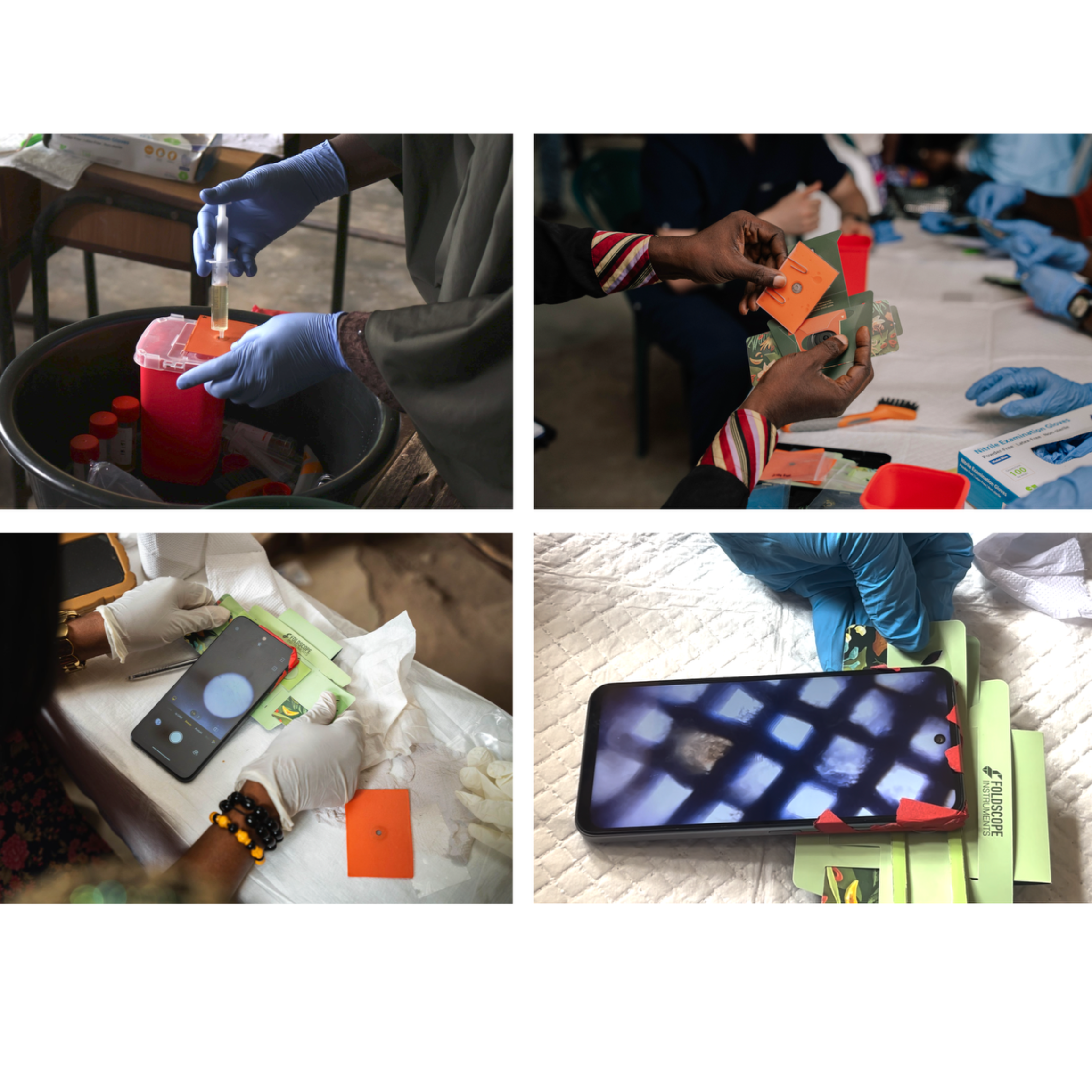
To use: draw urine into a syringe and expel slowly through the filtration membrane, trapping Schistosoma
eggs. Secure the coverslip with paperclips and assemble into the Foldscope by pulling down the backflap
and inserting the sample into the stage. Position a phone (via Foldscope magnetic pairing ring) or eye
over the lens to view. To reuse, remove the SchistoFilter from the Foldscope, wash the filtration
membrane with three syringe flushes of water from the reverse side of the central aperture, and dry with a
paper towel or alcohol wipe.
Development and bench validation of the SchistoFilter
We developed the SchistoFilter as a point-of-care filtration device to concentrate Schistosoma eggs from
urine without requiring electricity or laboratory consumables. Bench validation used schistosome eggs
previously isolated from rodent models of schistosomiasis. By suspending 50–200 eggs in 10 mL of
water, drawing the liquid into a syringe, and pushing the fluid through the membrane, the team was
consistently able to image trapped eggs on the SchistoFilter's stainless steel mesh using both a confocal
microscope (Revolve ECHO) and a Foldscope, confirming the device's efficacy across a range of egg
concentrations.
Sample size calculation
Sample size was calculated to estimate sensitivity and specificity within ±5 percentage points at 95%
confidence. Based on a 52% prevalence estimate from prior epidemiological surveys in Oyan River
communities [2,5,19] and an assumed sensitivity of 80%, the standard diagnostic accuracy formula
indicated 473 participants were needed. Applying a finite population correction for the study communities
(population ≈1,600), the adjusted target was 365 participants across five communities. We recruited 418
participants to account for potential dropout; 237 completed the full diagnostic protocol including CHEW
point-of-care assessment.
The standard formula applied was: N = [Z² × Se × (1 − Se)] / [d² × P], where N is the minimum required
number of infected individuals, Z is the standard normal deviate at 95% confidence (1.96), Se is the
expected sensitivity (0.80), d is the desired precision (0.05), and P is the estimated prevalence (0.52). This
yielded a target of 365 participants.
Additional participants and CHEW recruitment
Following documented verbal consent, CHEWs participated in eight days of combined training and data
collection. Six CHEWs participated for all eight days; one began on day four and another withdrew after
day four due to external commitments.
Laboratory scientists recruited from the Federal University of Agriculture, Abeokuta, and the Ministry of
Health conducted reference standard microscopy at field sites. Following field data collection, one
participating laboratory scientist trained by the research team subsequently cascade-trained a team of
students to carry out blinded secondary analysis of stored field samples using the Foldscope with gold
standard (SterliTech) filtration.The research team collaborated closely with officials from the local and state Ministries of Health
throughout. Recruited members from the Department of Neglected Tropical Disease provided clinical
oversight and assisted with community sensitization but were not directly involved in data collection or
analysis. At the conclusion of the study, these individuals were recruited and consented as key informant
interview participants.
Participant compensation
Community member participants did not receive the results of their reference standard screening;
however, 404/418 participants were administered praziquantel in an age- and height-appropriate dose by
the resident Ministry of Health doctor as preventive chemotherapy, in line with MDA protocol.
Community members also received approximately $1.50 USD (₦2,000) and a pack of snacks and
beverages for their participation. Eight CHEWs received a total of $140 USD (₦200,000). Laboratory
scientists were compensated $229 USD (₦320,000). Ministry of Health counterparts received between
$140–229 USD (₦200,000–320,000) for their clinical oversight, assistance, and interview participation.
Training of the CHEWs
Training sessions emphasized technical use of the tools and morphological identification of parasite eggs
by size, color, and shape. CHEWs were considered ready to assess clinical samples once they could
successfully identify eggs on glass slides and correctly operate each Foldscope component (focus ramp,
light module orientation, and slide loading). Urine filtering and SchistoFilter assembly were demonstrated
with a clinical sample aliquot before the study period began. Sample size for the CHEW validation subset
is limited to n=237 due to the timing and practical constraints of operating within a small schoolhouse
transient clinic.
Detailed sample collection and preparation
Urine samples were collected at midday (10:00–14:00). Each participant provided a minimum of 30 mL
of urine into a sterile container (CLINSAM 90 mL sterile urine collection cups). Each sample then
underwent two parallel filtration processes: (1) conventional gold standard urine filtration and (2)
SchistoFilter filtration.
For the gold standard technique, a 10 mL aliquot was drawn into a Luer-lock syringe (BH Supplies
BH110L) and expelled through a 20 µm polycarbonate membrane filter (Sterlitech) in a filter holder
(Sterlitech). The membrane was placed on a glass slide and examined under a compound microscope
(AmScope B120, 10–40× objectives) by an Ogun State Ministry of Health NTD laboratory scientist. The
whole slide was scanned and total egg counts recorded as eggs per 10 mL urine on a de-identified paper
chart. Any sample with at least one identified S. haematobium egg was classified positive; samples with
no identified eggs were classified negative. After counting, a drop of 10% formalin was added to each
filter, covered with a glass coverslip, and sealed with clear nail polish for long-term ambient storage.
For the SchistoFilter condition, a separate 10 mL aliquot from the same collection cup was expelled
through the SchistoFilter membrane by a trained CHEW. The filter was inserted into a Foldscope and
examined via a Tecno Pop 10 smartphone attachment. Egg counts were recorded using the same
classification criteria. After visualization, the SchistoFilter was washed in two sequential bowls of sterile
sachet water, dried on clean paper towels, and prepared for reuse.
Foldscope-based smartphone imagingDuring a preliminary meeting in Abeokuta with Ogun State clinicians, NTD coordinators, and research
collaborators, the team evaluated several locally available smartphones to identify a cost-effective option
for image capture. The Tecno Pop 10, widely owned by collaborators, provided the best balance of
camera quality and affordability. Two used Tecno Pop 10 phones were purchased from a local
marketplace for field use. Tape was applied to each phone to secure the Foldscope magnetic pairing ring
and ensure stable alignment.
Sources & Appendix
What is your age at your last birthday? ____________________________________
What is your gender? a. Male b. Female
What is your highest level of education? a. No formal education b. Primary school (completed/not completed) c. Secondary school (completed/not completed) d. Tertiary education (completed/not completed)
What is your primary occupation? a. Farming b. Fishing c. Artisan d. Civil servant e. Student f. Housewife g. Other (please specify): ___________________________________________________________
How many people live in your household? ___________________________________________________________
What is your estimated monthly household income? _______________________
Do you own your house? a. Yes b. No
Do you own land? a. Yes b. No II. Knowledge about Schistosomiasis
Have you ever heard about schistosomiasis (or its local name, e.g., "Atosi Aja" in Southwest Nigeria for urinary schistosomiasis)? a. Yes b. No (If yes, proceed to Q10. If no, skip to Section III).
What are some of the signs or symptoms of schistosomiasis that you are aware of? (Select all that apply) a. Blood in urine (hematuria) b. Abdominal pain c. Diarrhea d. Blood in stool e. Fever f. Fatigue/Malaise g. Pain during urination (dysuria) h. Itching after water contact i. Abnormal vaginal discharge (for females) j. Pain during/after coitus (for females) k. Sores on private parts l. Swollen abdomen (ascites) m. Don't know n. Other (please specify): ___________________________________________________________
How do people get infected with schistosomiasis? (Select all that apply) a. Swimming in rivers/open water b. Washing clothes in open water c. Bathing in open water d. Drinking dirty water e. Mosquito bite f. Contact with worms g. Through spiritual forces/curses h. Don't know i. Other (please specify): ___________________________________________________________
Do you believe schistosomiasis is treatable with drugs? a. Yes b. No c. Don't know
What problems can schistosomiasis cause if not treated? (Open-ended, then categorize responses): ___________________________________________________________
Where did you get information about schistosomiasis from? (Select all that apply) a. Health facility/health workers b. Community health workers/volunteers c. Friends/family d. School e. Radio f. Television g. Internet h. Traditional healers i. Unspecified sources j. Other (please specify): ___________________________________________________________ III. Attitudes towards Schistosomiasis Prevention and Control
How concerned are you about schistosomiasis affecting your household/community? a. Very concerned b. Somewhat concerned c. Not concerned
Do you believe that schistosomiasis can be prevented? a. Yes b. No c. Don't know
What do you think are the most effective ways to prevent schistosomiasis? (Open-ended, then categorize responses): ___________________________________________________________
How willing are you to participate in community programs aimed at controlling schistosomiasis? a. Very willing b. Somewhat willing c. Not willing IV. Practices related to Water Contact and Hygiene
What are your main sources of drinking water? (Select all that apply) a. Piped water (tap) b. Borehole/Well (protected) c. Unprotected well d. River/Stream e. Dam/Lake f. Rainwater g. Packaged/Sachet water h. Other (please specify): ___________________________________________________________
Do you treat your drinking water? a. Yes (boil, filter, add chlorine) b. No
What are your main sources of water for domestic activities (bathing, washing clothes, cooking, etc.)? (Select all that apply) a. Piped water (tap) b. Borehole/Well (protected) c. Unprotected well d. River/Stream e. Dam/Lake f. Rainwater g. Other (please specify): ___________________________________________________________
How often do you or members of your household come into contact with open freshwater bodies (rivers, streams, lakes, ponds)? a. Daily b. Several times a week c. Weekly d. Monthly e. Rarely/Never
What activities do you primarily engage in when in contact with open freshwater bodies? (Select all that apply) a. Bathing b. Washing clothes c. Fishing d. Farming/irrigation e. Swimming f. Recreation g. Fetching water h. Other (please specify): ___________________________________________________________
What is the main place you go to urinate? a. Toilet/Latrine b. Bush c. Open freshwater d. Other (please specify): ___________________________________________________________
What is the main place you go to defecate? a. Toilet/Latrine b. Bush c. Open freshwater d. Other (please specify): ___________________________________________________________
How close do you currently live to a source of fresh water (river or lake)? a. Less than 5 minutes' walk b. 5-10 minutes' walk c. 11-20 minutes' walk d. 21-30 minutes' walk e. More than 30 minutes' walk V. Healthcare Access and Treatment Practices
Have you or any member of your household ever been diagnosed with schistosomiasis? a. Yes b. No c. Don't know
If yes, did you receive treatment for schistosomiasis? a. Yes b. No
Where did you get the treatment from? (Select all that apply) a. Health facility b. Community drug distribution program (Mass Drug Administration) c. Local shop/pharmacy d. Traditional healer e. School f. Other (please specify): ___________________________________________________________
Did you complete the full course of treatment as advised? a. Yes b. No (If no) What were the reasons for not completing the treatment? (e.g., side effects, cost, feeling better, forgetting): ___________________________________________________________
Are schistosomiasis drugs readily accessible in this community? a. Yes b. No c. Don't know
What barriers do you face in accessing healthcare services for schistosomiasis? (Select all that apply) a. Distance to health facility b. Cost of treatment c. Lack of transportation d. Lack of knowledge about where to go e. Long waiting times f. Poor quality of services g. Cultural beliefs/preference for traditional medicine h. Fear of side effects i. Stigma j. Other (please specify): ___________________________________________________________
Has there been any schistosomiasis control programs or health education campaigns in your community? a. Yes b. No
Have you participated in any schistosomiasis control programs or health education campaigns in your community? a. Yes b. No 2. CHEW/CLINICIAN IN-DEPTH STRUCTURED INTERVIEW GUIDE IN-DEPTH INTERVIEW GUIDE: FEASIBILITY AND ACCEPTABILITY OF USING THE FOLDSCOPE IN THE DIAGNOSIS OF SCHISTOSOMIASIS AMONG CHEW’s AND CLINICIANSIntroduction:"Good morning/afternoon. Thank you for participating in this discussion. We are interested in your experiences and opinions about diagnosing schistosomiasis in your communities. We want to gather your thoughts on a new diagnostic approach that involves a urine microfilter and a small, portable microscope called the 'Foldscope.' Your honest insights are valuable to us, and there are no right or wrong answers."
Current Diagnostic Practices and Challenges
Can you describe the steps you currently take when a community member presents with symptoms suggestive of schistosomiasis? How do you typically confirm a diagnosis?
Can you describe the steps you currently take in community diagnosis of schistosomiasis?
What are the main challenges you encounter with the current diagnostic methods for schistosomiasis in your rural setting? Probe particularly in terms of availability of facilities for testing, accessibility, time, cost, or required skills.
From your experience, what are the most common reasons why suspected schistosomiasis cases might not get a confirmed diagnosis in your community?
How do community members currently react to or perceive the existing diagnostic procedures for schistosomiasis?
Feasibility of Microfluidics and Foldscope use in Schistosomiasis Diagnosis
We are exploring a new diagnostic approach – The Foldscope which you have used in the past few weeks to detect eggs in urine samples in the diagnosis of schistosomiasis. Can you describe the methods you used during test? Probe; How urine was collected, how the urine sample was prepared for viewing and how the Foldscope was used.
What are your initial thoughts or feelings about the Foldscope?
Please comment on the ease of use of the Foldscope and your level of confidence with using the foldscope. Probe on how easy or difficult to: assemble and use the Foldscope, hold the Foldscope steady and focus on the sample while observing for parasite eggs, especially in a community setting, identify specific schistosomiasis eggs through the Foldscope, considering their size and appearance.
Regarding preparing the urine specimen for viewing on the microfilter, how easy or difficult do you think it is to: a. transfer urine into the microfilter b. Perform the filtration process using this microfilter? c. Handle and dispose of the used microfilters safely and hygienically in your setting?
Tell us about the training you received on using the foldscope. Was it easy to understand how to use the foldscope? What aspects of the training were you satisfied with and what aspects could be improved upon.
What kind of training do you believe would be essential to ensure community health extension workers and clinicians confidently and accurately use both the urine microfilter and the Foldscope? What should be the content of such training. Under what conditions should such training be held.
What potential advantages do you see in using this microfilter and Foldscope combination in your daily work, especially compared to current methods?
What potential disadvantages or concerns come to mind when you consider using these devices for diagnosis?
What potential advantages do you see in using this microfilter and Foldscope combination in field settings?
What potential disadvantages do you see in using this microfilter and Foldscope combination in field settings?
How would you ensure the cleanliness and prevention of contamination when using these microfilters in the field?
How durable do you think a device like the Foldscope would be in the typical field conditions you work in.
Acceptability of Foldscope use in Schistosomiasis Diagnosis
Do you have any concerns about the accuracy of results obtained using the Foldscope? Please explain.
Would you be able to rely on a diagnosis made using a Foldscope for treating schistosomiasis cases? Please explain.
What do you feel regarding the acceptability of community members or your patients regarding diagnosis of schistosomiasis using the Foldscope? Please explain.
How do you envision this new diagnostic procedure fitting into your current work routine and existing schistosomiasis control activities, including Mass Drug Administration (MDA)?
What logistical support would you require to successfully implement this combined diagnostic approach in your community (e.g., consistent supply of devices and consumables, dedicated storage, regular supervision, technical support)?
Who do you think should be responsible for providing ongoing support and quality control for this diagnostic method at the community level? Perceived Impact, Challenges, and Recommendations
How might the use of the urine microfilter and Foldscope impact the early detection and management of schistosomiasis cases in your community?
Do you believe this approach could help identify cases that are currently missed or misdiagnosed due to limitations of existing methods? Please explain.
What are the biggest challenges you foresee in widely implementing this diagnostic approach across other rural communities in Ogun State?
What steps or support would be most important to ensure the successful and sustainable adoption of the urine microfilter and Foldscope by CHEWs and clinicians in the long term?
Is there anything else you would like to share about the feasibility or acceptability of using a urine microfilter and Foldscope for schistosomiasis diagnosis, or any other relevant insights from your experience as a CHEW? 3. KEY INFORMANT INTERVIEW GUIDEKEY-INFORMANT INTERVIEW GUIDE FOR KEY STAKEHOLDERSIntroduction:"Good morning/afternoon. Thank you for taking the time to participate in this discussion. We are conducting a study to understand the current challenges in schistosomiasis diagnosis and control in your LGA, and to gather your expert opinions on a novel, low-cost diagnostic tool called the 'Foldscope.' Your insights as health officials are crucial for informing future health interventions. Please feel free to share your honest perspectives."I. Current Schistosomiasis Control Landscape in the LGA
From your vantage point as a LGA/State health official, how would you describe the current burden of schistosomiasis in this LGA?
What are the existing strategies and programs for schistosomiasis control and elimination within your LGA? Probe on: Mass Drug Administration - MDA, health education, WASH interventions)
What are the primary diagnostic methods currently utilized for schistosomiasis within the LGA's health facilities and at the community level?
What are the most significant operational and resource challenges you face in diagnosing and managing schistosomiasis cases within the LGA? II. Initial Perceptions and Acceptability of the Foldscope
We are exploring the potential of a new diagnostic device, the 'Foldscope.' It's a very low-cost, paper-based microscope that can be assembled quickly and used with filtered urine samples to identify schistosomiasis eggs directly in the field and in the PHCs. What are your initial thoughts or impressions about such a device, particularly given its portability and low cost? Probe further on use in field settings and in clinic settings.
As a key health stakeholder in the LGA/State, what potential advantages do you see in having such a device available for schistosomiasis diagnosis?
Conversely, what potential disadvantages or concerns immediately come to mind regarding the use of the Foldscope at the LGA or community level and in PHCs/clinic settings?
How acceptable do you believe the Foldscope would be to the various health cadres within your LGA (e.g., medical doctors, nurses, CHEWs, laboratory technicians)? Please explain your reasoning.
From a public health standpoint, how do you anticipate the community would perceive and accept a diagnostic approach using a simple, portable device like the Foldscope, especially if it's used by community-level health workers? III. Feasibility for LGA-Wide Implementation (Operational & Systemic)
Considering the health system structure in your LGA/in Ogun State, how feasible would it be to integrate the Foldscope into existing schistosomiasis diagnostic and control programs?
What human resources (e.g., CHEWs, lab technicians) in your LGA do you believe would be best suited to use the Foldscope, and what would be the implications for their workload and existing responsibilities?
What level of training and supervision would be required to ensure accurate and consistent use of the Foldscope across the LGA? Who would be responsible for delivering this training?
From a supply chain perspective, what logistical considerations would be critical for the effective procurement, distribution, and maintenance of Foldscopes and associated consumables (e.g., urine collection cups, filters, slides)?
What existing infrastructure could support or hinder the widespread use of the Foldscope in your LGA?
How would you ensure the quality assurance and external validation of results obtained using the Foldscope at decentralized levels? IV. Policy, Resources, and Sustainability
What policy adjustments or guidelines might be necessary at the LGA or state level to formally recognize and integrate the Foldscope as a diagnostic tool for schistosomiasis?
How do you foresee the financial implications of adopting the Foldscope for LGA/State-wide use? What potential funding sources (e.g., government budget, donor funds, community contributions) could support its rollout and long-term sustainability?
What existing monitoring and evaluation frameworks could be adapted or developed to track the performance and impact of Foldscope implementation in the LGA/State?
How might the use of the Foldscope influence current data collection and reporting mechanisms for schistosomiasis in the LGA/state?
In your opinion, what is the long-term potential for the Foldscope to contribute to the sustained control and eventual elimination of schistosomiasis in this LGA? V. Challenges, Mitigating Strategies, and Recommendations
What major barriers or challenges do you anticipate in scaling up the use of the Foldscope from a pilot phase to LGA/state-wide implementation? (e.g., resistance to change, lack of sustained funding, technical issues, community trust).
What strategies or interventions would you recommend to mitigate these anticipated challenges?
What role do you see for inter-sectoral collaboration (e.g., WASH, education, community leaders) in supporting the successful adoption of the Foldscope?
Based on this discussion, what specific recommendations would you offer to the developers and implementers of the Foldscope for its successful deployment in rural LGAs like yours?
Is there anything else you would like to add regarding new diagnostic tools for schistosomiasis or the health system in your LGA/State?
Instructions & IMAGES
Foldscope Introduction Video with Manu Prakash: (A favorite among many): https://www.youtube.com/watch?v=ky-cqSI5mwE
SchistoFilter Introduction Video:
SCHISTOFILTER CONSTRUCTION:
The SchistoFilter device consists of four primary components assembled in a layered configuration. The coverslip layer (60 mm × 50 mm) was fabricated from 0.5 mm thick clear PETG sheet (McMaster-Carr, part #4076N11). The base layer (65 mm × 55 mm) was cut from 0.25 mm thick clear, scratch- and UV-resistant acrylic film (McMaster-Carr, part #85815K211) with a 5 mm diameter aperture positioned centrally.
The filtration membrane was constructed by excising an 8 mm diameter circle from a 38 mm circular stainless steel filter gasket containing 550-mesh (25-micron pore size) 304 stainless steel screen (USA Lab). The circular mesh segment was affixed to the base layer using two-part epoxy adhesive (Seal-All), positioned to completely occlude the 5 mm aperture, and cured for 24 hours at room temperature.
To complete assembly, a 65 mm × 55 mm vinyl adhesive backing layer (Cricut Smart Vinyl, permanent) with a central 5 mm aperture was applied to the reverse side of the base, creating a sandwich configuration with the metal mesh. This design produced a 3 mm overlap between the 8 mm mesh perimeter and the 5 mm vinyl aperture on all edges, securing the filtration membrane and creating a sealed sample chamber.
DEVELOPMENT AND TESTING OF THE SCHISTOFILTER:
We developed a point-of-care filtration device to concentrate Schistosoma eggs from urine samples that does not require electricity or laboratory consumables. To use, urine is filtered through the mesh with a syringe, trapping Schistosoma eggs. A plastic coverslip is then secured with paperclips, and the sample is viewed under the Foldscope. Scissors or a razor blade, laminate sheets, and a hole puncher can be used to construct the device from basic supplies.
See the next pages for graphical summary, side by side between foldscope and standard microscope, and field images from use in real scenarios in rural Ogun State, Nigeria.
Below is how the eggs are trapped in the Schistofilter:
STILL NEEDED:
Visual instructions on how to use / graphics for reference on the outside of the waste cup, instructions are cyclical, can wrap-around the waste cup
Box to fit all kit components
Cleaning instructions diagram
Protocol references
Ezeh et al., 2019; Archer et al., 2024; Faust et al., 2021; Mthethwa et al., 2015; WHO, 2023; Chatterji et al., 2024; Van et al., 2020; Dawaki et al., 2015; Ephraim et al., 2015; Oluwole et al., 2022; Xiao et al., 2016; Akinwale, 2010; Ekpo, 2012.
Acknowledgements
Collaborators/Co-Investigators
Institutional Affiliation: Health In Your Hands Team: https://healthinyourhands.my.canva.site/our-leadership
Dr. Olubodun (Senior Investigator), Dr. Adebayo, Dr. Adebayo - Federal Medical Center Abeokuta, Ogun State; Prof Ekpo - Federal University of Agriculture, Abeokuta (FUNAAB), Nigeria; Dr. Oluwole - Sightsavers; Dr. Adelakun - Oyo State College of Agriculture and Technology, Igboora, Nigeria; Dr. Mogaji - Federal University Oye-Ekiti, Nigeria; Dr. Stafford, Dr. Lee, Dr. Tebo, Dr. Darkoh, Dr. Hwang - UTHealth Science Center at Houston; Dr. Cybulski, Dr. Prakash - Stanford University, Foldscope Instruments; Dr. Morgan Jibowu - Baylor College of Medicine. In collaboration with Dr. Soneye Ismayilat, the Neglected Tropical Disease Coordinator of Ogun State.
We want to thank the Community Health Workers of Odeda and Abeokuta North for being co-Investigators on this project, and all the Ministry of Health leaders and staff who helped make this possible.