Jan 14, 2022
Protocol 3D Gait Analysis using Treadmill Approach (CAREN) - MUMC+
- Rachel Senden1,
- Rik Marcellis1,
- Paul Willems2,
- Jeroen Vermeulen3,
- Adhiambo Witlox4,
- Kenneth Meijer2
- 1Department of Physical Therapy, MUMC+, The Netherlands;
- 2Department of Nutrition and Movement Sciences, NUTRIM School of Nutrition and Translational Research in Metabolism, MUMC+, The Netherlands;
- 3Department of Neurology, MUMC+, The Netherlands;
- 4Department of Orthopaedic Surgery, MUMC+, The Netherlands
- MUMC


Protocol Citation: Rachel Senden, Rik Marcellis, Paul Willems, Jeroen Vermeulen, Adhiambo Witlox, Kenneth Meijer 2022. Protocol 3D Gait Analysis using Treadmill Approach (CAREN) - MUMC+. protocols.io https://dx.doi.org/10.17504/protocols.io.b2brqam6
License: This is an open access protocol distributed under the terms of the Creative Commons Attribution License, which permits unrestricted use, distribution, and reproduction in any medium, provided the original author and source are credited
Protocol status: Working
We use this protocol and it’s working
Created: November 24, 2021
Last Modified: January 14, 2022
Protocol Integer ID: 55377
Keywords: 3D gait analysis, Caren, Treadmill approach, Human body model-II, virtual environment, protocol 3d gait analysis, based 3d gait analysis, using treadmill approach, treadmill approach, step, caren, protocol
Abstract
This protocol describes the steps that are conducted when performing a treadmill based 3D gait analysis at MUMC+.
Materials
Computer Assisted Rehabilitation ENvironment (CAREN, Motek Medical, BV), comprising:
An instrumented dual-belt treadmill (force plates, 1000Hz) build on 6 degrees of freedom motion platform
Twelve motion capture cameras (100 Hz, Bonita, Vicon Motion Systems, Oxford, UK) surrounding the treadmill, and motion capture software (Nexus 2.7)
Three 2D video cameras
A 180° semi-cylindric screen in front of the treadmill were a virtual reality environment is projected on
D-Flow control software
Marker set (26 markers) according the Lower limb Human Body Model (HBM-||)
Matlab (R2016a, mathworks, Natick, MA, USA) and custom made algorithms to calculate gait parameters
Description 3D gait analysis set-up
The treadmill approach for 3D gait analysis involves walking at the Computer Assisted Rehabilitation Environment (CAREN, Motek Medical BV, Amsterdam, The Netherlands).
The CAREN is a rehabilitation system, using an instrumented dual belt treadmill for gait and balance training
and assessments. A large screen is used to provide real-time biofeedback to provide the subject with an interactive environment.
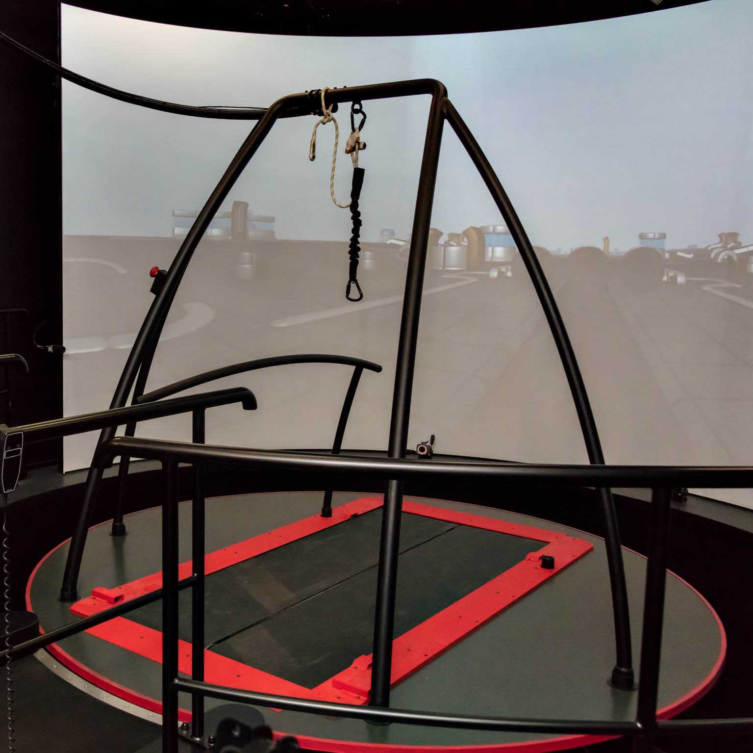
The CAREN consists of an instrumented dual-belt treadmill (force plates 1000Hz) with PC to control, on a 6 degrees of freedom motion platform. It includes 12 motion capture cameras (100 Hz, Bonita, Vicon Motion Systems, Oxford, UK) and motion capture software to collect 3D data of human movement, three 2D video cameras and an immersive Virtual Reality (industrial) environment that is projected on a 180° semi-cylindric screen, providing visual and/or auditory feedback. The hardware components (treadmill speed, 6 DoF motion, visual feedback and auditory feedback) are controlled by Motek software, specifically D-Flow control software.
Figure: The Caren system comprising a dual belt treadmill, build on a movable platform, surrounded by a 180° semi-cylindric screen where a virtual environment is projected on.
Preparation system
Before the subject enters the lab, the operator sets up the Caren system which includes:
- the configuration of the Vicon system hardware
- setting-up the D-flow software
- calibrate the Vicon system
- calibrate the forceplates
- the preparation of a database in Nexus 2.8.1 and Dflow
These steps are fully described in the CAREN user manual (Motek Medical BV, December 8, 2021).
Subject enters lab
After the subject has entered the lab, the Caren system is introduced to the subject by the operator.
In addition, the operator gives a brief explanation of the measurement that is performed to the subject (and parents/guardians in case of children < 17 years),
Subject preparation
1. The subject is asked to change clothes (underwear is advised, but tight, non-reflective sportswear is also allowed)
2. The subject gets standard gymnastic shoes from the researcher/operator.
3. Twenty six reflective markers are attached directly onto the skin at specific bony landmarks according to the lower limb Human Body Model2 plus Trunk (HBM-II). The figure belows shows the exact marker position, which are also described in the HMB2-Reference Manual HBM-gait (Motek Medical, August 27, 2018, p.9-10).
Markers are placed by two experienced examiners (RS, RM).
Figure: Front, side and rear view of the marker set used in HBM-||. Green markers are anatomical markers used to define the skeleton during initialization.
4. The subject is put on a safety harness.
5. The person is guided on the caren system by the operator and is fastened to an overhead frame on the Caren.
6. The operator checks that the harness is properly fitted, and then leaves the belt.
Calibration
Static initialization of the model is done by recording an initialization pose. In addition, a functional hip and knee joint calibration is performed. For all the calibration steps, the medial markers (knee and ankle) are used.
Labeling calibration
A standing static and dynamic subject calibration is conducted and recorded in Nexus 2.8.1.
The subject has to stand still for 5 seconds in T-pose: feet at shoulder width and in parallel to the Z-axis of the coordinate system in D-flow (or Y-axis in Nexus), each foot positioned at one belt (and thus force plate) and arms spread at shoulder height. After 5 seconds, the treadmill starts to run at a random slow speed, the subject starts walking and a few steps are recorded.
The calibration trial is directly processed in Nexus 2.8.1.
HBM-II model calibration
1. A functional knee calibration is performed and recorded in Dflow and Nexus 2.8.1:
The subject stands upright, thereafter the subject has to flex and extend the knee about five times through a range of approximately 0 to 45° of flexion. This is performed for each limb individually. In this way the knee axis can be estimated.
Detailed description in the HMB2-Reference Manual HBM-gait (Motek Medical, August 27, 2018, p. 11-12).
2. A functional hip calibration is performed and recorded in Dflow and Nexus 2.8.1:
The subject stands upright, thereafter the subject has to perform 5 different movements: 1) hip flexion, then back to neutral, 2) Combination of hip flexion and abduction, then back to neutral, 3) hip abduction, then back to neutral, 4) combination of hip extension and abduction, then back to neutral and finally 5) a full circumduction movement. This is performed for each leg individually. In this way the joint center of the hip is determined. Detailed description can be found in the HMB2-Reference Manual HBM-gait (Motek Medical, August 27, 2018, p. 12)
3. The static calibration is done again (step 1), but now the recording is done in the Dflow software.
After these calibration steps, the medial markers are removed.
Body weight of the subject and the knee and ankle width for left and right leg individually are determined by the Dflow software.
Determine comfortable walking speed
The RAMP protocol is used to determine the comfortable walking speed of the subject:
the subject starts walking at 0.5m/s. Every second, the speed is increased with 0.01m/s untill the subject states that his/her comfortable walking speed is reached. This is repeated three times and the average is taken as the comfortable walking speed.
In some cases, comfortable walking speed is determined in advance during at least five overground walking trials, using two movement detection portals placed 4 meter apart.
Familiarisation period
To get used to the CAREN system, subjects walk for at least six minutes at comfortable walking speed (determined in step 6) at the system. The six minutes is based on the study of Meyer C et al. who demonstrated that six minutes of familiarisation is needed to adapt a normal gait pattern (Sci Rep. 2019; 9(1): 5232).
Measurement
Subsequently, the measurements are started and recordings are made in Dflow and Nexus 2.8.1.
The measurements are performed while walking at different walking speeds:
- walking at comfortable walking speed (measured in step 6 using the RAMP protocol)
- walking at slow walking speed, which is 30% slower than the comfortable speed
- walking at fast walking speed, which is 30% faster than the comfortable speed
- walking at fixed slow speed of 0.5 m/s
The sequence of these conditions varies between subjets and is randomly choosen by the operator.
For every condition, 250 steps (125 cycles) are recorded in Dflow and Nexus 2.8.1.
After completion of measurement
1. After the desired measurements have been recorded, the person is removed from the system by the operator.
2. The harness is removed by the operator
3. The markers are removed by the operator
4. The subject gets dressed
5. The subject leaves the lab
Data recording and processing
The raw data obtained by the D-flow software (.mox and .txt files) are used for analysis.
First the quality of the data is checked and good kinematic and kinetic steps are identified using custom made algorithms programmed in Matlab (R2016a, mathworks, Natick, MA, USA).
For instance, strides where both feet are placed on one force plate are removed for kinetic steps, gaps in marker detection (>30 samples) are identified and these strides are removed (gaps <30 samples are interpolated using cubic spline fitting), the range of interest for analysis is determined.
Step detection (heelstrikes and toe off) is done, based on a combination of heel marker kinematics and force plate data (threshold of 50N) as described by JA Zeni et al. (JA Zeni, JG Richards, JS Higginson. Gait Posture 2008; 27(4):710-4.).
Afterwards, gait parameters are calculated using custom made algorithms programmed in matlab (R2016a, Mathworks, natick, MA, USA). These calculations rely on published formulas and principles, producing:
- spatiotemporal parameters: e.g. walking speed (m/s), step length (cm) and cadence (steps/min)
- 3D kinematics: joint angles in sagittal, frontal and transversal plane
- 3D kinetics: joint moments in sagittal, frontal, transversal plane and anterior-posterior, medio-lateral and vertical ground reaction forces.
The calculation of spatiotemporal parameters is based on all selected valid steps. The calculation of kinematics and kinetics is based on the valid kinematic (criteria based on van der Krogt, VUMC) and kinetic steps respectively.